JSM Clinical Cytology and Pathology
-
Case ReportPrimary Hydatid Cyst in the Subcutaneous Tissues of the Neck in the Sub-Occipital Area: An Unusual Site of Hydatid CystManucher Aghajanzadeh1*, Sharokh Yousefzadeh Jabock2, Hossein Hemmati1, Mohammad Sadegh Esmaili Delshad2, and Piroze Samidost11Department of Thoracic and General Surgery, Guilan University of Medical Sciences, Iran
2Department of Neurosurgery, Guilan University of Medical Sciences, Iran*Corresponding author: Manucher Aghajanzadeh, Department of Thoracic and General Surgery, Guilan University of Medical Sciences, Rasht, Iran, Tel: +98-9113-311-881; Email: maghajanzadeh2003@yahoo.comSubmitted: 08 April 2015; Accepted: 30 April 2015; Published: 04 May 2015 -
Introduction: Hydatid cyst is a condition commonly affecting liver and lungs caused most commonly by Echinococcus granulosus where as musculoskeletal or subcutaneous hydatidosis is very rare. and Usually are secondary and resulting from the spread of cysts from other organs, either spontaneously rupture or after spreading from operations for hydatidosis in other regions.Case Presentation: We present an unusual case of a primary hydatid cyst found in the subcutaneous scalp tissue under occipital region in a 36 year man. Clinical presentation of patient was a huge cystic mass. Ultrasound findings revealed a cyst lesion in the posterior aspect of neck under the occipital region. MRI show a cystic lesion. We removed the entire part of cyst lesion surgically. Macroscopic and microscopic histopathological examinations confirmed the diagnosis of subcutaneous hydatid cyst.Conclusion: In regions where hydatid disease is endemic, a cystic lesion in any part of the body should be considered a hydatid cyst. The best treatment is surgery and total evacuation of the cyst elements without any spillage of cyst contained and postoperative albendasol therapy.
-
Hydatid disease is a parasitic infection caused by the tapeworm Echinococcus granulosus. This parasitic diseaseis a significant public health concern in endemic country [1]. Hydatid disease is endemic in the Middle East, India, Africa, South America, New Zealand, Iran, Australia, Iran, Turkey and Southern Europe [1-4].Life cycle of Echinococcus involves two hosts, one definitive carnivore host (dogs, cats and certain wild carnivores) and the other intermediate herbivore host (sheep, goats, swine, small rodents and other wild herbivores) [5]. Humans act as an accidental intermediate host is infected after ingesting viable oncosphere-containing eggs, which have been shed in the feces of the definitive host [2]. But the liver and lungs are the main locations [2,3]. Various parts of body may be involved with hydatid cyst but Hydatid cyst develops most frequently in the liver (65%), the lungs (25%), and the remaining 10% occurs in muscle, spleen, bones, kidneys, brain, eye, heart, subcutaneous and pancreas [2,6,7]. Occurrence of hydatid cyst is extremely rare in the head and neck region even in endemic areas wherehydatid cyst infestation is frequent. Only a few cases of hydatid cyst located in head and neck have been reported in the literature [5,7]. We will present our experience in treating a case of hydatid cyst located in the subcutaneous neck area, which is considered one of the few cases published due to the relative rarity of the disease in the fore mentioned anatomical location.A 36 year old man, who is not known to have any medical history, presented to the head, neck and maxillofacial Surgery department of Poorsina and Razi hospital, with a chief complaint of a posterior under occipital and neck swelling, noted about 4 month back. The general condition of the patient was good; he had no history of fever or weight loss. The patient lived almost in a city near the mountain. He reported that the swelling was gradually increasing in size with pain.Clinical examination revealed a well-defined, non-tender posterior and upperneck under occipital region swelling of about 8 cm in diameter, other part of neck and scalp was normal. Systemic examination of chest, abdomen and extremities was unremarkable, On the ultrasonography (USG), a complex, cystic lesion (10x4x5 mm) with septet and debris in the subcutaneous adipose tissue was detected. On CT-scs in sagittal and coronal view, a cystic lesion was presented in the base of skull posterior ofneck (Figures 1-3). Anti-faciola tests was negative. Upon the clinical diagnosis of the subcutaneous cyst, the patient underwent surgery in prone position under general anesthesia. After prep and derep and aspiration 30 CC clear fluid from cyst cavity (Figure 4). 30CC hypertonic salin was injected in the ruminant cavity. After 10 minuet cyst was opened, a white laminated membranes was exposed and removed intraoperative with presentation of laminated membrane (Figure 4), our diagnosis was hydatid cyst. The cavity was irrigated with providing-idon and was closed with a external drainage. Histopathological examination confirmed the diagnosis of the hydatid cyst. Serological tests were negative for the Echinococcal antigens. No pathology was seen onabdominal andother organs in the CT-scnnand USG. The patient was started on an oral albendazole (800mg/daly) treatment for 28 days with 14 day interval in three periods. On follow-up, no recurrence was observed.
-
 Figure 1: MRI of neck (T1 weighted, axial view) showing cystic lesion
the in sub-occipital area base of skull. View Figure
Figure 1: MRI of neck (T1 weighted, axial view) showing cystic lesion
the in sub-occipital area base of skull. View Figure
-
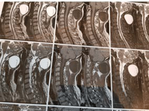 Figure 2: MRI of neck (T2 weighted, axial view) showing cystic lesion
in the neck just sub-occipital area. View Figure
Figure 2: MRI of neck (T2 weighted, axial view) showing cystic lesion
in the neck just sub-occipital area. View Figure
-
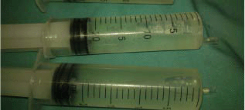 Figure 3: Showing intraoperative aspiration of cystic lesion fluid. View Figure
-
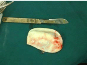 Figure 4: Showing Laminated membrane of cystic lesion, postoperative. View Figure
Hydatid disease is caused by the larval tapeworm of the genus Echinococcus granulosus, E.multilocularis. E.granulosus is the most common cause of hydatid disease [6]. the liver (50-70%) and lung (20-30%) are the most common organ which involve but may be found in any other organ of the body , including brain, heart and bones and adrenal gland (<10%) [7]. It can also affect the kidney, ureter, spleen, uterus, Fallopiantube, mesentery, pancreas, diaphragm and muscles and subcutaneous [8]. Muscular hydatid cysts may be primary, but may also occur secondarily when cysts spread from other areas, either spontaneously or after previous operations for hydatidosis in other regions of the body [8,9]. Hydatid cysts in soft tissues are an unusual presentation of hydatidosis [8]. Primary intramuscular hydatidosis is rare because the intramuscular growth of cysts is hindered by the muscle’s contractility, and by the lactic acid in the muscle. Parasitic cysts tend to grow around the muscles of the neck, trunk and roots of the limbs, perhaps because there is greater vascularization and less muscular activity in these regions [2,8]. They develop very slowly and act as space-occupying lesions, producing symptoms related to pressure on the surrounding tissues [5]. Other possible primary locations of the disease must be excluded by careful clinical and radiological examination of the patient, and these examinations must form the basis of the diagnosis. In particular, MRI images may identify various patterns of intramuscular hydatidosis such as its peripheral rim, peripheral edema, peripheral enhancement with gadolinium and multilocular cystic lesion (daughter cysts) [2,3,8]. In our case we used U&S and MRI for diagnosis. Percutaneous needle or Open biopsy in this solid and cystic lesion risk for spreading must be avoided [1,2]. Echinococcosis should be always suspected in differential diagnosis of cystic lesions in soft tissue until proven otherwise. Once the diagnosis is established, the surgeon should consider performing a radically [1,2,8].The treatment of choice is complete surgical excision of the cyst mass and thorough irrigation of the surrounding soft tissues with hypertonic saline to prevent recurrence [5,10]. Before the surrounding of cyst should de walling of with hypertonic saline solution.Administration of benzimidazole derivatives (albendasol) preoperatively and after surgery is advocated by some authors [2,8]. In this case we use albendazole (800mg/ daily) after surgery. The patient must be kept under clinical evaluation after surgery, for recurrence. Once the diagnosis is established, the surgeon should consider performing a radical procedure aiming in minimizing the possibility of a recurrence. The patient must be kept under clinical evaluation after surgery, for recurrence.In regions where hydatid disease is endemic, Echinococcosis should be always suspected in differential diagnosis of cystic lesions in soft tissue until proven otherwise. The best treatment is the total excision of the cyst with an intact wall. -
-
- Mohammad Sadegh Eamaeili Delshad, Manouchehr Aghajanzadeh, Hossein Hemmati, Siamakrimaz, Reza Shojaee. Complicated primary Hydatid Cyst in brachioradialis An extremely unusual site. MRJMMS. 2016; 4: 76-78.
- Manoucheher Aghajanzadeh, Mehdi Karimian, Zahra Sadat Segatoleslami, Shirin Manshori, Rassol Hassanzadeh, Tahereh Marasi. Primary Isolated Hydatid Cyst in Trapezes Muscle: A Extremely Rare Site. 2017; 5: 1-3.
- Reem Khalifa, Firas Nasser, Ahmed Elsetouhy, Ismail Farag. Case report: Hydatid cyst of the neck. A case report and literature review. Egypt J Ear Nose Throat Allied Sci. 2016; 17: 103-105.
- Metanat M, Sharifi-mood B, Sandoghi M, Alavi-Naini R. Osseous hydatid disease. Iran J Parasitol. 2008; 3: 60-64.
- Akal M, Kara M. Primary hydatid cyst of the posterior cervical triangle. J Laryngol Otol. 2002; 116: 153-155.
- Katilmis H, Ozturkcan S, Ozdemir I, Adadan I. Guvenc, Ozturan S. Primary hydatid cyst of the neck. Am J Otolaryngol. 2007; 28: 205-207.
- Goyal P, Ghosh S, Sehgal S, Panda I, Kumar A, Singh S. Primary Multilocular Hydatid Cyst of Neck with Unique Presentation: A Rare Case Report and Literature Review. Head Neck Pathol. 2014; 8: 334-338.
- Manouchehr Aghajanzadeh, Omid Mosafayi, Babak Karimi, Hossein Torabi, Mostafa Ziabari, Mahdi Pursafar. Isolatedextra Hepatic and Extra Pulmonary Hydatid Cyst: Report of 33 Rare cases. Ann Clin Pathol. 2018; 6: 1129.
- Combalia A, Sastre-Solsona S. Hydatid cyst of gluteus muscle. Two cases. Review of the literature. Joint Bone Spine. 2005; 72: 430-432.
- Sultana N, Hashim TK, Jan SY, Khan Z, Malik T, Shah W. Primary cervical hydatid cyst: a rare occurrence. Diagn Pathol. 2012; 7: 157.
Search Articles
-
Review Article
Screening for Depression among Medicyation Overuse Headache Patients and Treatment Could Be Useful for Improving their Quality of Life
Ljubisavljevic Srdjan1,2*#, Todorovic Stefan1# and Djokovic Filip1 | 2024-04-19- Short Communication
Why Multi-Drug Antiviral Therapy is Needed for COVID-19
Sibasish Dolai1 , Sabine Hazan2 , Christelle Pagonis1 , Sabrina Liu3 , Thomas J Borody1* and Robert R Clancy1 | 2024-04-11- Review Article
Elja: An R Package to Perform Environment-Wide Association Studies (Ewas / Envwas) Analysis
Marwan El Homsi1*and Isabella Annesi-Maesano1,2 | 2024-03-25- Case Report
Diagnosis of a Neck Mass in the Emergency Department
Schmidt S¹,²*, Lorenz KJ¹, Matthias C², Diekmeyer B2 and Müller G?,? | 2024-03-14- Case Study
The Impact of Neuropsychiatric Symptoms of Alzheimer’s Disease on Family Caregiver’s Distress
ATomasello L1,2*, Ranno1 , Raffaele2 , Laganà3 , Pitrone3 and Alibrandi4 | 2024-02-28News Feeds

Seth J. Worley, MD, FHRS, FACC
Director, Interventional Implant Program
Biography Paper Presentations
MedStar Heart & Vascular Institute,
Washington, DC, USACollaborations
Indexing


 © Copyright - JSM Central
 This work is licensed under a Creative Commons Attribution 4.0 International License.
- Short Communication