JSMC Nanotechnology and Nanomedicine
-
Review ArticlePolyethylene glycol – Indocyanine green Nanoparticles for Photodynamic Therapy TechniqueMohammadreza Saboktakin*Department of Nanomedicine, NanoBMat Company, Germany
*Corresponding author: Mohammadreza Saboktakin, NanoBMat Company, GmbH, Hamburg, Germany, Email: saboktakin123@gmail.comSubmitted: 13 May 2019; Accepted: 12 June 2019; Published: 15 June 2019 -
Nanoparticles formulated from biodegradable polymers such as polyethylene glycol (PEG) with molecular weight 6000 and 15,000 respectively are being extensively investigated as drug (Indocyanine green, ICG) delivery system due to their controlled release characteristics and biocompatibility. PEG nanoparticles for ICG delivery are mainly formulated by a routine technique using PVA as a stabilizer generating negatively charged particles and heterogeneous size distribution. The objective of the present study is to formulate cationic PEG nanoparticles with defined size and shape that can efficiently bind ICG. This technique to make cationic nanoparticles with very low size composed of biodegradable and biocompatible. PVA-chitosan blend was used to stabilize the PEG nanoparticles.Keywords: Nanoparticles; Polyethylene glycol; Indocyanine green
-
Nanoparticles (NPs) are synthetic materials with dimensions from one to hundreds of nanometers, and remarkable applications in biomedicine due to the unique way in which they interact with matter [1,2]. There are currently more than 35 US FDA-approved NPs often incorporating polyethylene glycol (PEG), with a larger number in preclinical studies for both imaging and therapy (Figure 1A) [1,3-9]. NPs have large payloads, stability, avidity, signal enhancement and the capacity for multiple, simultaneous applications owing to their unique size and high surface area: volume ratio [10]. While they are bigger than molecules and many proteins, yet smaller than cells, they behave differently to other therapies and imaging agents, affecting their in vivo applications. For example, in cancer tissue, NPs not only extravasate from the leaky tumor vasculature to a higher degree than healthy tissue, but also remain in the area by the enhanced permeability and retention (EPR) effect [11]. NPs lodged in the tumor can then perform signaling and/or therapy [10]. Despite these advantages, some fundamental challenges hamper NP deployment to the clinic. These include uptake by the reticuloendothelial system (RES), in which NPs are rapidly shuttled out of circulation to the liver, spleen or bone marrow, and nonspecific binding of NPs to nontargeted or nondiseased areas. Concerns about NP toxicity often arise because of this RES accumulation. Aggregation can lead to NP entrapment in the liver, lungs or elsewhere due to capillary occlusion [12]. The addition of PEG to the NP surface (PEGylation) can reduce many of these challenges (Figure 1B). PEG is a coiled polymer of repeating ethylene ether units with dynamic conformations (Figure 1C). In both drug-delivery and imaging applications, the addition of PEG to NPs reduces RES uptake and increases circulation time versus uncoated counterparts [13]. Aggregation decreases owing to passivated surfaces, and association with nontargeted serum and tissue proteins is diminished, resulting in so-called ‘stealth’ behavior. The PEG chains reduce the charge-based contact typical of proteins and small-molecule interactions. Solubility in buffer and serum increases due to the hydrophilic ethylene glycol repeats and the EPR effect is modulated due to NP size changes via addition of a PEG coat [14,15]. Due to these attributes, PEGylated NPs generally accumulate in the liver a half to a third of the amount of non- PEGylated NPs and demonstrate higher tumor accumulation versus background [16]. PEG is inexpensive, versatile and FDA approved for many applications [12]. All NPs contain at least two fundamental spatial components: the core and the corona that interact with the environment or solvent. While core/shell, core/multishell systems add further complexity, for example [18], all still possess an area in which NP interfaces with the solvent (Figure 1B). PEG chains modify this interface layer and increase circulation time. Circulation half-time (t½) describes blood pool residence and is the period over which the concentration of circulating NPs remains above 50% of the injected dose, analogous to a drug’s half-life [19]. NP efficacy requires sufficient t½ to not only reach the target, but also remain in the affected area (at concentrations sufficiently above background tissue) long enough for image capture or drug delivery. The RES system prevents site-specific accumulation because it removes the NPs from circulation, acting as a competitor to the intended target site [20]. In addition, the NPs must clear from the nontargeted area to produce imaging contrast or dosing efficiency. The ideal t½ is dependent on application. In imaging, 2-6 h is optimal for injection, accumulation at targeted site, clearance from nontargeted areas and data collection. The ideal circulation time for therapeutic NPs is longer (days) to allow repeated exposure to affected area. Unfortunately, this can also expose healthy organ systems to the drug and is the motivation for targeted NPs, as such systems preferentially accumulate in the diseased area. Approaches to measuring t½ vary with NP type. When labeled with radionuclides, g counting of either specific organ systems or blood aliquots determines NP circulation time. One limitation is dissociation of radionuclide from NPs; however, radioactivity measurements may always be carried out noninvasively [21].
-
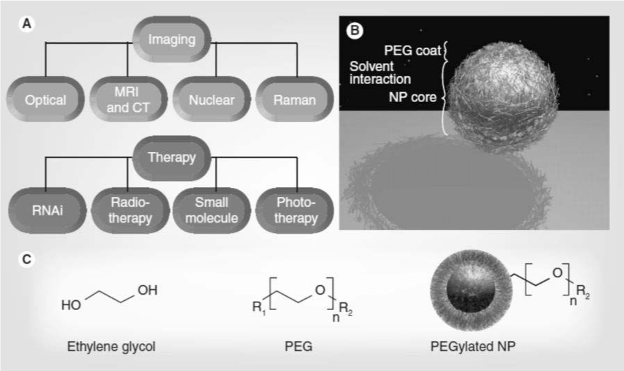 Figure 1: Nanoparticle applications of Polyethylene glycol. View Figure
Measurement of t½ via fluorescence, Raman, inductively coupled plasma or chromatography/mass spectrometry is very specific to the NP, but requires sequential sampling of the blood pool. The RES is an immune system component, utilizing circulating macrophages and monocytes, liver Kupffer cells and spleen and other lymphatic vessels to remove foreign material, such as bacteria and viruses, from the body [20]. Figure 2 illustrates how opsonin proteins associate with foreign bodies and coat its surface [22]. As bacteria and viruses have the same negative surface charge as phagocytic cells, opsonins are critical to reducing the charge repulsion between the two systems [13]. Next, phagocytic cells engulf the material and transport it to the liver or spleen for degradation and excretion (Figure 2 A3–A4). Additional phagocytic macrophages are permanently located in the liver. Known as Kupffer cells, these cells serve as a major filter for many types of NPs and are a major interference with long t½ [23]. The PEG polymer on a NP surface increases t½ by reducing this opsonization process (Figure 2B2), thus preventing recognition by monocytes and macrophages, allowing the NPs to remain in the blood pool [13,22]. Hydrophobic particles are also more vulnerable to the RES and hydrophilic PEG reduces these complications [22]. In addition to NP-RES interactions, poor t½ can also result from NP-NP interactions (i.e., aggregation). NPs aggregate primarily because the attraction between particles is stronger than the attraction for solvent [13,24]. NPs with a high surface energy have a greater tendency to aggregate as described by the Derjaguin-Landau- Verwey-Overbeek (DLVO) theory [25,26]. For spherical NPs, the interaction potential is related to the electrostatic repulsive potential and the van der Waals attraction potential [26]. PEG decreases the surface energy of NPs and minimizes van der Waals attraction [27-29]. Aggregation can be induced by solvents of high (>100mM) ionic strength (shielding of solvent from NP), highly concentrated solutions of NPs (less distance between the NPs), time from synthesis, or NP preparations with a very neutral (~±5 mV) zeta potential [30]. PEG decreases the amount of attraction between NPs by increasing the steric distance between them and increasing hydrophilicity via ether repeats forming hydrogen bonds with solvent. Other benefits to PEGylation include modifying the size of the particle. The reduced renal filtration of particles larger than 10 nm increases t½; however, at too large a size (>100 nm), liver uptake increases and EPR extravasation may decrease [31]. PEG modifies the NP flexibility and the NP can become ‘softer’ after PEGylation than the underlying material, influencing extravasation.-
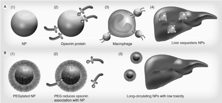 Figure 2: Polyethylene glycol prevents uptake by the reticuloedothelial system. View Figure
Prior to NP applications, PEG was used as a nontoxic, watersoluble dispersant/stabilizer. Also known as Carboxwax®, it is present in health and beauty aids, including laxatives, toothpastes and eye drops, and is an excipient in tablet formulations [32]. PEG stabilizes organ and blood donations. Early work with PEGylated NPs stemmed mostly from drug delivery [16,33-36]. One of the first reports on PEGylation was described by Davis and Abuchowski [37,38], where they covalently attached methoxy- PEGs (mPEGs) of 1900 and 5000 Da to bovine serum albumin and to liver catalase. Later, acrylic microspheres functionalized with PEG-modified human serum albumin increased t½ in vivo [39]. Li and colleagues found that 75-nm latex particles remained in rat circulation 40-times longer (half-life 20 min vs 13 h) when coated than uncoated with PEG larger than 5000 kDa [33]. Klibanov and Huang found that incorporation of dioleoyl N-(monomethoxy polyethyleneglycol succinyl) phosphotidylethanolamine (PEGPE) into posphatidylcholine: cholesterol liposomes (1:1) increased t½ from 30 min to 5 h without increasing leakage of the liposome interior [35]. In the mid- 1990s, Doxil® (liposomal delivery vehicle for doxorubicin) and oncospar (PEG-l- asparaginase) became the first FDA-approved NP therapeutics [40]. Doxil increases doxorubicin bioavailability nearly 90-fold at 1 week from injection of PEGylated liposomes versus free drug [41]. The use of PEG on the doxorubicin carrier yields a drug half-life of 72h with circulation half-life of 36h [42,43]. Later, Abraxane® was introduced as an albumin-functionalized NP for delivery of taxane without cremphor to enhance drug efficiency [44]. Thadakapally et al., developed a novel serum stable long circulating polymeric nanoparticles for curcumin with a modification to the well known and novel nanoparticle albumin bound technology. polyethylene glycolalbumin- curcumin nanoparticles were prepared using serum albumin and poly ethylene glycol using desolvation technique. Nanoparticles were characterized for encapsulation efficiency, particle size and surface morphology. Drug excipient compatibility was determined using fourier transform infrared spectroscopy. Physical state of the drug in the formulations was known by differential scanning colorimetry. In vitro release and solubility of the drug from nanoparticles were determined. In vivo Drug release, tissue uptake and kupffer cell uptake was determined with optimized nano formulation in rats after intravenous administration. Cell viability assay was determined using breast cancer cell line MD-MB-231. Entrapment efficiency for prepared nanoparticle was above 95%. The polyethylene glycol-albumin-curcumin nanoparticles exhibited an interesting release profile with small initial burst followed by slower and controlled release. Solubility of the drug from the formulation was increased. A sustained release of drug from nanoparticles was observed for 35 days in both in vitro and in vivo studies with the optimized formulation. Polyethylene glycol-albumincurcumin nanoparticles showed lesser liver and kupffer cell uptake as compared to that of curcumin-albumin nanoparticles suggesting the bestowment of stealthness to nanoparticles with pegylation. Also, the antiproliferative activity of polyethylene glycol-albumin- curcumin nanoparticle formulation was more as compared to native curcumin. Polyethylene glycol-albumincurcumin nanoparticles thus developed can be conveniently used in breast cancer with improved efficacy compared to conventional therapies and as an alternate to nanoparticle albumin bound technology which is used in producing Abraxane, albumin based breast cancer targeting nanoparticles of paclitaxel [45]. Photothermal therapy (PTT) and photodynamic therapy (PDT) are emerging physical tumor treatments utilizing near infrared (NIR) light-absorbing agents which lead to thermal ablation of cancer cells or generate highly reactive oxygen species (ROS) via photosensitizer to ablate tumors [46,47]. PTT and PDT possess several advantages, such as minimal invasion, high therapeutic efficacy, limited side-effects, selective localized treatment and reproducible properties [48,49], and hence have received much attention in recent years [50,51]. Until now, a variety of materials has been explored as PTT or PDT agents due to their high absorption in the tissue- transparent NIR wavelength range, including organic fluorescent dyes [52], gold nanorods [53], CuS nanoparticles (NPs) [54], polymer NPs [55], carbon nanomaterials [56], etc. [57,58]. However, fluorescent dyes may be removed rapidly from the systemic circulation and lack specificity to a tumor, and inorganic photothermal agents have potential long-term toxicity due to the difficulty of degrading in the body [59]. Therefore, exploiting biocompatible and targeted therapeutic nanoagents with enhanced photothermal conversion capability and ROS generation ability to amplify PTT and PDT treatments remains challenging. Indocyanine green (ICG) is a clinical infrared imaging agent approved by the U.S. Food and Drug Administration (FDA), and has been applied in optical imaging of human vasculature, tissue and cells due to its biocompatibility and unique optical properties [60]. Due to strong absorption at 780 nm, ICG can effectively convert absorbed NIR optical energy into heat for PTT [61], and produce ROS for PDT [62], under NIR laser irradiation [63]. Nevertheless, the application of ICG in tumor phototherapy is limited by its tendency to aggregate, rapid degradation in aqueous solution [64], poor photo-stability and non-specific binding to proteins [65]. To overcome those limitations, various nanoparticle delivery systems have been developed to encapsulate ICG [66]. Lv et al., used a mesoporous silica (mSiO2) matrix to load ICG molecules, and demonstrated that loaded ICG displayed a more enhanced photothermal effect than pure ICG [67]. ICG-loaded mesoporous silica NPs also could not only limit the degradation of ICG, but reach and stay at a tumor for a long period of time due to an enhanced permeability and retention (EPR) effect [68]. Hence, loading of ICG within targeting nanocarriers with high efficiency is shown to be an effective way to promote the application of ICG in PTT and PDT treatment.To improve the efficacy of PTT, polyethylene glycol (PEG) nanoparticles (NPs), indocyanine green (ICG) for NIR laserinduced PTT. ICG after administered intravenously will be readily bound with blood proteins and hence leads to only 2 ± 4 min of plasmatic half-life. Among various pharmaceutical polymers, PEG is one of the best defined biomaterials with FDA approval for drug encapsulation due to its biocompatibility, biodegradability, and controllability for drug release. -
-
- Wagner V, Dullaart A, Bock A, Zweck A. The emerging nanomedicine landscape. Nat Biotechnol. 2006; 24: 1211-1218.
- Kim BY, Rutka JT, Chan WC. Nanomedicine. N Eng J Med. 2010; 363: 2434-2443.
- Jacobson GB, Gonzalez-Gonzalez E, Spitler R, Shinde R, Leake D, Kaspar RL, et al. Biodegradable nanoparticles with sustained release of functional siRNA in skin. J Pharm Sci. 2010; 99: 4261-4266.
- O’Brien ME, Wigler N, Inbar M, Rosso R, Grischke E, Santoro A, et al. Reduced cardiotoxicity and comparable efficacy in a Phase III trial of pegylated liposomal doxorubicin HCl (CAELYX/doxil) versus conventional doxorubicin for first-line treatment of metastatic breast cancer. Ann Oncol. 2004; 15: 440-449.
- Davis ME, Zuckerman JE, Choi CH, Seligson D, Tolcher A, Alabi CA, et al. Evidence of RNAi in humans from systemically administered siRNA via targeted nanoparticles. Nature. 2010; 464: 1067-1070.
- Scranton R, Cincotta A. Bromocriptine – unique formulation of a dopamine agonist for the treatment of Type 2 diabetes. Expert Opin Pharmacother. 2010; 11: 269-279.
- Lim WT, Tan EH, Toh CK, Hee SW, Leong SS, Ang PC, et al. Phase I pharmacokinetic study of a weekly liposomal paclitaxel formulation (Genexol-PM) in patients with solid tumors. Ann Oncol. 2009; 21: 382-388.
- Winter PM, Morawski AM, Caruthers SD, Fuhrhop RW, Zhang H, Williams TA, et al. Molecular imaging of angiogenesis in early-stage atherosclerosis with avb3-integrin-targeted nanoparticles. Circulation. 2003; 108: 2270-2274.
- Keren S, Zavaleta C, Cheng Z et al. Noninvasive molecular imaging of small living subjects using Raman spectroscopy. Proc Natl Acad Sci USA. 2008; 105: 5844-5849.
- Jain PK, Huang X, El-Sayed IH, El-Sayed MA. Noble metals on the nanoscale: optical and photothermal properties and some applications in imaging, sensing, biology, and medicine. Acc Chem Res. 2008; 41: 1578-1586.
- Maeda H, Wu J, Sawa T, Matsumura Y, Hori K. Tumor vascular permeability and the EPR effect in macromolecular therapeutics: a review. J Control Release. 2000; 65: 271-284.
- Knop K, Hoogenboom R, Fischer D, Schubert US. Poly (ethylene glycol) in drug delivery: pros and cons as well as potential alternatives. Angew Chem Int Ed Engl. 2006; 49: 6288-6308.
- van Vlerken LE, Vyas TK, Amiji MM. Poly(ethylene glycol)-modified nanocarriers for tumor-targeted and intracellular delivery. Pharm Res. 2007; 24: 1405-1414.
- Kanaras AG, Kamounah FS, Schaumburg K, Kiely CJ, Brust M. Thioalkylated tetraethylene glycol: a new ligand for water soluble monolayer protected gold clusters. Chem Commun. 2002; 20: 2294-2295.
- Kwon GS. Polymeric micelles for delivery of poorly water-soluble Crit Rev Ther Drug Carrier Syst. 2003; 20: 357-403.
- Gref R, Minamitake Y, Peracchia MT, Trubetskoy V, Torchilin V, Langer R, et al. Biodegradable long-circulating polymeric nanospheres. Science. 1994; 263: 1600-1603.
- Roberts MJ, Bentley MD, Harris JM. Chemistry for peptide and protein PEGylation. Adv Drug Deliv Rev. 2002; 54: 459-476.
- Li D, He Q, Li J. Smart core/shell nanocomposites: intelligent polymers modified gold nanoparticles. Adv Colloid Interface Sci. 2009; 149: 28-38.
- Prencipe G, Tabakman SM, Welsher K, Liu Z, Goodwin AP, Zhang L, et al. PEG branched polymer for functionalization of nanomaterials with ultralong blood circulation. J Am Chem Soc. 2009; 131: 4783-4787.
- Saba Physiology and physiopathology of the reticuloendothelial system. Arch Intern Med. 1970; 126: 1031-1052.
- Peracchia MT, Fattal E, Desmaële D, Besnard M, Noël JP, Gomis JM, et al. Stealth PEGylated polycyanoacrylate nanoparticles for intravenous administration and splenic targeting. J Control Release. 1999; 60: 121-128.
- Owens D 3rd, Peppas N. Opsonization, biodistribution, and pharmacokinetics of polymeric nanoparticles. Int J Pharmaceut. 2006; 307: 93-102.
- Decker K. Biologically active products of stimulated liver macrophages (Kupffer cells). Eur J Biochem. 1990; 192: 245-261.
- Zolnik BS, Sadrieh N. Regulatory perspective on the importance of ADME assessment of nanoscale material containing drugs. Adv Drug Deliv Rev. 2009; 61: 422-427.
- Guzman K, Finnegan M, Banfield J. Influence of surface potential on aggregation and transport of titania nanoparticles. Environ Sci Technol. 2006; 40: 7688-7693.
- Yang P, Ando M, Murase N. Various Au nanoparticle organizations fabricated through SiO2 monomer induced self-assembly. Langmuir. 2010; 27: 895-901.
- Jun Y, Casula M, Sim J, Kim SY, Cheon J, Alivisatos AP. Surfactant-assisted elimination of a high energy facet as a means of controlling the shapes of TiO2 nanocrystals. J Am Chem Soc. 2003; 125: 15981-15985.
- Förster S, Antonietti M. Amphiphilic block copolymers in structure-controlled nanomaterial hybrids. Adv Mat. 1998; 10: 195-217.
- Zhao W, Brook M, Li Y. Design of gold nanoparticle based colorimetric biosensing assays. Chem Bio Chem. 2008; 9: 2363-2371.
- Sze A, Erickson D, Ren L, Li D. Zeta-potential measurement using the Smoluchowski equation and the slope of the current–time relationship in electroosmotic flow. J Colloid Interface Sci. 2003; 261: 402-410.
- Alexis F, Pridgen E, Molnar LK, Farokhzad OC. Factors affecting the clearance and biodistribution of polymeric nanoparticles. Mol Pharm. 2008; 5: 505-515.
- Strickley RG. Solubilizing excipients in oral and injectable formulations. Pharm Res. 2004; 21: 201-230.
- Tan JS, Butterfield DE, Voycheck CL, Caldwell KD, Li JT. Surface modification of nanoparticles by PEO/PPO block copolymers to minimize interactions with blood components and prolong blood circulation in rats. Biomaterials. 1993; 14: 823-833.
- Mueller BG, Kissel T. Camouflage nanospheres: a new approach to bypassing phagocytic blood clearance by surface modified particulate carriers. Pharmaceut Pharmacol Lett. 1993; 3: 67-70.
- Klibanov AL, Maruyama K, Torchilin VP, Huang L. Amphipathic polyethyleneglycols effectively prolong the circulation time of liposomes. FEBS Lett. 1990; 268: 235-237.
- Kataoka K, Harada A, Nagasaki Y. Block copolymer micelles for drug delivery: design, characterization and biological significance. Adv Drug Deliv Rev. 2001; 47: 113-131.
- Abuchowski A, McCoy JR, Palczuk NC, Vanes T, Davis FF. Effect of covalent attachment of polyethylene-glycol on immunogenicity and circulating life of bovine liver catalase. J Biol Chem. 1977; 252: 3582-3586.
- Abuchowski A, Vanes T, Palczuk NC, Davis FF. Alteration of immunological properties of bovine serum-albumin by covalent attachment of polyethylene- glycol. J Biol Chem. 1977; 252: 3578-3581.
- Arturson P, Laakso T, Edman P: Acrylic microspheres in vivo. Blood elimination kinetics and organ distribution of microparticles with different surface characteristics. J Pharm Sci. 1983; 72: 1415-1420
- Petros RA, DeSimone JM. Strategies in the design of nanoparticles for therapeutic applications. Nat Rev Drug Discov. 1999; 9: 615-627.
- Laginha KM, Verwoert S, Charrois GJ, Allen TM. Determination of doxorubicin levels in whole tumor and tumor nuclei in murine breast cancer tumors. Clin Cancer Res. 2005; 11: 6944-6949.
- Ahmed M, Lukyanov AN, Torchilin V, Tournier H, Schneider AN, Goldberg SN. Combined radiofrequency ablation and adjuvant liposomal chemotherapy: effect of chemotherapeutic agent, nanoparticle size, and circulation time. J Vasc Interv Radiol. 2005; 16: 1365-1371.
- Gabizon A, Shmeeda H, Barenholz Y. Pharmacokinetics of PEGylated liposomal Doxorubicin: review of animal and human studies. Clin Pharmacokinet. 2003; 42: 419-436.
- Moreno-Aspitia A, Perez EA. Nanoparticle albumin-bound paclitaxel (ABI- 007): a newer taxane alternative in breast cancer. Future Oncol. 2005; 1: 755-762.
- Thadakapally R, Aafreen A, Aukunuru J, Habibuddin M, Jogala S. Preparation and Characterization of PEG-albumin-curcumin Nanoparticles Intended to Treat Breast Cancer. Indian J Pharm Sci. 2016; 78: 65‑
- Gao S, Wang G, Qin Z, Wang X, Zhao G, Ma Q, Zhu L. Oxygen- generating hybrid nanoparticles to enhance fluorescent/photoacoustic/ultrasound imaging guided tumor photodynamic therapy. Biomaterials. 2017; 112: 324-335.
- Luo T, Zhang Q, Lu QB. Combination of near infrared light-activated photodynamic therapy mediated by indocyanine green with etoposide to treat non- small-cell lung cancer. Cancers. 2017; 9: 63.
- Zhang S, Guo W, Wei J, Li C, Liang XJ, Yin M. Terrylenediimide- based intrinsic theranostic nanomedicines with high photothermal conversion efficiency for photoacoustic imaging-guided cancer therapy. ACS Nano. 2017; 11: 3797-3805.
- Dong Z, Gong H, Gao M, Zhu W, Sun X, Feng L, et al. Polydopamine nanoparticles as a versatile molecular loading platform to enable imaging-guided cancer combination therapy. Theranostics. 2016; 6: 1031-1042.
- Song S, He S, Tao Y, Wang L, Han F, Chen H, et al. Indocyanine green loaded magnetic carbon nanoparticles for near infrared fluorescence/magnetic resonance dual-modal imaging and photothermal therapy of tumor. ACS Appl Mater Interfaces. 2017; 9: 9484-9495.
- Li X, Xing L, Hu Y, Xiong Z, Wang R, Xu X, et al. An RGD-modified hollow silica@Au core/shell nanoplatform for tumor combination therapy. Acta Biomater. 2017; 62: 273-283.
- Chu CK, Tu YC, Hsiao JH, Yu JH, Yu CK, Chen SY, et al. Combination of photothermal and photodynamic inactivation of cancer cells through surface plasmon resonance of a gold nanoring. Nanotechnology. 2016; 27: 115102.
- Jia Q, Ge J, Liu W, Liu S, Niu G, Guo L, et al. Gold nanorod@silica-carbon dots as multifunctional phototheranostics for fluorescence and photoacoustic imaging-guided synergistic photodynamic/photothermal therapy. Nanoscale. 2016; 8: 13067-13077.
- Peng S, He Y, Er M, Sheng Y, Gu Y, Chen H. Biocompatible CuS-based nanoplatforms for efficient photothermal therapy and chemotherapy in vivo. Biomater Sci. 2017; 5: 475-484.
- Wang W, Wang L, Liu S, Xie Z. Metal-organic frameworks@polymer composites containing cyanines for near-infrared fluorescence imaging and photothermal tumor therapy. Bioconjugate Chem. 2017; 28: 2784-2793.
- Poland CA, Duffin R, Kinloch I, Maynard A, Wallace WAH, Seaton A, et al. Carbon nanotubes introduced into the abdominal cavity of mice show asbestos-like pathogenicity in a pilot study. Nat Nanotechnol. 2008; 3: 423-428.
- Liu H, Liu T, Wu X, Li L, Tan L, Chen D, et al. Targeting gold nanoshells on silica nanorattles: A drug cocktail to fight breast tumors via a single irradiation with near-infrared laser light. Adv Mater. 2012; 24: 755-761.
- Li X, Xing L, Zheng K, Wei P, Du L, Shen M. Formation of gold nanostar-coated hollow mesoporous silica for tumor multimodality imaging and photothermal therapy. ACS Appl Mater Interfaces. 2017; 9: 5817-5827.
- Liu Z, Fan AC, Rakhra K, Sherlock S, Goodwin A, Chen X, et al. Supramolecular stacking of doxorubicin on carbon nanotubes for in vivo cancer therapy. Angew Chem Int Ed. 2009; 48: 7668-7672.
- Jian WH, Yu T, Chen CJ, Huang WC, Chiu HC, Chiang WH. Indocyanine green-encapsulated hybrid polymeric nanomicelles for photothermal cancer therapy. Langmuir. 2015; 31: 6202-6210.
- Wu M, Wang Q, Zhang D, Liao N, Wu L, Huang A, et al. Magnetite nanocluster@poly(dopamine)- PEG@ indocyanine green nanobead with magnetic field-targeting enhanced MR imaging and photothermal therapy in vivo. Colloids Surf B. 2016; 141: 467-475.
- Guan S, Weng Y, Li M, Liang R, Sun C, Qu X, et al. An NIR- sensitive layered supramolecular nanovehicle for combined dual-modal imaging and synergistic therapy. Nanoscale. 2017; 9: 10367-10374.
- Liu Y, Zhi X, Yang M, Zhang J, Lin L, Zhao X, et al. Tumor-triggered drug release from calcium carbonate- encapsulated gold nanostars for near-infrared photodynamic/photothermal combination antitumor therapy. Theranostics. 2017; 7: 1650-1662.
- DeDora DJ, Suhrland C, Goenka S, Chowdhury SM, Lalwani G, Mujica-Parodi LR, et al. Sulfobutyl ether _-cyclodextrin (captisol®) and methyl _-cyclodextrin enhance and stabilize fluorescence of aqueous indocyanine green. J Biomed Mater Res. Part B. 2016; 104: 1457-1464.
- Cai W, Gao H, Chu C, Wang X, Wang J, Zhang P, et al. Engineering phototheranostic nanoscale metal-organic frameworks for multimodal imaging-guided cancer therapy. ACS Appl Mater Interfaces. 2017; 9: 2040-2051.
- Ren S, Cheng X, Chen M, Liu C, Zhao P, Huang W, et al. Hypotoxic and rapidly metabolic PEG-PCL-C3-ICG nanoparticles forfluorescence-guided photothermal/photodynamic therapy against OSCC. ACS Appl Mater Interfaces. 2017; 9: 31509-31518.
- Lv R, Wang D, Xiao L, Chen G, Xia J, Prasad PN. Stable ICG-loaded upconversion nanoparticles: Silica core/shell theranostic nanoplatform for dual- modal upconversion and photoacoustic imaging together with photothermal therapy. Sci Rep. 2017; 7: 15753.
- Wang HQ, Hu P, Zheng Y, Zhao Z, Zheng B, Chang J, et al. Construction of ICG encapsulated W18O49@MSN as a fluorescence carrier for real-time tracked photothermal therapy. Mater Sci Eng C. 2017: 80: 102-109.
Search Articles
-
Review Article
Screening for Depression among Medicyation Overuse Headache Patients and Treatment Could Be Useful for Improving their Quality of Life
Ljubisavljevic Srdjan1,2*#, Todorovic Stefan1# and Djokovic Filip1 | 2024-04-19- Short Communication
Why Multi-Drug Antiviral Therapy is Needed for COVID-19
Sibasish Dolai1 , Sabine Hazan2 , Christelle Pagonis1 , Sabrina Liu3 , Thomas J Borody1* and Robert R Clancy1 | 2024-04-11- Review Article
Elja: An R Package to Perform Environment-Wide Association Studies (Ewas / Envwas) Analysis
Marwan El Homsi1*and Isabella Annesi-Maesano1,2 | 2024-03-25- Case Report
Diagnosis of a Neck Mass in the Emergency Department
Schmidt S¹,²*, Lorenz KJ¹, Matthias C², Diekmeyer B2 and Müller G?,? | 2024-03-14- Case Study
The Impact of Neuropsychiatric Symptoms of Alzheimer’s Disease on Family Caregiver’s Distress
ATomasello L1,2*, Ranno1 , Raffaele2 , Laganà3 , Pitrone3 and Alibrandi4 | 2024-02-28News Feeds
Seth J. Worley, MD, FHRS, FACC
Director, Interventional Implant Program
Biography Paper Presentations
MedStar Heart & Vascular Institute,
Washington, DC, USACollaborations
Indexing


 © Copyright - JSM Central
 This work is licensed under a Creative Commons Attribution 4.0 International License.
- Short Communication