JSM Obstetrics and Gynecology
-
Case ReportCondylomata Lata on the VulvaMilan Bjekić1 and Hristina Vlajinac2*1City Institute for Skin and Venereal Diseases, Serbia
2Institute of Epidemiology, University of Belgrade, Serbia*Corresponding author: Hristina Vlajinac, Institute of Epidemiology, University of Belgrade, Kursulina 6-8, 11000 Belgrade, Serbia, Tel: 381 11 2439-659; Fax: 381 11 3249-192; Email: kristiv@eunet.rsSubmitted: 20 May 2019; Accepted: 06 June 2019; Published: 08 June 2019 -
We report condylomata lata on the vulva in 25-year-old woman as an isolated manifestation of secondary syphilis who responded favorably to treatment with penicillin. Gynecologists must stay alert and consider syphilis in the differential diagnosis of unusual papular and warts-like lesions of the vulva.Keywords: Condylomata lata; Secondary syphilis; Vulva
-
Syphilis is a venereal disease caused by Treponema pallidum. In 2012, there were estimated 5.6 million new syphilis cases in women and men aged 15-49 years globally [1]. Untreated, syphilis passes through four stages: primary, secondary, latent and tertiary. With the patient’s consent, we present a case of condylomata lata, one of extremely broad spectrum of skin and mucosal lesions seen in patients with secondary syphilis [2].A 25-year-old woman is presented with a four-week history of painless verrucous lesions in her genital area. She was referred by a gynecologist to an outpatient clinic for skin and venereal diseases. Physical examination revealed the warty, non-painful numerous lesions on the vulva (Figure 1). Further examination showed no other mucous membrane lesions or cutaneous manifestations. She was HIV-negative and otherwise healthy. Laboratory findings, including complete blood count and blood chemistry were within normal limits.
-
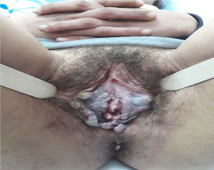 Figure 1: Multiple flesh-colored and whitish verrucose nodules and
plaques on the vulva. View Figure
Figure 1: Multiple flesh-colored and whitish verrucose nodules and
plaques on the vulva. View Figure
Patient had unprotected vaginal sex with an unknown partner 3 months before the onset of the lesion. Serological results included positive nontreponemal reaction - Venereal Disease Research Laboratory (VDRL) test titer was 1:64, with specific Treponema Pallidum Haemagglutination Assay (TPHA) test being positive as well. Clinical manifestations and positive serologic tests for syphilis confirmed the diagnosis of secondary syphilis, and condylomata lata were the only symptom of the disease.The patient was treated with a single dose of 2.4 million units of intramuscular benzathine penicillin G and the lesions completely subsided three weeks after the treatment. Six months later, the VDRL titer significantly declined (1:4).Condylomata lata are characterized by the flat moist papules or elevated verrucous or cauliflower-like papules or plaque usually located at sites where two body surfaces are in apposition such as anogenital areas. Characteristics of this site are constant moisture, friction and maceration facilitating coalescence and growth of these highly contagious lesions.Condylomata acuminata or bowenoid papulosis which are associated with human papillomavirus infection are common mimickers of condylomata lata. The diagnosis of condylomata lata is based on typical skin lesions and positive serologic tests for syphilis.Gynecologists must stay alert and consider syphilis in the differential diagnosis of unusual papular and warts-like lesions of the vulva. -
-
- Newman L, Rowley J, Vander Hoorn S, Wijesooriya NS, Unemo M, Low N, et al. Global estimates of the prevalence and incidence of four curable sexually transmitted infections in 2012 based on systematic review and global reporting. PLoSONE. 2015; 10: e0143304.
- Balagula Y, Mattei PL, Wisco OJ, Erdag G, Chien AL. The great imitator revisited: the spectrum of atypical cutaneous manifestations of secondary syphilis. Int J Dermatol. 2014; 53: 1434-1441.
Search Articles
-
Review Article
Screening for Depression among Medicyation Overuse Headache Patients and Treatment Could Be Useful for Improving their Quality of Life
Ljubisavljevic Srdjan1,2*#, Todorovic Stefan1# and Djokovic Filip1 | 2024-04-19- Short Communication
Why Multi-Drug Antiviral Therapy is Needed for COVID-19
Sibasish Dolai1 , Sabine Hazan2 , Christelle Pagonis1 , Sabrina Liu3 , Thomas J Borody1* and Robert R Clancy1 | 2024-04-11- Review Article
Elja: An R Package to Perform Environment-Wide Association Studies (Ewas / Envwas) Analysis
Marwan El Homsi1*and Isabella Annesi-Maesano1,2 | 2024-03-25- Case Report
Diagnosis of a Neck Mass in the Emergency Department
Schmidt S¹,²*, Lorenz KJ¹, Matthias C², Diekmeyer B2 and Müller G?,? | 2024-03-14- Case Study
The Impact of Neuropsychiatric Symptoms of Alzheimer’s Disease on Family Caregiver’s Distress
ATomasello L1,2*, Ranno1 , Raffaele2 , Laganà3 , Pitrone3 and Alibrandi4 | 2024-02-28News Feeds

Seth J. Worley, MD, FHRS, FACC
Director, Interventional Implant Program
Biography Paper Presentations
MedStar Heart & Vascular Institute,
Washington, DC, USACollaborations
Indexing


 © Copyright - JSM Central
 This work is licensed under a Creative Commons Attribution 4.0 International License.
- Short Communication