Case Report | Volume 6 - Issue 1 | Article DOI :
Download PDF
Kalina Kurtova MD, MSc, Alexandra Konstantinidou MD, MSc, Dr. Charalampos Triantafyllidis MD, MSc, PhD, ACCP
1st Pulmonary Department & Clinic, Kavala General Hospital, Greece
Corresponding Author:
Kalina Kurtova, 1st Pulmonary Department & Clinic, Kavala General Hospital, Greece
Keywords
Tuberculosis; Multidrug-resistant tuberculosis; Tuberculosis treatment regimen
Abstract
Tuberculosis is a disease caused by the Mycobacterium tuberculosis complex. It typically involves the lungs and can manifest as latent tuberculosis or as an active infection. While it is treatable and preventable, according to the WHO, it remains one of the leading causes of death in the world among infectious diseases. The guidelines suggest new pulmonary tuberculosis to be treated with a 6-month multidrug regimen. In some cases an inappropriate regimen or poor patient compliance can lead to drug resistance and consequent relapse of symptoms. This has caused two types of resistant tuberculosis to emerge: multidrug-resistant tuberculosis and extensively drug-resistant tuberculosis. We present a 29-year-old female with a medical history of tuberculosis that had not undergone adequate treatment and presented to our Emergency Department with persistent cough and hemoptysis. An initial work up was performed and the sputum smear microscopy for Acid Fast Bacilli confirmed a reactivation of tuberculosis. Subsequent tests revealed that the specimen was resistant to isoniazid, rifampicin and streptomycin, which allowed us to diagnose the relapse as multidrug-resistant tuberculosis. Despite the in vitro resistance the patient continued her treatment course with rifampicin, isoniazid, ethambutol and pyrazinamide. That led to a full resolution of her symptoms and after three consecutive negative sputum smears the patient was discharged from our clinic.
Citation
Kurtova K, Konstantinidou A, Triantafyllidis C (2023) A Case Study of Multi Drug-Resistant Tuberculosis (MDR-TB) in a Patient with Re activation of Tuberculosis. SM J Pulm Med 6: 9.
Abbreviations
LTBI: Latent Tuberculosis Infection, TB: Tuberculosis, MDR TB: MultiDrug-Resistant Tuberculosis, XDR-TB: Extensively Drug-Resistant Tuberculosis
Introduction
Tuberculosis (TB) is a bacterial infection that typically affects the lungs. It is caused by the Mycobacterium Tuberculosis complex, which includes the bacteria M. Tuberculosis, M. Bovis, M. Africanum and M. Microti (1). It is an important cause of morbidity and mortality worldwide and according to the WHO it remains one of the leading causes of death among infectious diseases (2). There are two types of TB - primary tuberculosis and reactivation tuberculosis. Primary tuberculosis could be latent (Latent Tuberculosis Infection, LTBI) or could lead to an active infection (in 1-5% of cases). LTBI is asymptomatic and not contagious, however, there is still a risk of eventual reactivation (5-10%) (3). Active tuberculosis is usually defined by night sweats,shivering, fever, weight loss and productive cough. Its treatment, according to WHO guidelines, consists of a 6-month multidrug regimen (4). The standard protocol includes a combination of Rifampin, Isoniazid, Ethambutol and Pyrazinamide. However, therapy is difficult because Mycobacterium Tuberculosis is an intracellular bacteria that can enter and survive in macrophages. That is a consequence of its ability to inhibit intracellular killing by blocking both phagosome maturation and phagolysosome fusion (5). Additionally, in some cases an inappropriate regimen or poor patient compliance can lead to drug resistance and consequent relapse of symptoms. There are two forms of resistant TB: MultiDrug-Resistant Tuberculosis (MDR-TB) resistant to rifampicin and isoniazid and Extensively Drug Resistant Tuberculosis (XDR-TB), which is resistant not only to rifampicin and isoniazid, but also to a fluoroquinolone and a second-line injectable like amikacin (6). MDR-TB emerges when M. Tuberculosis becomes resistant to two of the first-line antituberculosis antimicrobial regimens¹. That can be a result of incomplete treatment of a TB infection, since the bacteria that survive after an incomplete course of antituberculosis antimicrobials are more likely to develop resistance. Another reason for resistance can be incorrect dosing or duration of the anti-tuberculosis drugs. If the dosage or duration of treatment is not enough, then resistance is more likely to appear due to incomplete elimination of the bacteria. Moreover, suboptimal drug concentrations may lead to the emergence of mutations that confer resistance to the drugs and since these strains are not successfully eliminated, more drug-resistant populations are likely to develop. That is also why poor adherence to the treatment protocol is a major reason for the development of MDR-TB. One more reason for MDR-TB is the transmission of drug-resistant strains from one patient to another. Research shows that the incidence of MultiDrug-Resistant Tuberculosis has been steadily rising and that is bound to become a great challenge for clinicians. Our case highlights the difficulties of treating MDR TB and that despite the in vitro multidrug-resistance first-line antituberculosis agents were able to lead to full resolution of clinical symptoms and negative sputum smears.
Case Report
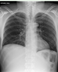
A 29-year-old Caucasian female with a medical history of Tuberculosis referred to our Emergency Department from a peripheral hospital complaining of persistent productive cough and hemoptysis. The patient disclosed a hospitalization three years earlier because of a TB infection for which she underwent a three-month therapy with first-line antibacterial medications. She presented an X-ray and a CT scan from her hospitalization (Figure 1, Figure 2). The imagining from her previous hospitalization revealed opacities in the upper lobes, a common finding among patients with active Tuberculosis.

Figure 1: Chest x-ray, 05/11/2020, during patient’s initial TB infection, revealing opacities in the upper lobes.

Figure 2: CT scan, 12/11/2020, reveals infiltrates in the upper lobes.
Additionally, the patient revealed that after her discharge she had not complied with the treatment regimen and had discontinued the antitubercular medications on her own. Upon presentation the patient was alert and hemodynamically stable (Heart Rate: 89/min, SpO2: 98% (FiO2=21%)). Auscaltation revealed decreased breath sounds in the left lung field. A Thorax CT scan revealed areas with consolidation in both lungs, as well as cavitation in the left upper and lower lobes (Figure 3).

Figure 3: Computed Tomography imaging findings on admission (25/5/2022) show consolidation with cavitation in the left upper and lower lobes.
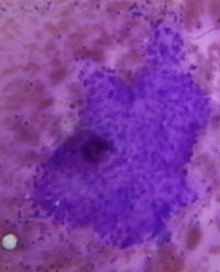
Sputum smear microscopy for Acid Fast Bacilli was requested and confirmed a TB relapse. Sputum smear microscopy is a valuable diagnostic tool for detecting pulmonary Tuberculosis. This test is used to detect the presence of Mycobacterium Tuberculosis in a patient’s sputum and is especially helpful because it provides results faster than a culture. While the results of a sputum smear test may be ready in one to two days, a culture may require up to 8 weeks to detect the growth of mycobacteria. Therefore, this test allows for quicker diagnosis of the disease and early beginning of treatment. Additionally, sputum smear microscopy is considered to have high specificity, meaning that if the test detects the presence of mycobacteria it is highly likely that the diagnosis is1 Tuberculosis. Figure 4 shows the presence of Acid Fast Bacilli on sputum smear microscopy (Ziehl-Neelsen stain) from samples obtained from our patient. The background remains blue while Acid Fast Bacilli like Mycobacterium Tuberculosis stain pink or red. This is due to the fact that these microorganisms have a lipoid capsule that resists discoloration and stains from the carbol-fuchsin from the solution (7).
In our case after the sputum smear tests were reported positive, a course of rifampin, isoniazid, ethambutol and pyrazinamide was initiated. In the meantime more sputum samples were collected, cultivated in a LJ-T culture and assessed with BACTEC MGIT 960. The result confirmed a Mycobacterium Tuberculosis culture-positive specimen resistant to isoniazid, rifampicin and streptomycin (sensitive to ethambutol and pyrazinamide). Sensitivity testing for rifampicin and isoniazid identified two mutations - mutation S531L in the rpoB gene and mutation S315T1 in the katG gene respectively. Despite the MDR TB diagnosis the regimen was continued because of the patient’s favorable response to the treatment and her significant clinical improvement. New recommendations from WHO, issued in 2022 for the treatment of MDR-TB, suggest the administration of a regimen consisting of bedaquiline, pretomanid, linezolid and moxifloxacin (recommendation with a very low certainty of evidence) (8). However, we could not initiate treatment with bedaquiline and pretomanid due to their unavailability in our country. Moreover, the results of the sputum culture that indicated resistance to isoniazid and rifampicin came out 44 days after her admission and while the patient was already on regimen with first-line antituberculosis antimicrobials. The hemoptysis resolved three days after her admission and treatment with tranexamic acid, the patient remained afebrile and the blood work performed continual improvement. The result of the initial Acid-Fast Bacilli smear was positive (4+). On day 60 the Acid-Fast Bacilli smear showed a result of 1+, while on day 78 the result of the smear was negative for the first time. Liver function was monitored routinely due to the antituberculous antimicrobials’ known and certified hepatotoxicity and remained inside normal range. The patient’s blood work in the end of her treatment revealed no signs of inflammation or liver damage (Table 1). The patient additionally received a vitamin B complex in order to prevent pyridoxine deficiency (a potential side effect of the use of isoniazid) and peripheral neuropathy (9).
|
Table 1: Relevant laboratory findings before the patient’s discharge show no elevated inflammatory markers or liver damage.
|
|
Investigation
|
Result
|
Normal range
|
|
WBC count
|
9,5
|
4,0-10,80 K/μl
|
|
Platelet count
|
292,00
|
150-350 K/μl
|
|
Hb
|
13,5
|
13,5-17,5 g/dL
|
|
Sodium
|
138
|
136-145 mmol/l
|
|
Potassium
|
4,1
|
3,5-5,1 mmol/l
|
|
Creatinine
|
0,5
|
0,7-1,3 mg/dl
|
|
C-reactive protein
|
0,1
|
<0,3 mg/dL
|
|
Lactate dehydrogenase
|
137
|
85-227 U/I
|
|
C.P.K.
|
39
|
39-308 U/I
|
|
C.P.K. MB
|
0,30
|
0-3,6 ng/mL
|
|
γ-GT
|
47
|
5-55 U/I
|
|
SGOT
|
30
|
15-37 U/I
|
|
SGPT
|
50
|
16-63 U/I
|
|
Sputum smear
|
Negative
|
-
|
Altogether, the patient was treated with a regimen of rifampicin (300mg 2x1), isoniazid (100mg 3.5x1), ethambutol (500mg 2x1) and pyrazinamide (500mg 2x1) throughout her hospitalization (99 days). The dosages remained constant throughout hospitalization and neither rifampicin nor isoniazid was discontinued. After three consecutive negative sputum smears the patient was discharged to continue a home-based course of antituberculosis treatment. The patient remained afebrile and reported full resolution of symptoms at the time of discharge. The patient did not attend any follow-up appointments in our hospital, but her pulmonologist informed us periodically of her condition and reported no recurrent hemoptysis, fever, weight loss or other symptoms indicating a relapse.
Conclusion
MDR-TB is an emerging global health problem. Mismanagement of TB treatment and lack of patient compliance is increasing the incidence of MDR-TB, leading to greater social and economic costs and exacting an enormous toll-burden for the national healthcare system. The management of our case shows that despite the in vitro multidrug resistance, the administration of first-line antituberculosis antimicrobial agents in vivo led to full resolution of the patient’s clinical symptoms and to negative sputum smears. It also highlights the importance of research into treatment regimens for MDR-TB. Additionally, the prevention of MDR-TB should be an essential part of managing this problem. Strategies should be implemented both at the individual and the community levels. Treatment courses should be completed fully as inadequate treatment is one of the biggest contributors to the emergence of drug resistance. Early detection and diagnosis of TB cases is another vital part of the fight against Tuberculosis. Healthcare facilities and laboratory services need to be widely available so that cases of drug resistance can also be detected early on and treated successfully. Another useful strategy is identifying and testing individuals who have been in contact with confirmed MDR-TB patients in order to detect potential new cases. Overall, the prevention of MDR-TB requires a multifaceted approach and the implementation of different social policies in addition to continued research and development of new treatments.
References
1. Shinnick TM, Good RC. Mycobacterial taxonomy. Eur J Clin Microbiol Infect Dis. 1994;13:884–901. 10.1007/BF0211148
2. World Health Organization. Tuberculosis [Internet]. www.who.int. World Health Organization; 2020. Available from: https://www.who. int/health-topics/tuberculosis#tab=tab_1
3. Fact Sheets: The Difference Between Latent TB Infection and TB Disease. [Internet]. 2019. Available from: https://www.cdc.gov/tb/ publications/factsheets/general/ltbiandactivetb.htm
4. World Health Organization. Tuberculosis - Treatment [Internet]. www.who.int. World Health Organization; 2020. Available from: https://www.who.int/health-topics/tuberculosis#tab=tab_1
5. Meena LS, Rajni. Survival mechanisms of pathogenic Mycobacterium tuberculosis H37Rv. FEBS J. 2010; 277(11): p.2416-2427. doi: 10.1111/j.1742-4658.2010.07666.x.
6. Centers for Disease Control and Prevention. Fact Sheets | Drug-Resistant TB | Extensively Drug-Resistant Tuberculosis (XDR TB) | TB | CDC [Internet]. www.cdc.gov. 2020. Available from: https://www.cdc.gov/tb/publications/factsheets/drtb/xdrtb. htm#:~:text=Extensively%20drug%2Dresistant%20TB%20(XDR
7. Yousif MQ, Qasem SA. Chapter 3 - Tissue Processing and Staining for Histological Analyses [Internet]. Albanna MZ, Holmes IV JH, editors. ScienceDirect. Boston: Academic Press; 2016. p. 49–59. Available from: https://www.sciencedirect.com/science/article/abs/pii/ B9780128016541000036
8. WHO operational handbook on tuberculosis. Module 4: treatment-drug-resistant tuberculosis treatment, 2022 update [Internet]. www.who.int. Available from: https://www.who.int/publications/i/ item/9789240065116
9. Snider DE. Pyridoxine supplementation during isoniazid therapy. Tubercle. 1980 Dec;61(4):191-6.
Citation
Kurtova K, Konstantinidou A, Triantafyllidis C (2023) A CaseStudy of Multi Drug-Resistant Tuberculosis (MDR-TB) in a Patient with Reactivation of Tuberculosis. SM J Pulm Med 6: 9.