Case Report | Volume 8 - Issue 1 | Article DOI :
Download PDF
Ahmad Behzadpour¹* and Joseph Durandis²
¹American University of the Caribbean, School of Medicine, FL, USA
²Borinquen Medical Centers, FL, USA
Corresponding Author:
Ahmad Behzadpour, American University of the Caribbean, School of Medicine. Tel: 312-5321098
Abstract
The most common parasitic central nervous system infection in humans is neurocysticercosis . It arises after the larval stage of the swine tapeworm Taenia solium is consumed. Taenia solium is endemic in under developing and developing countries in Latin America, SubSaharan Africa, Southeast Asia, the Indian subcontinent, and China. As reported by World Health Organization, there are about 50 million cases of cysticercosis worldwide where the mortality rate is said to be 100 in every 100000 cases due to NCC or the seizure as the result of NCC. While NCC is a rare occurrence in the United States, it is the most common cause of secondary seizure disorder, so it should be on a physician’s differential. This case report explores patients’ clinical manifestations of a patient with neurocysticercosis who presented to the trauma center due to a motor vehicle accident due to a generalized tonic-clonic seizure.
One major observation is that whether a medical intervention with antiparasitic drugs or a surgical approach is warranted, treating the symptoms should come before or along with the treatment plan. Lastly, general practitioners should consider Tinea solium infection as one of their differential diagnoses when patients present with chronic headache and or seizure.
Citation
Behzadpour A, Durandis J (2022) A Common Underdeveloped and Developing Countries Disease Known as Neurocysticercosis (NCC) in the US as a Vigilant Case for General Practitioners. SM J Case Rep 8: 3.
Abbreviations
NCC: Neurocysticercosis, CNS: Central nervous system, MRI: Magnetic Resonance Imaging.
INTRODUCTION
The most common parasitic central nervous system (CNS) infection in humans is neurocysticercosis (NCC)1. It arises after the larval stage of the swine tapeworm Taenia solium (Cysticercus cellulosae) is consumed. NCC is the most prevalent cause of secondary seizure disease, and patients with it may require long-term anti-epileptic treatment2. Taenia solium is endemic in under developing and developing countries in Latin America, Sub-Saharan Africa, Southeast Asia, the Indian subcontinent, and China. In addition to the above, due to immigration and frequent traveling, developing countries such as the United States are not spared from this incident1.
As reported by World Health Organization, there are about 50 million cases of cysticercosis worldwide where the mortality rate is said to be 100 in every 100000 cases due to NCC or the generalized tonic-clonic seizure as the result of NCC. There are approximately 2000 cases of yearly hospitalization due to NCC, and it is thought to be responsible for around 2% of emergency room admissions for seizures. While 85% of the death due to NCC were infected outside the United States, the remaining 15% has never left the country3,4.
CASE DISCRIPTION
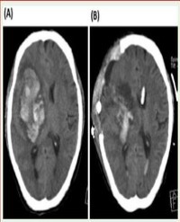
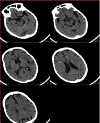
A 55-year-old African American male presented to the trauma center due to a motor vehicle accident. He had a seizure while he was driving, which resulted in the accident. While in the ED, he was seizing with gaze preference, so he was intubated due to airway protection. Head CT was performed, showing calcified heterogenous hyperdensity in the left posterior parietal lobe as seen in figure 1. There was some sign of edema with no mass effect.

Figure 1: Axial CT illustrates calcified heterogenous hyperdensity in the left posterior parietal lobe.
After the patient recovered from his postictal state, he was assessed further by the Neurosurgery team. The patient was wide-awake, briskly following commands without any focal presentations. He reports that he has had seizures and headaches for the past two years, but the episodes were transient and improved in a matter of hours. He is an immigrant who came to the US in 1981, and he works in the airport. The patient was admitted for further observation, and he was prescribed Levetiracetam 500mg PO BID for the seizure episodes. All of his lab values were within the normal limits except LFTs and TEGs, which were elevated. Further assessment was warranted due to elevated LFTs and TEGs, and the Neurosurgery team requested EEG and brain MRI with and without the contrast. MRI revealed a partially calcified 2.7 x 3.4 cm lesion in the left posterior parietal lobe as seen in figure 2.

Figure 2: Axial T1 contrast positive MRI illustrating a partially calcified 2.7 x 3.4 cm lesion in the left posterior parietal lobe.
Since the patient was stabled and did not experience any further seizure, he was discharged home after three days of hospitalization.
The patient agreed to come back for resection of the brain lesion via elective craniotomy. Intraoperatively, it was found that the lesion was a solid thin-walled cystic mass which was confirmed to be a Neurocysticercosis NCC utilizing the frozen section. The whole cystic lesion was resected, and he was put on anticonvulsants and steroids. Since the lesion was taken out completely, there was no need for antiparasitic agents.
Follow up
After one month, the patient visited his primary care physician, and he was healthy, reporting no more seizure episodes or headaches since the surgery. The patient needs to take Keppra per guideline recommendation.
DISCUSSION
The incidence of NCC in the United States is estimated to be between 0.2 and 0.6 cases per 100,000 people5. There are two subcategories of NCC known as extraparenchymal and parenchymal NCC. While patients with large cysts may experience some focal deficits6 and some may encounter cognitive issues or psychiatric symptoms like depression7,8, these are not the significant reasons that patients entreat for aid. However, a common denominator for all of them is that patients with NCC experience chronic headaches and seizure episodes.
The process of focal inflammation progressing into cystic degeneration and finally becoming calcified occur as cysts progress from a viable, dormant state. Moreover, degeneration is the main reason for worsening neurological symptoms due to perilesional inflammation9. The border between a viable cyst with inflammation and a degenerated parenchymal cyst, as seen in our patient MRI is poorly defined. Some scholars classify cysts with perilesional contrast enhancement and edema as either cyst with inflammation or degenerating cysts, whereas others point to variations in the density of cyst contents10.
The local inflammation enclosing the cyst or the calcification resulting from chronic inflammation is one of the primary causes of subsequent seizure relapses in patients with NCC11. Furthermore, scarring caused by high glial cell turnover, which results in hypertrophy and hyperplasia around the lesion, increases the incidence of seizure recurrence12. This warrants the long-term usage of anticonvulsants like Keppra as in our patient care.
A recent study about Tinea Solium by Garcia et al. came up with a revised diagnostic criterion developed based on preceding research by Brutto, 2017. Both reports agree that in patients with NCC who have clinical manifestations such as seizures or cysticercosis exposure, a conclusive diagnosis cannot be made without the information presented by neuroimaging13,14.
As with any other disease or pathology, the management and treatment can offer both medical and surgical approaches. In the case of NCC, depending on the location of the cyst/s, one can decide how to manage it. It is essential to understand that symptomatic care can take precedence over antiparasitic therapy. Moreover, antiparasitic drugs in NCC have an unusual feature, whereas no substantial increase is anticipated in the first few days or weeks. On the other hand, its use causes local perilesional inflammation, which may exacerbate or intensify neurological symptoms; therefore, steroids or other agents are used at the same time to counteract this negative impact15. On the other hand, until initiating antiparasitic drug therapy, symptomatic treatment is vital and should be well-established; moreover, symptomatic medicine, such as analgesics, antiepileptic agents, mannitol, and steroids, is generally prescribed as it would be for seizures, headache, or intracranial hypertension provoked by some other trigger16. If the lesion site allows, a surgical operation should be performed, followed by symptomatic therapy for gliosis caused by scarring in the surgical field, as in our patient. A ventricular shunt is allowed in some other situations where the lesions are intraventricular, and there is a risk of hydrocephalus. Excision of the mass is also desirable if there is no risk of ventricular wall injury17.
CONCLUSION
This case report explores patients’ clinical manifestations of a patient with neurocysticercosis. While NCC is a rare occurrence in the United States, it is the most common cause of secondary seizure disorder, so it should be on a physician’s differential. One major observation is that whether a medical intervention with antiparasitic drugs or a surgical approach is warranted, symptoms management should come before or along with the treatment plan.
Acknowledgements
We would like to thank Samira Jasmhidi-Shal, Farshid Tabari and Morteza Momenzadeh for their outstanding mentorship and guidance.
This manuscript is being submitted on behalf of all authors listed. The authors declare that this manuscript is solely being submitted to JSMC Clinical Case Reports and is not currently under review by any other journal.
REFRENCES
1. Garcia HH, Gonzalez AE, Gilman RH; Cysticerosis Working Group in Peru. Diagnosis, treatment and control of Taenia solium cysticercosis. Curr Opin Infect Dis. 2003;16(5):411-419. doi:10.1097/00001432-200310000-00007
2. Del Brutto OH, Santibañez R, Noboa CA, Aguirre R, Díaz E, Alarcón TA. Epilepsy due to neurocysticercosis: analysis of 203 patients. Neurology. 1992;42(2):389-392. doi:10.1212/wnl.42.2.389
3. O’Neal SE, Flecker RH. Hospitalization frequency and charges for neurocysticercosis, United States, 2003-2012. Emerg Infect Dis. 2015;21(6):969-976. doi:10.3201/eid2106.141324
4. Sorvillo FJ, DeGiorgio C, Waterman SH. Deaths from cysticercosis, United States. Emerg Infect Dis. 2007;13(2):230-235. doi:10.3201/ eid1302.060527
5. Serpa JA, White AC Jr. Neurocysticercosis in the United States. Pathog Glob Health. 2012;106(5):256-260. doi:10.1179/204777321 2Y.0000000028
6. Ghasemi R, Rowe A, Shah R, Venkatesan P, England TJ. Neurocysticercosis presenting as a ‘Stroke Mimic’. Acute Med. 2016;15(2):79-83.
7. Ramirez-Bermudez J, Higuera-Calleja J, Espinola-Nadurille M, CoronaT. Neuropsychiatric disorders in patients with neurocysticercosis. Asia Pac Psychiatry. 2017;9(2):10.1111/appy.12250. doi:10.1111/ appy.12250
8. Garcia HH, Gonzales I, Lescano AG, et al. Efficacy of combined antiparasitic therapy with praziquantel and albendazole for neurocysticercosis: a double-blind, randomised controlled trial. Lancet Infect Dis. 2014;14(8):687-695. doi:10.1016/S1473-3099(14)70779-0
9. Nash TE, Mahanty S, Garcia HH; Cysticercosis Group in Peru. Corticosteroid use in neurocysticercosis. Expert Rev Neurother. 2011;11(8):1175-1183. doi:10.1586/ern.11.86
10. Dumas JL, Visy JM, Belin C, Gaston A, Goldlust D, Dumas M. Parenchymal neurocysticercosis: follow-up and staging by MRI. Neuroradiology. 1997;39(1):12-18. doi:10.1007/s002340050358
11. Herrick JA, Maharathi B, Kim JS, et al. Inflammation is a key risk factor for persistent seizures in neurocysticercosis. Ann Clin Transl Neurol. 2018;5(5):630-639. Published 2018 Apr 10. doi:10.1002/acn3.562
12. de Souza A, Nalini A, Kovoor JM, Yeshraj G, Siddalingaiah HS, Thennarasu K. Perilesional gliosis around solitary cerebral parenchymal cysticerci and long-term seizure outcome: a prospective study using serial magnetization transfer imaging. Epilepsia. 2011;52(10):1918-1927. doi:10.1111/j.1528-1167.2011.03189.x
13. Garcia HH, Gonzalez AE, Gilman RH. Taenia solium Cysticercosis and Its Impact in Neurological Disease. Clin Microbiol Rev. 2020;33(3):e00085-19. Published 2020 May 27. doi:10.1128/ CMR.00085-19
14. Del Brutto OH, Nash TE, White AC Jr, et al. Revised diagnostic criteria for neurocysticercosis. J Neurol Sci. 2017;372:202-210. doi:10.1016/j. jns.2016.11.045
15. Singh AK, Singh SK, Singh A, et al. Immune response to Taenia solium cysticerci after anti-parasitic therapy. Int J Parasitol. 2015;45(12):749-759. doi:10.1016/j.ijpara.2015.05.007
16. Bustos JA, García HH, Del Brutto OH. Antiepileptic drug therapy and recommendations for withdrawal in patients with seizures and epilepsy due to neurocysticercosis. Expert Rev Neurother. 2016;16(9):1079-1085. doi:10.1080/14737175.2016.1194757
17. Psarros TG, Krumerman J, Coimbra C. Endoscopic management of supratentorial ventricular neurocysticercosis: case series and review of the literature. Minim Invasive Neurosurg. 2003;46(6):331-334. doi:10.1055/s-2003-812470