Research Article | Volume 2 - Issue 2 | Article DOI :
Download PDF
Jefferson M Homem1 , Aline S DeMaman3 , Denise Lachat1 , Ariane Zamarioli2 , José A Thomazini1 and João-José Lachat1 *
1 Department of Surgery and Anatomy, Ribeirão Preto Medical School, University of São Paulo, Brazil
2 Department de Biomechanics, Medicine and Rehabilitation of the Locomotor System, Ribeirão Preto Medical School, University of São Paulo, Brazil
3 Departamento de Biologia, Centro de Ciências Biológicas e da Saúde, Universidade Estadual da Paraíba, Brazil
Corresponding Author:
João-José Lachat, Department of Surgery and Anatomy, Ribeirão Preto Medical School, University of São Paulo, Av. Bandeirantes 3900, 14049-900 Ribeirão Preto, São Paulo, Brazil, Tel: +55 1633153245; Email: jjlachat@fmrp.usp.br
Keywords
Glenoid cavity; Anatomical
variation; Sexual dimorphism;
Anthroposcopy; Anthropometry
Abstract
The glenoid cavity of the scapula shows high morphological variability that has not been clearly elucidated in the literature. Its morphological aspects knowledge can be used in the sex determination, to improve the development of more functional prostheses and in the accurate image diagnostic. The purpose of the present study was to analyze and compare sex and dominance differences in the articular surface of the human glenoid cavity. Anthropometric measurements of the glenoid cavities of scapula were taken of 200 specimens, besides we developed a morphological classification system according the shape of inferior and superior poles and the presence of the glenoid notch in the anterior margin. For all anthropometric parameters, the male scapula showed higher values than the measures in female scapula both in right and left sides, but we did not find a significant difference between the left and right scapula in either sex. On the other hand, taking into consideration the anthroposcopic aspects, 50% of male and 58% of female were classified in a different morphological group when compared to the contralateral glenoid in the same individual. The present study revealed an evident sexual dimorphism and provides information about the articular surface of normal glenoid cavities to improve the diagnosis of orthopedic lesions, to produce better prosthesis and to improve the medico-legal identification
Citation
Homem JM, DeMaman AS, Lachat D, Zamarioli A, Thomazini JA and Lachat JJ. Anatomical and Anthropological Investigation of the Articular Surface of the Human Glenoid Cavity in Brazilian Corpses. SM J Clin Anat. 2018; 2(2): 1010
Introduction
Biological anthropology is related to the study of the normal human body and the variations that may affect anatomical structures. This broader topic is divided into zoological anthropology, racial anthropology, anthropological typology, and anthropogenesis [1]. Anthropological typology refers to the study of biotypes according to the features of ancestry groups, sex, age, and profession; it allows the identification of individuals by forensic medicine and in police identification and investigation procedures. There are 2 main study types within anthropological typology: anthroposcopy which analyzes qualitative descriptive features and anthropometry, a quantitative analysis that measures angles and dimensions in different segments of the body [2].
Anatomical analyses, over the long term, help investigators to obtain consistent information and provide unmistakable anatomical characteristics. In addition, important information may be added to some specific fields of medicine such as orthopedic prosthesis fixation and the correction of fractures and dislocations.
The glenoid cavity of the scapula demonstrates a high morphological variability that has not been clearly elucidated in the literature. Differences may be observed between males and females, and even between the 2 scapulas of a single individual. There is no consensus on the classification of the glenoid cavity shape [3], but in most anatomy texts the glenoid cavity is described as a shallow and oval depression in the lateral angle of the scapula, 3 to 4 cm in length and 2 to 3 cm in width [4-7].
The glenoid cavity of the scapula is seated on an anatomical neck of bone and takes on a pear shaped structure due to the presence of 2 poles: the lower pole is larger than the upper pole. A notch is present at the anterior margin. The structure has a concave articular surface,with a vertical long axis and a superiorly and anterior-laterally oriented articular face. Additionally, the glenoid margin serves as the fixation point of the glenoid labrum, a fibro cartilaginous rim of tissue that serves the function of increasing the depth and size of the articular surface of the cavity. There is consequent increase in articular congruency and stability [8].
Knowledge of the geometry and the morphological aspects of the articular surface of the glenoid cavity can be used in sex estimation. This knowledge also has clinical importance to the analysis of glenohumeral joint stabilization, the performance of arthroplasty, the development of more functional and precise prostheses, and to the accuracy of diagnoses using plain radiography, computed tomography (CT), and magnetic resonance imaging (MRI) of the shoulder [9-13]. The present study analyzed and compared sex differences and laterality (right versus left) in the articular surface of the human glenoid cavity, devoid of articular cartilage, using both anthropometric and anthroposcopic analysis.
Materials and Methods
This study examined the glenoid cavities of scapula collected from the Cemetery Bom Pastor in Ribeirão Preto, São Paulo, Brazil, and from dried specimens in the Human Anatomy Laboratories of both the Faculty of Medicine, University of São Paulo, Ribeirão Preto, Brazil, and the Faculty of Medicine, University of São Paulo, Barão de Mauá, Brazil. We analyzed both the left and right glenoid cavities obtained from 100 Brazilian adult human corpses of unknown ethnicity and of both sexes (50 female and 50 male), for a total of 200 specimens that were without lesions or evidence of degenerative disease
The glenoid cavities were cleaned of necrotic material and a first template was made using odontological moldable silicone (Vigodent SA, Rio de Janeiro, Brazil). In order to obtain samples resembling the original specimens, the template was then filled with a plaster paste to provide the first replica. Afterward, the first replicas were placed in a 10% solution of formaldehyde for 24 hours for sterilization and were then stored for future use. In order to obtain a second replica, a second mold was made with odontological moldable silicone using the first replica rather than the original anatomical specimen, and the second replica was created with acrylic resin in order to provide safe handling for the researchers
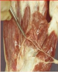
The anthropometric and anthroposcopic analysis took into consideration the side of the body and the cadaver’s sex,as reported by the Cemetery Administration and the Anatomy Laboratories. For each specimen, we demarcated the articular face of the cavity with a perimetric line to identify the bony area normally in contact with the head of the humerus in the shoulder joint (Figure 1).

Figure 1: Schematic of the perimetric line used to delineate the articular surface, the maximal height (A-B) and the maximal width (C-D) of the glenoid cavity
The resin replicas were scanned and the images were printed and analyzed by a semi-automatic image analyzer system (MiniMop®,Kontron Bildanalyse GmBH, Ehing, Germany). The surface area, perimeter, the maximal height and maximal width or the maximal and minimal diameters respectively of the articular surface of the glenoid cavity were measured (Figure 1).
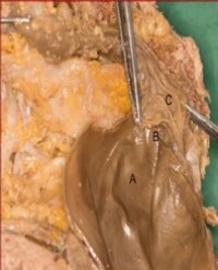
To measure the volume of the cavity, the articular surfaces of the resin acrylic replicas were filled with moldable silicone; this material is resistant to humidity and therefore has a constant density of 1.543 g/cm3. A silicone sphere (Figure 2A) was compressed into the acrylic replica, filling it completely (Figure 2B).

Figure 2: Schematic of the procedure used to measure the volume of the articular surfaces. A-E, referred in the text.
A uniform, cylindrical baton was used to remove excess silicone from the surface of the replica (Figure 2C). The quantity of silicone that was removed (Figure 2D) was then molded into a sphere (Figure 2E) and evaluated in a precision weighing machine. The volume of the articular surface was calculated based on the value of both the weight remaining in the cavity and the density of the silicone.
In order to measure the deepest point of the articular surface, the margins of the superior and inferior poles were positioned adjacent to a straight surface. A conical-tipped ruler, graded in millimeters, was then used to measure the maximal depth of the cavity. The deepest point of the articular cavity was assessed by sliding the ruler along the concave surface between the superior and inferior poles, and noting the maximal depth (Figure 3).

Figure 3: Procedure used to assess the deepest point of the articular surface of the glenoid cavity.
Statistical Analysis
An unpaired, 2-tailed t-test was used to detect statistical differences in the glenoid measurements between sexes and between the right and left sides in each sex. A p-value of < 0.05 was considered significant. Statistical analyses were conducted using GraphPad Prism software, version 5.02 (GraphPad Software, Inc. La Jolla, CA, USA)
Results
Anthropometric analysis
For all parameters of interest, the male scapula showed higher values than the female scapula, on both the right and left sides (p<0.05). We did not find a significant difference between the left and right scapula in either sex. The data are shown graphically, expressed by the mean and standard error.
Surface area: In male specimens, the right-sided articular surface area ranged from 3.34 to 7.70 cm2, with a mean of 5.76 ± 0.14 cm2; the left-sided articular surface area ranged from 4.17 to 8.19 cm2, with a mean of 5.98 ± 0.12 cm2. In female specimens, the right-sided surface area ranged from 2.94 to 7 cm2, with a mean of 4.60 ± 0.12 cm2; the left-sided surface area ranged from 3.49 to 6.60 cm2, with a mean of 4.58 ± 0.10 cm2 (Figure 4).

Figure 4: Morphometrical parameters of the glenoid cavities of the right and left sides in female and male scapula. Data are expressed as the mean with standard error; n=50 for each category; *p < 0.05.
The male scapula had higher surface area measurements than the female scapula, with a significant difference in both the right-sided specimens (p < 0.001; t = 6.368) and the left sided specimens (p < 0.001; t = 8.765).
Minimal diameter: The minimal diameter of the right articular surface in males ranged from 1.59 to 2.74 cm, with a mean of 2.25 ± 0.04 cm; on the left, the minimal diameter ranged from 1.57 to 2.90 cm, with a mean of 2.30 ± 0.04 cm. In the female specimens, the right sided minimal diameter ranged from 1.40 to 2.64 cm, with a mean of 1.99 ± 0.03 cm; on the left, the minimal diameter ranged from 1.60 to 2.44 cm, with a mean of 1.98 ± 0.03 cm (Figure 4). The male scapula had higher minimal diameters than the female scapula, with significant differences in both the right-sided specimens (p < 0.001; t = 4.765) and the left-sided specimens (p < 0.001; t = 7.225)
Volume: The volume of the right articular surface in males ranged from 0.06 to 1.30 cm3, with a mean of 0.49 ± 0.04 cm3; on the left, the volume ranged from 0.06 to 1.35 cm3, with a mean of 0.45 ± 0.04 cm3. In females, the right-sided volume ranged from 0.07 to 1.48 cm3, with a mean of 0.31 ± 0.04 cm3; on the left, the volume ranged from 0.08 to 0.95 cm3, with a mean of 0.28 ± 0.02 cm3 (Figure 5).

Figure 5: Volume and depth of the glenoid cavities of the right and left sides in female and male scapula. Data are expressed as the mean with standard error; n=50 for each category; *p < 0.05.
The male scapula had higher volumes than the female scapula, with significant differences in both the right-sided specimens (p < 0.0054; t = 2.847) and the left-sided specimens (p < 0.001; t = 4.562).
Depth: The deepest point of the articular surface in the right-sided male glenoid cavity ranged from 0.20 to 0.65 cm, with a mean of 0.44 ± 0.01 cm; on the left side, the deepest point ranged from 0.20 to 0.60 cm, with a mean of 0.45 ± 0.01 cm. In females, the deepest point in the right side ranged from 0.25 to 0.65 cm, with a mean of 0.38 ± 0.01 cm; on the left, the deepest point ranged from 0.20 to 0.65 cm, with a mean of 0.37 ± 0.01 cm (Figure 5). The male scapula had greater depth than the female scapula, with a significant difference seen in both the right-sided specimens (p < 0.0021; t = 3.152) and the left sided specimens (p < 0.001; t = 4.338).
Anthroposcopic analysis
The results of anthroposcopic analysis showed that the glenoid cavity had variable geometry in both males and females: the cavity took on an ovoid shape when the lower pole was larger than the upper pole and an ellipsoid shape when both poles were similar. We classified the cavities into 5 morphological groups, taking into consideration the shape of the inferior and superior poles, the shape of the articular surface, and the presence or absence of a glenoid notch in the anterior margin. These differences were recorded according to sex and laterality.
Group 1 consisted of glenoid cavities with articular surfaces presenting a well-defined notch in the anterior margin and having a superior pole thinner than the inferior pole, characterizing a ovoid shape (Figure 6).

Figure 6: Schematic showing the 5 glenoid cavity morphological groups with the percentage values on anthroposcopic analysis.
This group contained 73 glenoid specimens (36.5%): 22 (11%) right male, 19 (9.5%) left male, 17 (8.5%) right female, and 15 (7.5%) left female.
Group 2 included glenoid cavities with articular surfaces presenting a less-evident notch in the anterior margin but still having a thinner superior than inferior pole, preserving the ovoid morphology (Figure 6). This group contained 63 glenoid specimens (31.5%): 14 (7%) right male, 18 (9%) left male, 13 (6.5%) right female, and 18 (9%) left female.
Group 3 was characterized by glenoid cavities with articular surfaces lacking a notch in the anterior margin and with superior poles thinner than the inferior poles, preserving the ovoid shape (Figure 6). This group contained 40 glenoid specimens (20%): 10 (5%) right male, 9 (4.5%) left male, 11 (5.5%) right female, and 10 (5%) left female.
Group 4 included glenoid cavities with articular surfaces lacking a notch in the anterior margin and with similar poles, resulting in an ellipsoid shape (Figure 6). This group comprised 20 glenoid specimens (10%): 3 (1.5%) right male, 2 (1%) left male, 8 (4%) right female, and 7 (3.5%) left female.
Group 5 contained glenoid cavities with articular surfaces presenting a well-defined notch in the anterior margin but with similar poles, also characterizing an ellipsoid shape (Figure 6). This group contained 4 glenoid specimens (2%): 1 (0.5%) right male, 2 (1%) left male, and 1 (0.5%) right female. We did not find a left female glenoid cavity with the characteristics of group 5.
Based on the analysis of the male scapula, 25 glenoid-cavity pairs (50%) contained more than 1 morphological group. In the right scapula of these male individuals, 11 samples (44%) were classified in group 1, 6 (24%) were in group 2, 5 (20%) in group 3, 2 (8%) in group 4, and only 1 (4%) was classified in group 5. On the left side, 8 glenoid cavities (32%) were in group 1, 10 (40%) in group 2, 4 (16%) in group 3, 1 (4%) in group 4, and 2 (8%) in group 5
In females, 29 glenoid-cavity pairs (58%) contained more than 1 morphological group. On the right side of these female individuals, 4 samples (13.8%) were classified in group 1, 9 (31%) in group 2, 9 (31%) in group 3, 6 (20.7%) in group 4, and only 1 (3.4%) was classified in group 5. On the left side, 2 glenoid cavities (6.9%) were in group 1, 14 (48%) in group 2, 8 (27.6%) in group 3, 5 (17%) in group 4, and none fell into group 5.
Discussion
Several authors have attempted to characterize the shape and the dimension of normal and pathologic glenoid cavities [4,12-17]. In the present study, we evaluated the anthropometric and anthroposcopic qualities of the articular surface of the human Brazilian scapula glenoid cavity. Similar to Merrill et al [8], our outcomes show that the lower poles are generally larger than the upper ones.
We quantified the area, perimeter, maximal diameter, minimal diameter, volume, and depth of these structures in both sexes and on both sides of the body. In all samples, the male glenoid cavities showed higher measurements than the female glenoid cavities, on both the right and left sides of the body. This finding is in agreement with those described in prior studies [4,8,10,13,18-20] and characterizes a clear sexual dimorphism. Checroun et al [10] found that male glenoid are approximately 10% larger than their female counterparts.
We demonstrated a mean value of 3.1 cm for the maximal glenoid diameter in females and 3.4 cm for the maximal diameter in males, with an overall mean of 3.25 cm. The maximal diameter can also be considered to be the length of the glenoid cavity. The minimal diameter, considered to be the width of the cavity, demonstrated a mean value of 1.9 cm in females and 2.3 cm in males, with an overall mean of 2.1 cm. Like the present study, Churchill et al [4], in a sample of both sexes and multiple ancestry groups in the United States, found that the female glenoid cavity is an average of 3.2 cm in length and 2.3 cm in width. Ebraheim et al [21] found a mean value of 1.3 cm to 2.3 cm for the width of the glenoid cavity in both men and women. Von Schroeder et al [5] performed a similar study in Canadians and found larger averages: 3.6 cm for length and 2.9 cm for width. The authors of a Turkish study found an average of 3.6 cm for length and 2.4 cm for width [6]. In 2007, Nakamura et al [7] concluded that the glenoid cavity in Japanese adults is smaller than in European adults: the average Japanese glenoid cavity is 2.8 cm in length and 2.1 cm in width, and in Europeans the measurements are 3.5 cm for length and 2.5 cm for width. Our results are in agreement with these previous assessments
In the present study, the glenoid depth had a mean value of 0.3 cm in women and 0.4 cm in men, similar to the results of the study by Merrill et al [8] that also obtained similarly significant differences between female and male specimens for each dimension measured.
The mean area of the glenoid cavity was 4.6 cm2 in women and 5.9 cm2 in men, with an overall mean of 5.25 cm2 The area of the glenoid cavity was also studied in German adults and ranged from 3.5 to 9.7 cm2 [22]. Prescher and Klümpen [18] found mean values of 7.18 cm2 for German females and 9.87 cm2 for German males, indicating the sexual dimorphism we also noted in the present study. Kwon et al [17] observed a mean area of 8.67 cm2; these measures were obtained from CT images of North Americans. Taken together, these previous studies and the present study also demonstrate the morphometrical differences among ancestry groups.
We found no significant difference when comparing the right and left glenoid cavities of a given individual. Our data are in agreement with those of Sugaya et al [16]. They investigated 75 normal glenoid cavities by means of 3-dimensional CT and observed that the width and length of the normal glenoid do not differ substantially between the right and left sides in the same patient. All glenoid cavities in that study demonstrated a pear-shaped morphology, with the inferior portion of the glenoid contour approximating a true circle.
We evaluated the presence of the glenoid notch in 200 specimens. We observed a notch in 70% of the glenoid cavities (groups 1, 2, and 5), with the notch absent in 30% (20% in group 3 and 10% in group 4). The prevalence of the glenoid notch in the present study is higher for males (76%) than females (64%), with a difference of 12%, differing from the results of Merril et al [8] showing that 80% of females have an anterior glenoid notch, compared with only 57% of males. Prescher and Klumpen [14] identified the notch in the anterior margin of the glenoid cavity in 55% of the evaluated scapula, noting that this notch resulted in a pear-shaped glenoid. The notch was absent in the remaining 45% of scapula, lending an oval shape to the glenoid, but no sex difference was found in the prevalence of the notch. A similar study in a Turkish population showed that only 28% of individuals had the glenoid notch and the glenoid pear-shape [6]. At present there is no clear explanation for the presence of a glenoid notch. Whether the position of the glenoid notch is related to that of the tendon of the subscapularis muscle, which crosses the shoulder joint at this point, remains speculative [14]. It is possible that handedness may influence glenoid morphology.
Although we did not find profound differences in the anthropometric measures of the glenoid cavity pairs in a single subject, we were able to recognize morphological asymmetry. In 25 men and 29 women, the glenoid cavity demonstrated a different morphological group in the right and the left side. Prescher and Klumpen [14] found that females were more likely than males to demonstrate this morphologic asymmetry. We were able to classify the articular surfaces into 5 distinct morphological groups based on anthroposcopic analysis. Group 1 morphology was more prevalent in the right side in both males and females, while Group 2 was more prevalent in the left side. In Group 3, the distribution was equal on both sides for both sexes. Group 4 was much more prevalent in females, while group 5 had a low prevalence in both sides and both sexes, although it was more likely to be seen in males
Conclusion
In the present study, we demonstrated that sexual dimorphism is present in the glenoid cavity. We also are able to provide information about the articular surface of normal glenoid cavities. This will help to establish anatomical and clinical standards, relevant to improving medico-legal identification, making the diagnosis and determining the extensiveness of orthopedic lesions, planning arthroplasty procedures, and developing more precise and functional articular prostheses.
References
1. Siegel JA, Saukko PJ, Knupfer G. Anthropometry in Encyclopedia of forensic sciences. 2nd edn. Academic Press: USA. 2000; 284-290.
2. Krishan K. Anthropometry in Forensic Medicine and Forensic Science ’Forensic Anthropometry’. Internet J Forensic Sci. 2007; 2: 1-14.
3. De Wild LF, Berghs BM, Audenaert E, Sys G, Van Maele GO, Barbaix E. About the variability of the shape of the glenoid cavity. Surg Radiol Anat. 2004; 26: 54-59.
4. Churchill RS, Brems JJ, Kotschi H. Glenoid size, inclination, and version: an anatomic study. J Shoulder Elbow Surg. 2001; 10: 327-332.
5. von Schroeder HP, Kuiper SD, Botte MJ. Osseous anatomy of the scapula. Clin Orthop Relat Res. 2001; 383: 131-139.
6. Coskun N, Karaali K, Cevikol C, Demirel BM, Sindel M. Anatomical basics and variations of the scapula in Turkish adults. Saudi Med J. 2006; 27: 1320 1325.
7. Nakamura T, Yamamoto N, Yamada K, Itoi E. Quantitative description of the japanese glenoid morphology. J Biomechanics. 2007; 40: 586.
8. Merrill A, Guzman K, Miller SL. Gender differences in glenoid anatomy: an anatomic study. Surg Radiol Anat. 2009; 31: 183-189.
9. Inui H, Sugamoto K, Miyamoto T, Machida A, Hashimoto J, Nobuhara K. Evaluation of three-dimensional glenoid structure using MRI. J Anat. 2001; 199: 323-328.
10. Checroun AJ, Hawkins C, Kummer FJ, Zuckerman JD. Fit of current glenoid component designs: an anatomic cadaver study. J Shoulder Elbow Surg. 2002; 11: 614-617.
11. Williams GR, Abboud JA. Total shoulder arthroplasty: glenoid component design. J Shoulder Elbow Surg. 2005; 14: 122-128.
12. Huysmans PE, Haen PS, Kidd M, Dhert WJ, Willems JW. The shape of the inferior part of the glenoid: a cadaveric study. J Shoulder Elbow Surg. 2006; 15: 759-763.
13. Bicknell RT, Patterson S, King G, Chess D, Johnson J. Glenoid vault endosteal dimensions: an anthropometric study with special interest in implant design. J Shoulder Elbow Surg. 2007; 16: 96-101.
14. Prescher A, Klumpen T. The glenoid notch and its relation to the shape of the glenoid cavity of the scapula. J Anat. 1997; 190: 457-460.
15. Couteau B, Mansat P, Darmana R, Mansat M, Egan J. Morphological and mechanical analysis of the glenoid by 3D geometric reconstruction using computed tomography. Clin Biomech. 2000; 15: 8-12.
16. Sugaya H, Moriishi J, Dohi M, Kon Y, Tsuchiya A. Glenoid rim morphology in recurrent anterior glenohumeral instability. J Bone Joint Surg Am. 2003; 85: 878-884.
17. Kwon YW, Powell KA, Yum JK, Brems JJ, Iannotti JP. Use of three dimensional computed tomography for the analysis of the glenoid anatomy. J Shoulder Elbow Surg. 2005; 14: 85-90.
18. Prescher A, Klumpen T. Does the area of the glenoid cavity of the scapula show sexual dimorphism? J Anat. 1995; 186: 223-226.
19. Macaluso PJ Jr. Sex determination from the glenoid cavity in black South Africans: Morphometric analysis of digital photographs. Int J Legal Med. 2011; 125: 773-778.
20. Matsumura N, Oki S, Ogawa K, Iwamoto T, Ochi K, Sato K, et al. Three dimensional anthropometric analysis of the glenohumeral joint in a normal Japanese population. J Shoulder Elbow Surg. 2016; 25: 493-501