Case Report | Volume 1 - Issue 1 | Article DOI :
Download PDF
Sreenivasa Rao Sudulagunta1 *, Shyamala Krishnaswamy Kothandapani2 , Mahesh Babu Sodalagunta3 , Hadi Khorram2 , Mona Sepehrar4 and Zahra Noroozpour1
- Columbia Asia Hospital, India
- Dr. B R Ambedkar Medical College, India
- KS.Hegde Medical College, India
- Baptist Hospital, India
Corresponding Author:
Sreenivasa Rao Sudulagunta, MD Hospitalist, Columbia Asia Hospital, Bangalore, India, Email: drssreenivasarao@gmail.com
Keywords
lung carcinoma, tuberculosis, lateral rectus palsy, trochanteric metastasis, fibrosis.
Abstract
We report a case of large cell undifferentiated lung carcinoma in a middle aged patient with previously treated tuberculosis and scarring. 20 pack years of smoking history was noted. He presented with metastasis at multiple sites including trochanter, liver and acute bilateral lateral rectus palsy. Chest radiography showed fibrosis of right upper zone with homogenous opacity. Sputum examination for AFB was negative. Computerized tomography of the thorax showed an irregular heterogeneously enhancing mass involving right upper lobe with cavitation, necrosis along with lymph node involvement and the erosion of the 4th rib with liver metastasis. Radiography of hip joint was suggestive of lesser trochanteric metastasis. MRI brain was suggestive of mass at base of brain in parasellar area. Fine needle aspiration cytology and CT guided biopsy confirmed undifferentiated large cell carcinoma of lung. Tuberculosis and smoking may increase the risk of lung scarring and malignancy and pulmonary scarring may be associated with increased lung carcinoma in ipsilateral lung. Clinician needs to be more sensitive to look for malignancy association particularly in patient with or previously treated for tuberculosis and even more emphasis to be laid if scarring of lung is observed.
Introduction
Fifth common cancer in India is lung carcinoma with 63,000 new cases detected per year [1]. Men and smokers have increased incidence [2,3]. Concurrent presence of tuberculosis and malignancy pose a diagnostic challenge [4]. Risk of malignancy increases with pulmonary tuberculosis diagnosed more than 20years as per studies [5]. Scarring was associated with increased lung cancer risk on the ipsilateral lung [6].
Tuberculosis (TB) presents a global threat in both developing and developed countries. Onethird of the world’s population is thought to have been infected with M. tuberculosis [7], and new infections occur in about 1% of the population each year [8]. In 2007, an estimated 13.7 million chronic cases were active globally [9], while in 2013, an estimated 9 million new cases occurred [10]. In 2013 there was between 1.3 and 1.5 million associated deaths [11], most of which occurred in developing countries [12]. Tuberculosis is the second-most common cause of death from infectious disease after HIV/AIDS [13]. The aim of the case report is to improve knowledge regarding malignancy in tuberculosis scar.
Case Report
A 40 year old male patient, chronic smoker (20 pack years of smoking) and alcoholic (80 g per day for 20 years) who was treated successfully for sputum positive pulmonary tuberculosis 4 years ago (Figure 1), labourer by occupation, presented with pain in lower limbs and difficulty in walking and significant weight loss over a period of 1 month, double vision over a period of 10 days with no chest symptoms. Patient was tested negative for tuberculosis (sputum for Acid Fast Bacilli) on 3 occasions over these 4 years. Patient described pain as severe grade 8/10 aching type associated with tingling, numbness and heaviness of lower limbs. Patient has taken treatment from local clinic did not relieve his pain and double vision. He was on medications tramadol and multivitamins.

Figure 1: Chest radiograph of patient 4 years ago after successful treatment of TB.
On physical examination patient was emaciated, anaemic (Figure 2), with enlarged axillary nodes on right side, clubbing of both lower and upper extremities (Figure 3). Eye examination showed bilateral lateral rectus palsy (Figure 4). No Horner’s syndrome was noted. Chest examination showed loss of lung volume in right supraclavicular, suprascapular, infraclavicular areas compared to left. Lower limb examination showed loss of power 4/5. Muscle tone was difficult to assess due to pain. All reflexes were elicitable in clinical examination.

Figure 2: Patient’s examination showing emaciation.

Figure 3: Clubbing of upper and lower limbs.
Patient had 4 enlarged lymph nodes in right axilla and neck, 2×1 cm in size, firm in consistency, mobile, borders well made out, smooth surfaced. Sputum for AFB (Acid Fast Bacilli) and HIV tests were negative. Chest radiography showed parenchymal opacity in right upper zone (Figure 5). Computerized tomography of thorax showed irregular heterogeneously enhancing mass involving right upper lobe with cavitation and necrosis along with lymphnode involvement and erosion of the 4th rib postero-laterally and postero medially abutting part of transverse process of adjacent vertebra, ribs and transverse process of T1 (Figure 6).

Figure 4: Bilateral lateral rectus palsy in patient

Figure 5: Chest radiography showing right upper zone opacity.

Figure 6: Computerized tomography of thorax showing lung malignancy.
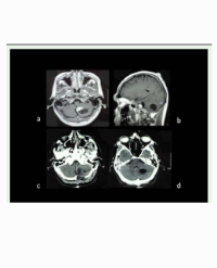
Antero-medially mass is found to be abutting superior vena cava and trachea with encasement right main bronchus. Irregular peripherally enhancing lesion in left lobe of liver noted (Figure 7). Computerized tomography of brain and MRI brain revealed mass in base of brain in parasellar area. Computerized tomography guided biopsy showed undifferentiated large cell carcinoma (Figures 8,9). Hip joint radiography showed metastasis in lesser trochanter (Figure 10). Immunohistochemistry was positive for CD45. Patient was referred to oncology centre for palliative treatment. Patient expired after 20 days.

Figure 7: Liver metastasis in CT scan.

Figure 8: CT guided fine needle aspiration cytology showing large cell carcinoma.

Figure 9: Large cell carcinoma.

Figure 10: Trochanteric metastasis in hip radiography.
Discussion
According to the previous studies [5], risk of malignancy increases with severe tuberculosis. There are 14 case reports described regarding tuberculosis associated with cancer development and 47 case reports regarding coexistence of tuberculosis and malignancy [5]. Non-Small Cell Lung Cancer (NSCLC) accounts for approximately 85% of all lung cancers. In most series, large cell carcinoma comprises between 5% and 10% of all lung cancers [14]. Estimated odds ratio associated with smoking two or more packs/day for current smokers is 37.0 in men and 72.9 in women [15]. Scarring was more common in current and former smokers, related to cumulative amount of tobacco use [6].
Scarring is associated with increased cancer risk in ipsilateral lung [6]. Adenocarcinoma is the commonest malignancy in peripheral lung scars. Our patient’s presentation with acute bilateral lateral rectus palsy due to brain metastasis and trochanteric metastasis was rare. Large cell carcinoma in peripheral scar of lung is unusual. Brain metastases occur in 30-50% of Non-Small Cell Lung Cancer patients with worse prognosis and quality of life [16].
Lung Scar Carcinoma (LSC) was first reported in 1939 by Friedrich. The majority of lesions were localised to the upper lobe of the lungs in the periphery and were smaller than 3 cm. Male predominance was observed and adenocarcinoma is commonest peripheral lung cancer [17-19]. In adenocarcinoma, fibrosis or scarring (hyalinosis) may be present at the centre of the tumour [18].
Carbon powder decomposition and intensive fibrotic degeneration will be visible on the cut surface. Adenocarcinomatous cells are arranged in a single layer or in small nests. Few malignant cells may be localised to the subpleural area or interlobar pleura. LSCs have a tendency for early lymph-node metastasis and smallvessel invasion [17], which results in a lower 5-year survival rate (5%) than the general 5-year survival rate of adenocarcinomas (22%) and adenosquamous carcinomas (28%).
LSC should be suspected when a new lesion emerges in a lung scar or a focal scar shows enlargement in follow-up radiographic images. In study by Bakris GL, et al. [20] the comparison revealed scar carcinomas possess distinctive features: A higher histologic distribution of adenocarcinoma (58% versus 15% in non-scars) and the frequent presentation (53%) with only non-pulmonary symptoms and signs related to metastasis. In scar carcinomas both bronchoscopy and sputum cytology were ineffective as initial diagnostic tools since chest findings were absent or minimal. Lung scars usually occur secondary to bronchiectasis, tuberculosis, organized pneumonia, pulmonary abscess, trauma or infarction, with tuberculosis being the commonest cause [18].
The pathogenesis of LSC is not clear. It may be the result of chronic inflammation of a lung scar. Repeated injury and repair process over a prolonged period stimulates bronchial and alveolar epithelium, causing atypical hyperplasia or canceration of focal lesions. Fibrosis can block or disturb lymphatic drainage, resulting in pooling of cancer cells in the scar. This causes more extensive vascular and lymphatic seeding in scar cancer compared to regular adenocarcinoma or squamous cell carcinoma.
We put forward following statements based on our case report. Tuberculosis and smoking may increase the risk of lung scarring and malignancy. Pulmonary scarring may be associated with increased lung carcinoma in ipsilateral lung. Clinician needs to be more sensitive to look for malignancy association particularly in patient with or previously treated for tuberculosis and even more emphasis to be laid if scarring of lung is observed.
References
1. Noronha V, Dikshit R, Raut N, Joshi A, Pramesh CS, George K, et al. Epidemiology of lung cancer in India: Focus on the differences between nonsmokers and smokers: a single-centre experience. Indian J Cancer. 2012; 49: 74-81.
2. Jemal A, Murray T, Samuels A, Ghafoor A, Ward E, Thun MJ. Cancer Statistics 2003.CA Cancer J Clin. 2003; 53: 5-26.
3. Lu C, Onn A, Vaporciyan AA, et al. “78: Cancer of the Lung”. Holland-Frei Cancer Medicine. 8th edition. People’s Medical Publishing House: 2010 ISBN 978-1-60795-014-1.
4. Martin Y, Artaz MA, Bornand-Rousselot A. Pyothorax associated lymphoma in an elderly woman with a history of lung tuberculosis. J Am Geriatr Soc. 2004; 52: 1226-1227.
5. Falagas ME, Kouranos VD, Athanassa Z, Kopterides P. Tuberculosis and malignancy. QJM. 2010; 103: 461-487.
6. Yu YY, Pinsky PF, Caporaso NE, Chatterjee N, Baumgarten M, Langenberg P, et al. Lung cancer risk following detection of pulmonary scarring by chest radiography in the prostate, lung, colorectal, and ovarian cancer screening trial. Arch Intern Med. 2008; 168: 2326-2332.
7. Tuberculosis Fact sheet N°104”. World Health Organization. 2010.
8. Tuberculosis. World Health Organization. 2002.
9. World Health Organization. “Epidemiology” (PDF). Global tuberculosis control: epidemiology, strategy, financing. 2009.
10. Improved data reveals higher global burden of tuberculosis. who. int. 2014.
11. GBD 2013 Mortality and Causes of Death Collaborators. Global, regional, and national age-sex specific all-cause and cause-specific mortality for 240 causes of death, 1990-2013: a systematic analysis for the Global Burden of Disease Study 2013”. Lancet. 2015; 385: 117-171.
12. Pranali P, Pradeep A, Rakesh T. Evaluation of Microscopy, Culture and PCR Methods in the Laboratory Diagnosis of Genito-urinary Tuberculosis. American Journal of Infectious Diseases and Microbiology. 2014; 2: 17-21.
13. Lawn SD, Zumla AI. Tuberculosis. Lancet. 378: 57-72.
14. Molina JR, Yang P, Cassivi SD, Schild SE, Adjei AA. Non-small cell lung cancer: epidemiology, risk factors, treatment, and survivorship. Mayo Clin Proc. 2008; 83: 584-594.
15. Muscat JE, Stellman SD, Zhang ZF, Neugut AI, Wynder EL. Cigarette smoking and large cell carcinoma of the lung. Cancer Epidemiol Biomarkers Prev. 1997; 6: 477-480.
16. Schouten LJ, Rutten J, Huveneers HA, Twijnstra A. Incidence of brain metastases in a cohort of patients with carcinoma of the breast, colon, kidney, and lung and melanoma. Cancer. 2002; 94: 2698-2705.
17. Freant LJ, Joseph WL, Adkins PC. Scar carcinoma of the lung. Fact or fantasy? Ann Thorac Surg. 1974; 17: 531-537.
18. Madri JA, Carter D. Scar cancers of the lung: origin and significance. Hum Pathol. 1984; 15: 625-631.
19. Balo J, Juhasz E, Temes J. Pulmonary infarcts and pulmonary carcinoma. Cancer. 1956; 9: 918-922.
20. Bakris GL, Mulopulos GP, Korchik R, Ezdinli EZ, Ro J, Yoon BH. Pulmonary scar carcinoma. A clinicopathologic analysis. Cancer. 1983; 52: 493-497.
Citation
Sudulagunta SR, Kothandapani SK, Sodalagunta MB, Khorram H, Sepehrar M and Noroozpour Z. Carcinoma in Tuberculosis scar. SM J Clin Med. 2015; 1(2): 1008.