Back to Journal

SM Journal of Clinical Anatomy

Gingival Biotype on Aesthetic Implant Zone: A Review

[ ISSN : 2578-6954 ]

Abstract Citation Introduction The Characteristics of Gingival Biotypes Methods for Determining Gingival Biotype Conclusion References
Details

Received: 24-Nov-2023

Accepted: 27-Dec-2023

Published: 29-Dec-2023

Arathi Anil, Arun KV*, Biju Balakrishnan, Rajesh Vyloppillil, Maya Rajan Peter, and Reshma Suresh

Amrita School of Dentistry, Department of Periodontics, India

Corresponding Author:

Arun KV, Department of Periodontics, Amrita School of Dentistry, Kochi, India

Keywords

Dental implant; Gingiva; Soft tissue biotype; Aesthetic dentistry.

Abstract

In the anterior region, dental implant treatment necessitates not only functional but also good aesthetic outcome. The gingival structure between the implant restoration and the neighboring natural dentition should be in harmony, balance, and continuity. In restoration and regenerative therapy gingival biotype plays an important part. Therefore, the identification of periodontal biotypes is important in clinical practice. This article focuses on the general characteristics of gingival biotypes associated with aesthetic.

Citation

Anil A, Arun KV, Balakrishnan B, Rajesh V, Maya Rajan P, et al. (2023) Gingival Biotype on Aesthetic Implant Zone: A Review. SM J Clin Med 3: 7.

Introduction

Anterior maxillary dental Implants are today considered as a feasible treatment option to replace missing teeth both for aesthetics and function. The deep-rooted success of aesthetic restoration depends on number of factors, including the gingival biotype, structure, and the position of the teeth in front. The morphology of the gingiva is crucial in the treatment of periodontitis, a vital effect on the aesthetic result in the end (1). Hence, when planning the treatment, it’s critical to understand the variations in gingival tissue morphology (2). A variety of gingival biotypes have different responses to inflammatory, restorative, traumatic, along with dysfunctional habits.

In oral cavity, esthetic zone is considered from canine to first and second premolar region. Thus, implant placement in aesthetic zone is often more complex and challenging than in other area of dental arch. Immediate implantation in the cosmetic zone and restoration of single implants has several benefits, such as a lower overall chair time, reduced surgical intervention. Patient satisfaction has improved as a result of less stressful surgery. This therapeutic procedure, on the other hand, has the following inherent drawbacks like difficulty to achieve stability, and substantial probability of implant failure, the necessity of soft and hard tissue levels grafting (2).

The Characteristics of Gingival Biotypes

The width of the gingiva in the faciopalatal dimension determines gingival biotype. It has a major impact on restorative,regenerative, and implant therapy outcomes. Gingival biotypes can be classified as follows in Table 1 (3).

Table 1:Classification of gingival biotype.

Some of the characteristics and disease response with pocket formation and infrabony defect, soft tissue is flat and bony layout, a denser and more granulation tissue soft tissue curtain, a big quantity of connected masticatory mucosa, resistance to acute stress, disease response with pocket development and infrabony defect, and a significant amount of attached masticatory mucosa). The teeth are also squarer in form, with flatter posterior cusps. Facially and incisogingivally, the contact regions of neighbouring teeth are greater. Gingival biotypes with thin gingiva are fragile, transparent and heavily scalloped. Soft tissues appear soft and brittle with a minimum of gingival adhesion. The bone underneath is the thin or a minimum of bone above the labial root, and fenestration and dehiscence may be present (4).

Patients having a biotype that is thin scalloped are regarded at risk because they’ve got a lower rate of soft tissue response after surgery and/or repair. Teeth are more triangular than scalloped biotypes and has a steep posterior tip (5). The contact surface of adjacent teeth is small on the lingual and gingival margins of the face, towards one-third incisal or occlusal gingival thickness can affect medication outcomes due to differences in blood supply to the underlying bone’s, sensitivity to absorption (6). It is more frequent to take place in patients with thin biotypes, and the post extraction process of remodelling makes alveolar resorption in the apical and gingival instructions are more dramatic (7). Non traumatic removal and alveolar preservation is a term that refers to the preservation of the alveolar plate when the location is being used for implantation (8).

Methods for Determining Gingival Biotype

To quantify tissue thickness, a variety of invasive and non-invasive approaches have been developed. The visual examination direct methods include transgingival probing, endodontic reamers and files, probe transparency (TRAN), ultrasonic methods, and a cone-beam computed tomography (CBCT) scan are some of these options listed in Table 2.

Table 2:Different techniques available for the assessment of gingival biotype

Gingiva is visually assessed and evaluated based on its overall look during a visual examination. Benefits include: The extent of soft tissue cannot be measured using this procedure, which is simple and non-invasive. Due to a substantial inter examiner variation, accuracy is low (9).

Presurgical Oral Examination

The following should be covered in a comprehensive oral evaluation prior to surgery (10).

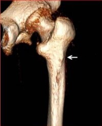
Quality and quantity of bone:The facial expression and labial position, and the grin can all be considerably influenced by the contour ,thickness, height, of the facial alveolar plate (11). The alveolar plate’s architecture varies greatly from one individual to another (12). It takes bone with a bucco-lingual diameter of 6 mm and a mesio-distal dimension of 5–6 mm to support the dwelling of a conventional 3.75–4 mm implant. Implant angulation affects the facial alveolar plate’s height and thickness. A broad, flat bone in the face that supports soft tissue in an abnormally more coronal position is connected to a lingual tilting of an implant. A thin, scalloped face alveolar bone that is frequently found in an apical position is linked to a labial implant inclination. The facial alveolar plate may need to be expanded both vertically and horizontally following implant insertion. For the overtime maintaining soft tissue height, this is crucial (13–15). The root positions of nearby teeth may contribute to boundaries in bone amount in the mesio-distal dimension. The required room for implant implantation may be created through orthodontic movement used to alter the position of the root. A smaller horizontal separation between teeth and an adjacent implant may negatively impact the level of bone on the tooth side(14).

Dental morphology

The anterior aesthetic area around the mouth is where this relationship between tooth form and periodontal biotype is most obvious. The tooth’s triangular form is connected to a narrow periodontium with scallops (Biotype I). The interproximal contact area of this biotype is connected to a long, thin papilla, and it is situated in the crown’s coronal third. The tooth’s connection to a thick, flat periodontium is square in shape (Biotype II)(2). A short, wide papilla is supported by the interproximal contact area, which is situated in the middle third of the crown (5).

Occlusion and occlusal forces: The linked bone density and thickness indicate the orientation, magnitude, and time of masticatory stresses on implants. The facial alveolar plate’s width and height appear to be negatively correlated with the angular implant forces. To focus as much of the masticatory forces as possible on an implant’s longitudinal shaft, occlusal correction may be necessary in order to achieve a favourable prognosis (16). To determine if an implant restoration would be in compliance with the demands of proper phonetics and occlusion, prosthodontic treatment planning is frequently necessary.

Adjacent periodontium: Nearby periodontally healthy teeth’s cemento-enamel junction and the crown-abutment junction mainly line up (17–19). The consequences of significantly reduced periodontium around teeth next to implants are (I) more substantial clinical implant crown and (ii) diminished papilla size or absence. To enable the restoration to blend with a natural profile, a conventional 3.75- or 4-mm-diameter implant should be positioned 3- 4 mm apical to the buccal soft tissue level of the neighbouring teeth. To provide allows a progressive change from the implant platform’s 4-mm diameter to the crown’s 7-8 mm size at the gingival edge, a vertical distance of 3-4 mm is required. If a maxillary lateral incisor needs to be replaced, the implant can be placed more coronally since there is less space needed for transition because the typical crown diameter at the gingival level is about 5 mm.

Lip position, lip mobility and smile line: The main anatomical aspect of the lips is their vermilion border, or the redness around the mouth. The vertical groove on the middle top lip, where the Vermilion-skin interface is most clearly marked, is referred to as the philtrum. The centre Vermilion beneath the philtrum is filled out by a clearly pronounced upper-lip tubercle. The site of the implants, the peri implant architecture, and the dental architecture all have a considerable impact on the tonus, form, and tubercle of the Vermilion, as well as the philtrum (20). In comparison to the maxilla, the mandible has less of a demand for an aesthetic result, allowing the dentist to place more of an emphasis on long-term implant health than on aesthetics. 

Texture, volume, colour, and structure of soft tissue: The oral mucosa normally consists of a coral-pink masticatory mucosa and a glossy red alveolar mucosa (21). Collagen fibres make up the moveable alveolar mucosa loosely, and the epithelium is lacking rete pegs, nonkeratinized, and is thin. The masticatory mucosa’s epithelium, in contrast, is immovable, thick, keratinized, and made of dense, well-organized collagen fibres. The masticatory mucosa is stippled, robust, and securely linked to the periosteum, making it resistant to thermal, chemical, and physical stress (22). The upper lip’s contour is often parallel to the gingival border of the maxillary teeth. The canines and central incisors of the maxilla share a mucosal margin that is equal in height. In comparison to the canine and central incisor, the mucosal border of the maxillary lateral incisor is 0.5–1.0 mm more coronal. When someone smiles normally, the labial tone and location cause to be visible: 75–100% of the maxillary anterior teeth and any associated soft tissue (20).

Tissue Reaction to Therapy: Tissue biotypes are a major factor influencing the outcome of aesthetic treatment. The findings show that inflammation, trauma, and surgery affect these two tissue biotypes injury in different ways. Thick bone plates linked with thick biotypes respond differently to extraction than thin bone plates associated with thin biotypes. Thick biotypes minimize atrophy of the coat of arms after extraction (23).

However, extraction-induced trauma can result in fractures of thin biotype, labial apical and lingual plates and traumatic ridge resorption sides. Bone and gingival tissues differ between biotypes of thick and thin tissue, and these differences have a substantial influence on Preparation of the position of the implant and the treatment preparing the width of the gingival and bone uniformity of the apex and soft tissue is instantly tied to tissue (24). To build an aesthetics each prosthesis not to mention matches size, shape, and location, and colour of the next tooth, but also the soft tissue all over the tooth implant and the surrounding gingiva and mucosa, resulting in an aesthetically acceptable replacement to establish compatibility. It also aids in the preservation of bone structure. To achieve the best aesthetic effects, enhancement of both soft and rigid tissues should be done at the same time, performed in addition to implantation (25).

Indices: The ICAI, PES/WES, and a novel index called the “Peri-Implant and Crown Index (PICI),” which the authors had developed, were all approved and employed in Table 3. The PES/WES (pink aesthetic score/white aesthetic score) criteria, which include five white and five pink parameters, were utilised to compare the pink (PES) and white (WES) of single implant reconstructions to the contralateral tooth.

The ICAI was also utilised to compare the pink and white aesthetics reconstructions with a single implant to those of neighbouring and contralateral teeth. Table 3 lists the four pink and five white criteria that were evaluated and rated

Finally, the new PICI was developed for this study in order to compare the pink and white aesthetic qualities to the appearance of the contralateral tooth using visual analogue scales. Three pink, white, and subjective total criteria make up the PICI (Table 3).

Table 3:Guidelines for the three Esthetic Indices and Comparative Esthetic Calculation

The extreme left of the visual analogue scale for pink and white aesthetics suggests that the implant restoration will be totally different from the contralateral tooth while the right indicates that the crown of the implant will be similar to the tooth in contralateral side (26)

CEI (Criteria for Esthetic Index) for Implant Supportedanterior Maxillary Restorations

The suggested CEI (Criteria for Esthetic Index) is summarised in Table 4. Index).

Table 4:CEI for an Anterior Maxillary Implant- supported restoration S, P and R Assessment Ratings and Evaluation Grades.

The soft tissue index (S), predictive index (P), and implant-supported restoration index (ISR) make up this aesthetic index (R). Specific criteria within each area were assessed and rated as adequate (rating 20%), compromised (rating 10%), or poor (rating 0%).

Soft tissue shape, colour and texture alterations, vertical deficiency, and mesial and distal papillae appearance are all characteristics of the S that have been previously identified. The P evaluated the proximal and horizontal contour inadequacy, gingival tissue biotype, implant apico-coronal location, and distal interproximal bone height are all factors to consider.

A periodontal probe was used to calculate from the dental implant’s centre to an imagined line drawn on the buccal bone flange surrounding teeth to determine the horizontal contour defect. Appropriate, compromised, and poor grades were determined as distances of 0, 1 to 3, and >3 mm, respectively.

The R looked at the Colour and translucency of implant supported restorations, surface roughness and ridges, implant/ crown incisal edge position, Table 1 shows, for each grade there is a variance in parameter ratings. Appropriate, compromised, or inadequate crown width/length ratios were determined as 0.85,0.85 to 1.0,>1.0.

Each of the indices’ components (S, P, and R) is portrayed separately to make the CEI more useful. As previously stated, each component comprised of five distinct characteristics that were classified as adequate (20%), compromised (10%), or poor (10%). (0 percent)(27).

Clinical Significance: When planning treatment, soft tissue biotypes should be considered because they have an impact on the final therapy outcome. The thickness and contour of the soft tissue are crucial diagnostic criteria that alter the cosmetic result of implant repair. The long-term stability of the implant and the gingival margin around the adjacent tooth depends on the proper facial bone height and thickness (Table 5)(28).

Table 5:Treatment for thick and thin gingival biotype

Biotype and Immediate Implant Placement: Wohrle first reported on the Immediate implantation and provisionalization technique in the aesthetic zone. It has since been used in a number of studies and has proven to have the ability of effective high-success/survival therapy rates and gingival health that is stable in structure. The width of the keratinized gingiva in the central buccal part of the socket should be at least 2 mm, and the biotype of the gingiva should be thick (more than 2 mm). To be able to maintain the stability in the primary implant, the apical bone behind the extraction socket must be at least 4 mm.

The tips among the interdental contact and the most coronal papillae should be the mesial and distal papillae. The gap between the extraction socket’s face bone and the implant should be at least 2 mm to keep the soft tissue profile of the implant for ideal aesthetics. The most difficult goal of implant treatment in the aesthetic field is to achieve Stability of soft and rigid tissues throughout time. Retraction of the mucosa in the central face is one among the most popular problems after a quick implantation.

The anatomical factors associated with the retraction of the central face after the gingival biotype and the keratinized mucosal width are two factors that influence rapid implantation. When implants were implanted in individuals with thin gingival biotypes and keratinized mucosa less than 2 mm broad, there was more recession. Despite simultaneous bone management, 8.3% of sockets with narrow V-shaped facial bone defects larger than 3 mm observed central facial recession after immediate implantation and provisionalization, larger than 1.5 mm after a year (29).

Limitations

Psychological factors: While helping to restore oral function and attractiveness, implant dentistry can occasionally fall short of exceedingly high patient expectations. It is critical to realise that not every patient receives a flawless treatment result from dental implants. It is occasionally possible to achieve equivalent or even better aesthetic outcomes by replacing anterior teeth with resin-bonded restorations or traditional fixed partial dentures. In order to avoid exaggerated expectations and misconceptions, proper patient-dentist communication and documentation are required (30,31).

Health vs. esthetics: The most successful objective of peri implant interference is to diminish and reduce morbidity, such as mucosal inflammation and peri-implantitis. While dental implants elevates a poor aesthetic outlook, which may hinder people and adversely affect sociability, lifestyle, and performance, this is not the main goal of these procedures (32,33). The health status surrounding teeth and dental implants is influenced by the interplay between prospective pathogens and the immunological reaction of the host. It is believed that local host-parasite interactions are influenced by the composition and design of dental implants. The aspects of layout of implant that influence the elevation of soft tissue and bone surrounding implants, meanwhile, are still not completely understood (34).

Interimplant anterior scalloped papilla: The alveolar bone crest serves as the basic guide for peri-implant mucosal height; nevertheless, the determinants of the development of interimplant papilla are complicated and cannot be entirely under the control of implant design characteristics or surgical treatments. Although in the morphology of tooth, contact point of the interdental area, and the quality and alignment of soft tissue fibres can all affect outlook of soft tissue, height and thickness of the bone. A significant barrier to the appearance and control of soft tissue around implants is the paucity of dento-gingivo alveolar circular, semi-circular, intergingival, transeptal, and interpapillary fibres. A key issue in dental implant aesthetics is the absence of interimplant papillae, which results in an interimplant black triangle (34).

Provisional phase: For optimum healing, the kind of temporary prosthesis utilised during the healing process is essential. The interim restoration’s design has to take into account comprehensive diagnostic data and aim to cause the soft tissues as little discomfort and pressure as possible after surgery (35). A suitable interim restoration can offer helpful recommendations regarding the aesthetic appeal (18)

Conclusion

It is vital to know gingival biotype prior to immediate implant placement because they show various pathological reactions when exposed to inflammatory, traumatic, or surgical injuries. These various reactions dictate distinct treatments. Present day periodontal surgery techniques have the ability to increase soft tissue quality and hence improve the restoration environment. Therefore, by considering the gingival tissue biotype in the course of treatment planning, better periodontal management action plan can be developed, leading to more predictable treatment outcomes.

References

1. Abraham S, Deepak KT, Ambili R, Preeja C, Archana V. Gingival biotype and its clinical significance – A review. Saudi J Dental Res. 2014; 5: 3-7.

2. Ochsenbein C, Ross S. A Reevaluation of Osseous Surgery. Dental Clinics of North America. 1969; 13: 87–102.

3. Shah HK, Sharma S, Shrestha S. Gingival Biotype Classification, Assessment, and Clinical Importance: A Review. J Nepalese Society Periodontol Oral Implantol. 2021; 4: 83-88.

4. Kao RT, Fagan MC, Conte GJ. Thick vs. thin gingival biotypes: a key determinant in treatment planning for dental implants. J Calif Dent Assoc. 2008; 36: 193-198.

5. Olsson M, Lindhe J. Periodontal characteristics in individuals with varying form of the upper central incisors. J Clin Periodontol .1991.

6. Anderegg CR, Metzler DG, Nicoll BK. Gingiva Thickness in Guided Tissue Regeneration and Associated Recession at Facial Furcation Defects. J Periodontol .1995.

7. Kois JC. Predictable single tooth peri-implant esthetics: five diagnostic keys. Europe PMC. 2001; 22: 199-206.

8. Romeo E, Lops D, Rossi A, Storelli S, Rozza R, Chiapasco M. Surgical and Prosthetic Management of Interproximal Region With Single Implant Restorations: 1-Year Prospective Study. J Periodontol. 2008; 79: 1048-1055.

9. Sarma M, Shenoy N, Bhandary R. Gingival Biotype: A Secret for Esthetic Success. J Health Allied Sci. NU. 2022; 12: 13-17.

10. Palacci P, Nowzari H. Soft tissue enhancement around dental implants. Periodontol. 2008; 47: 113-132.

11. Katranji A, Misch K, Wang HL. Cortical bone thickness in dentate and edentulous human cadavers. J Periodontol. 2007; 78: 874-878.

12. Becker W, Ochsenbein C, Tibbetts L, Becker BE. Alveolar bone anatomic profiles as measured from dry skulls. Clinical ramifications. J Clin Periodontol. 1997; 24: 727-731.

13. Bengazi F, Wennström JL, Lekholm U. Recession of the soft tissue margin at oral implants. A 2-year longitudinal prospective study. Clin Oral Implants Res. 1996; 7: 303-310.

14. Esposito M, Ekestubbe A, Gröndahl K. Radiological evaluation of marginal bone loss at tooth surfaces facing single Brånemark implants. Clin Oral Implants Res. 1993; 4: 151-157.

15. Grunder U. Stability of the mucosal topography around single tooth implants and adjacent teeth: 1-year results. Int J Periodontics Restorative Dent. 2000; 20: 11-17.

16. Hsu ML, Chen FC, Kao HC, Cheng CK. Influence of off-axis loading of an anterior maxillary implant: a 3-dimensional finite element analysis. Int J Oral Maxillofac Implants. 2007; 22: 301-309.

17. Berglundh T, Lindhe J. Dimension of the periimplant mucosa. Biological width revisited. J Clin Periodontol. 1996; 23: 971-973.

18. Jemt T. Regeneration of gingival papillae after single-implant treatment. Int J Periodontics Restorative Dent. 1997; 17: 326-333.

19. Kan JYK, Rungcharassaeng K, Umezu K, Kois JC. Dimensions of peri implant mucosa: an evaluation of maxillary anterior single implants in humans. J Periodontol. 2003; 74: 557-562.

20. Davis NC. Smile design. Dent Clin North Am. 2007; 51: 299-318.

21. Katafuchi M, Matsuura T, Atsawasuwan P, Sato H, Yamauchi M. Biochemical characterization of collagen in alveolar mucosa and attached gingiva of pig. Connect Tissue Res. 2007; 48: 85-92.

22. Strub JR, Gaberthüel TW, Grunder U. The role of attached gingiva in the health of peri-implant tissue in dogs. 1. Clinical findings. Int J Periodontics Restorative Dent. 1991; 11: 317-333.

23. Denissen HW, Veldhuis AH, Waas AJV. Anatomic Considerations for Preventive Implantation. JOMI.1993; 8: 191-196.

24. Wennström JL, Lindhe J,Sinclair F, Thilander B. Some periodontal tissue reactions to orthodontic tooth movement in monkeys. J Clin Periodontol. 1987.

25. Wennström JL. Lack of association between width of attached gingiva and development of soft tissue recession. A 5-year longitudinal study. J Clin Periodontol. 1987; 14: 181-184.

26. Tettamanti S, Millen C, Gavric J, Buser D, Belser UC, Brägger U, et al. Esthetic Evaluation of Implant Crowns and Peri-Implant Soft Tissue in the Anterior Maxilla: Comparison and Reproducibility of Three Different Indices. Clin Implant Dentist Related Res. 2016; 18: 517-526.

27. Juodzbalys G, Wang HL. Esthetic index for anterior maxillary implant supported restorations. J Periodontol. 2010; 81: 34-42.

28. Levine RA, Ganeles J, Kan J, Fava PL. 10 Keys for Successful Esthetic Zone Single Immediate Implants: Importance of Biotype Conversion for Lasting Success. 2018; 39: 522-529.

29. Meng HW, Chien EY, Chien HH. Immediate Implant Placement and Provisionalization in the Esthetic Zone: A 6.5-Year Follow-Up and Literature Review. Case Rep Dent. 2021; 2021: 4290193.

30. Parkerson GR, Broadhead WE, Tse CK. Quality of life and functional health of primary care patients. J Clin Epidemiol. 1992; 45: 1303-1313.

31. Sullivan RM. Perspectives on esthetics in implant dentistry. Compend Contin Educ Dent. 2001; 22: 685-692.

32. Park LE. Appearance-based rejection sensitivity: implications for mental and physical health, affect, and motivation. Pers Soc Psychol Bull. 2007; 33: 490-504.

33. Nowzari H, Botero JE, DeGiacomo M, Villacres MC, Rich SK. Microbiology and cytokine levels around healthy dental implants and teeth. Clin Implant Dent Relat Res. 2008; 10: 166-173.

34. Marques Filho JS, Gobara J, da Silva Salomao GV, Sumita LM, Shibli JA, Viana RG, et al. Cytokine Levels and Human Herpesviruses in Saliva from Clinical Periodontal Healthy Subjects with Peri-Implantitis: A Case-Control Study. Mediators Inflamm. 2018; 2018: 6020625.

35. Cho SC, Shetty S, Froum S, Elian N, Tarnow D. Fixed and removable provisional options for patients undergoing implant treatment. Compend Contin Educ Dent. 2007; 28: 604-608

Other Articles

Article Image 1

Cannieu-Rich

Objective: The aim of this paper is to report the incidences and provide a new classification of the various types of Cannieu-Riché Anastomosis (CRA).

Materials and methods: The anatomical dissections of 80 limbs from 40 fresh adult cadavers were performed in the Department of Anatomy of the Medical School of the Catholic University of São Paulo. Sixty hands were dissected from 1979 to 1983, and 20 hands were dissected from 2011 to 2015. In all subjects, both hands were studied. Careful dissections were performed under high magnification (with a surgical microscope) with special reference to the incidence of CRA

Results: CRA was found in all of the dissected hands (100%). We propose a new classification of CRA: Type I until Type XI, describing the communication between the recurrent branch of the median nerve and the deep branch of the ulnar nerve to the deep head of flexor pollicis brevis, which is the most common type of anastomosis and our others observations.

Conclusion: According to our study, CRA should be viewed as a normal anatomical neural connection and not as an anatomical variation. The knowledge of this anastomosis is essential because the presence of such neural communication can cause confusing clinical, surgical and electromyographical findings in cases of complete or incomplete median or ulnar nerve lesions or entrapment neuropathies

Edie Benedito Caetano*, Yuri da Cunha Nakmichi, Maico Minoru Sawada, Renato Alves de Andrade, Mauricio Tadeu Nakasone and Luiz Angelo Vieira


Article Image 1

A Long Rectal Duplication Cyst in an Adult Cadaver: A Rare Anatomical Variation

This report describes a long rectal duplication cyst found during routine dissection of an 82 year old male cadaver. The cyst was tubular, blind-ending and non-communicating. It emerged from the posterior rectal wall approximately 13cm superior to the anal verge and was 12cm in length. Long duplication cysts such as that described may cause complications such as perforation, bleeding and chronic pain. Hence duplication cysts should form part of the differential work up in a clinical setting.

Aneesh Dave*, Rohan Dalal, Bardia Aryaie and Bahadir Cem Demirdes


Article Image 1

Low-Cost Models for Simulation as a Tool for Increasing Medical Student Interest in and Understanding of Otolaryngology

Objectives: We sought to determine the utility of a low-cost simulator and brief educational session to help students understand midline neck anatomy and increase exposure to otolaryngology with an easily reproducible model.

Design: Pilot study including survey with lecture and hands on instructional lesson.

Methods: As part of the first-year medical student curriculum at our university, the otolaryngology department offers a workshop on head and neck anatomy and related components of the physical exam. Students participated in five 20-minute stations. One was devoted to neck anatomy with models of midline structures and a tracheostomy. Students completed a survey after the head and neck station.

Results: 182 students responded to the survey. Over 85% of participants felt the head and neck station and models helped them understand anatomy of the neck. Eighty-five percent of students responded the models helped them visualize what a tracheostomy would look like in a patient. Before the head and neck station, 16% of students agreed or strongly agreed that they had interest in ENT, Ear Nose and Throat surgery, as a career. After the station, that increased to 73% (p<0.05). Sixty-nine percent of students stated the tracheostomy models specifically increased their interest in otolaryngology

Conclusion: Simulation is a useful adjunct for teaching head and neck anatomy to medical students. Our workshop’s head and neck station, with lecture and simulation, strongly increased participant interest in otolaryngology and solidified relevant anatomy.

Lara Reichert1 *, Kevin Lin2 , Daniel Farishta2 , Harold Pine1 and Susan McCammon1


Article Image 1

Micro-Anatomy of the Brachial Plexus in a Cadaver with Ultrasound Correlation and Surface Videography in a Live Model

We colorized 1860 saggital slices from the brachial plexus of a cadaver including the fascicles, blood vessels and bones. These slices were compiled into a continuous video and correlated with continuous sonography of a live model. The video shows cadaver slices and ultra-sound videos of:

(1) The nerve roots and transverse processes

(2) The intercostal nerves and paravertebral space

(3) The plexus trunks and scalene muscles

(4) The plexus divisions, subclavian artery and pleura

(5) The plexus cords and axillary artery

(6) The formation of peripheral nerves and the brachial artery

Paul E Bigeleisen1 *, Jeremy Kaplowitz1 , JooYeon Ha2 , Gerbrand Groen3 and Nizar Moayeri4


Article Image 1

Assessment of an Anatomic Variant That May Mimic Prefracture Findings of Drug-Associated Atypical Femoral Fractures on Conventional Radiographs: The Third Trochanter

Introduction: The study objective was to assess lateral femoral cortex variants that may mimic prefracture findings of drug-associated atypical femoral fractures (AFF) among hip radiographs.

Materials and Methods: Bilateral hip radiographs of 1493 consecutive patients (mean age 67.7, 804 women) were reviewed. Hips were positive if localized lateral subtrochanteric femoral cortical thickening (LSFCT) was present. Positive studies were divided into a medication group if history of bisphosphonate or denosumab use was present or a variant group. The medication group was subcategorized into a prefracture group if classing beaking LSFCT or a contralateral AFF was presentor a non-prefracture group. The LSFCT width, femoral head and lesser subtrochanteric distances were measured. Analysis of Variance (ANOVA) was performed (p <0.01) to compare the three groups, with post hoc Tukey HSD evaluation. Cross-sectional imaging for each group was reviewed.

Results: Of the1493 exams, 1079 were included. In the 24 patients with LSFCT, 8 patients were assigned to the medication group and 16 to the variant group. Of the 8, 3 met criteria for the prefracture group and 5 were subcategorized to the non-prefracture group. Differences among the prefracture versus the non-prefracture and variant groups were statistically significant (p<0.01). Cross-sectional imaging of the correlated LSFCT with the third Trochanter posterolaterally in the variant group and laterally for the prefracture group

Conclusions: The third trochanter is an anatomic variant that may mimic prefracture findings of drugassociated AFF. The third trochanter can be differentiated radiographically from prefracture AFF findings with a < 3 mm width and < 3mm lesser subtrochanteric distance.


Troy H Maetani1 *, Stacy E Smith2 and Barbara N Weissman2


Article Image 1

Branching Pattern of Collateral Branches of the Subclavian Artery

Introduction: The arteries of the posterior cervical triangle (lateral cervical region) are frequently used for flap design in plastic and reconstructive surgery. In particular, the musculocutaneous flaps, such as the flap of the lower trapezius and the dorsal scapular flap, are based on the integrity and functional availability of the dorsal scapular artery. Likewise, the cervico-dorsal and cervico-scapular flaps are based on the superficial cervical artery. We can assume that the mode of birth of these arteries - and the possibility of their origin directly from the subclavian artery - will have consequences on their vascular dynamics. These consequences can result in good or bad viability of the irrigated flaps. The objective of the present study was to highlight the birth modalities of the subclavian artery branches

Material and Methods: Dissection was performed in 58 anatomical regions from 32 non-formalin fixed cadavers. The cadavers had no history of surgery or deformity in the areas targeted for dissection (supraclavicular and dorsal regions). They were embalmed using a glycerin-rich, formalin-free solution to preserve tissue suppleness.

Results: The average age of the donors was 72.32 years (range, 34–90) and there were 21 males and 11 females. We find some similarities, with a predominance for types D (Cervico-scapular trunk from the thyrocervical trunk: 28%), A (Separate origin of superficial cervical, dorsal scapular, and suprascapular arteries: 19%), and C (Cervico-dorsal trunk from the subclavian artery: 15%).

Conclusion: There are many anatomical variations concerning the birth modalities of the collateral branches of the subclavian artery. Surgical teams should consider these variations when exploring the region of the lateral triangle of the neck.

Philippe Manyacka Ma Nyemb1,2*, Christian Fontaine3, Xavier Demondion3, Maurice Demeulaere3, Fabien Descamps3 and Jean-Marc Ndoye4


Article Image 1

Anatomical Apps and Smartphones: A Pilot Study with 100 Graduation Students

Background: Anatomical teaching is going through several changes. Several studies observed that the students are deeming anatomy as outdated and irrelevant. The rates of lectures attendance and interest are declining. In this context, tools such as body painting, board games and team quizzes are appearing in the classroom to aid and motivate the student. Often, these complementary tools showed positive results towards their goals in the learning process of several branches of medicine. The smartphone has also been used for these purposes, although the literature lacks studies regarding anatomy. The study conducted herein aimed to create a pilot version of an anatomical application for the smartphone and disseminate its usage in nursing students

Methods: A smartphone application was self-produced with the tool known as Fábrica de Aplicativos© (https://fabricadeaplicativos.com.br) and consisted of anatomical terms and their meaning (Dictionary of Anatomy). The students had the opportunity to use this application during the semester and by the end of the period they answered a questionnaire with questions about the quality and efficacy of the app, with the option of submitting suggestions for its improvement.

Results: 100 graduation students were enrolled in this study. The overall evaluation of the dictionary was excellent/great (65%), while critics such as the lack of better images, the lack of a search resource and the lack of sections were pointed out.

Conclusion: As such, it was observed that the smartphone may have a positive effect in the learning process of anatomy, as demonstrated by our study

Lucas Alves Sarmento Pires1 , Tulio Fabiano de Oliveira Leite2 , Albino Fonseca Junior1 , Marcio Antonio Babinski1 and Carlos Alberto Araujo Chagas1


Article Image 1

A Symptomatic Multiseptate Gallbladder without Gallstones

Multiseptate Gallbladder is a rare congenital abnormality, which can be totally asymptomatic or responsible for biliary pain in the absence of cholelithiasis or cholecystitis. In case of persistent symptomatology, cholecystectomy represents the treatment of choice

Damien Dousse1 *, Laura Marcu2 and Francesco Martini1


Article Image 1

Evidence for Endogenous Neurosteroid Production in the Mammalian Olfactory Mucosa: Immunocytochemical Localization of Cytochrome P450 SideChain Cleavage Enzyme

Steroid hormones and their metabolizing enzymes have previously been identified in the olfactory mucosa. Enzymes of the cytochrome P450 family that are thought to play a role in the metabolism or activation of airborne toxins have also been identified in mammalian olfactory mucosae. In the synthesis of steroid hormone, cholesterol is transported to the mitochondria where the side-chain cleavage enzyme (cytochrome P450scc) converts cholesterol into pregnenolone. Conversion of cholesterol to pregnenolone is an obligate step in steroid hormone production. The specific aim of this study is to identify cytochrome P450scc in the mammalian olfactory mucosae. Using polyclonal antibodies to cytochrome P450scc, we found immunoreactivity for cytochrome P450scc in the rat olfactory mucosa. Within the olfactory epithelium, the supranuclear region of sustentacular cells was immunoreactive for cytochrome P450scc. Olfactory neurons, basal cells, olfactory nerve axons and acinar cells of Bowman’s glands were unstained. In positive control tissue (adrenal glands), staining for cytochrome P450scc was seen in all layers of the adrenal cortex. The localization of cytochrome P450scc to sustentacular cells is consistent with a functional role for mitochondrial cytochrome P450scc in the production of olfactory mucosa-specific neurosteroids. These endogenous neurosteroids are most likely involved in the modulation of olfactory function

James D Foster*


Article Image 1

Anatomical and Anthropological Investigation of the Articular Surface of the Human Glenoid Cavity in Brazilian Corpses

The glenoid cavity of the scapula shows high morphological variability that has not been clearly elucidated in the literature. Its morphological aspects knowledge can be used in the sex determination, to improve the development of more functional prostheses and in the accurate image diagnostic. The purpose of the present study was to analyze and compare sex and dominance differences in the articular surface of the human glenoid cavity. Anthropometric measurements of the glenoid cavities of scapula were taken of 200 specimens, besides we developed a morphological classification system according the shape of inferior and superior poles and the presence of the glenoid notch in the anterior margin. For all anthropometric parameters, the male scapula showed higher values than the measures in female scapula both in right and left sides, but we did not find a significant difference between the left and right scapula in either sex. On the other hand, taking into consideration the anthroposcopic aspects, 50% of male and 58% of female were classified in a different morphological group when compared to the contralateral glenoid in the same individual. The present study revealed an evident sexual dimorphism and provides information about the articular surface of normal glenoid cavities to improve the diagnosis of orthopedic lesions, to produce better prosthesis and to improve the medico-legal identification

Jefferson M Homem1 , Aline S DeMaman3 , Denise Lachat1 , Ariane Zamarioli2 , José A Thomazini1 and João-José Lachat1 *