Research Article | Volume 2 - Issue 3 | Article DOI :
Download PDF
Owembabazi Elna1 *, Ninsiima Herbert Izo2 , Keneth Iceland Kasozi2 , Abili Sadrax3 , Ssempijja Fred1 , Echoru Isaac1 and Bukenya Edmund1
1 Department of Anatomy, Kampala International University-Western Campus, Uganda
2 Department of Physiology, Kampala International University-Western Campus, Uganda
3 Department of Pharmacology, Kampala International University-Western Campus, Uganda
Corresponding Author:
Owembabazi Elna, Department of Anatomy, Kampala International University-Western Campus, P.O. Box 71 Ishaka Bushenyi, Uganda, Tel: +256703640027; Email: lenaoet@gmail.com
Keywords
Ethambutol; Optic Nerve;
Visual Acuity; Lantana Trifolium; Optic
Neuropathy
Abstract
Introduction: Ethambutol (EMB), an important drug in treatment of multidrug resistant tuberculosis, has been associated with severe side effects including visual impairment.
Purpose: To establish the protective potential of Lantana trifolium ethanolic extract against EMB induced changes in visual acuity.
Materials and methods: Experimental design involving 25 male adult Wistar rats of 110-130g average weight, divided into five groups each comprising five animals. Group A, the negative control received distilled water. Group B, the positive control was treated with EMB 100 mg/kg/day. Test groups C, D, and E were treated with 25, 50, and 100 mg/kg/day of Trifolium Extract (TE) respectively, one hour before administering 100 mg/ kg/day of EMB for five weeks. Visual acuity was determined by the mean escape latencies obtained using a modified Morris water maze
Results: Lantana trifolium ethanolic extract had a dose dependent protective potential against EMB induced changes in visual acuity. This was shown by the significant increase in the escape latencies of positive control group when compared with those of the group A (4.35±0.50), D (4.85±0.65), and E (3.6±0.38). This effect is likely due to the ability of Latana trifolium to prevent inflammation and accumulation of anti-oxidants in the optic nerve.
Conclusion: Lantana trifolium ethanolic extract has a dose dependent protective potential against EMB induced changes in visual acuity. Studies to determine the exact phytochemical component and mechanism of action responsible for this effect should be conducted.
Citation
Elna O, Izo NH, Kasozi KI, Sadrax A, Fred S, Isaac E, et al. Lantana Trifolium Ethanolic Extract has a Protective Potential against Ethambutol Induced Changes in Visual Acuity. SM J Clin Anat. 2018; 2(3): 1017.
Introduction
Ethambutol (EMB) is an antibiotic belonging to the amino-alcohol group [1]. It is an efficacious f irst line antituberculosis agent [2]. EMB is known to cause changes in the optic nerve that result into toxic optic neuropathy (TON) [3]. This was established soon after its discovery in 1960s. In the early 1960’s, studies reported EMB induced optic neuropathy in 50% of patients receiving 60–100 mg/kg/d of EMB [4]. This toxicity is generally described as dose and duration dependent [3], but there is no effective dose that is entirely free from the threat of toxicity [5]. The toxicity is observed at a dose as low as 12.3 mg/kg [6]. According to World Health Organization (WHO) the initial treatment dose should be 15mg/kg daily or 25 mg/kg three times weekly for the first 2 months of treatment and a retreatment dose of 25 mg/kg daily. Although the onset of EMB induced toxic optic neuropathy symptoms are usually delayed [7], occurring at least 1.5 months after therapy initiation [8]. A number of cases of toxicity occurring as early as after less than six days of treatment have been reported [9].
Ethambutol induces changes in the optic nerve that result into visual impairment. This is a progressive painless condition that is mostly diagnosed when recovery is impossible [10]. Although EMB induced changes are often reversible when EMB is discontinued, in some patients recovery is incomplete [11,12]. Moreover, permanent visual impairment has also been reported in some patients [11]. In Uganda TB burden is still high [13], the use of EMB is frequent but the eye tests before and during its use as recommended by WHO are rarely done due to limited resources [14]. Therefore, means of protecting the optic nerve against EMB toxicity need to be sought.
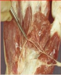
Traditional eye medicines (TEM) are commonly used to treat different eye diseases worldwide [15]. Lantana trifoliuma three leafed, scrambling, evergreen shrub (Figure 1),

Figure 1: Lantana trifolium Plant.
is widely used to treat many eye conditions such as blindness, glaucoma, conjunctivitis, and bacterial and viral eye infection [16]. It is also used to treat epilepsy, infant cerebral malaria, asthma, sinusitis, menstrual pains, mental illiness, sickle cell anaemia, stomach ache and skin conditions [17]. Phytochemical composition of Lantana trifolium has not been studied extensively. However previous studies have reported that the plant contains phenols, phenylpropanoids, flavonoids and terpenoids [18]. Previous ethno-medicinal studies about this plant have reported that its extract inhibited carrageenan and histamine induced rat paw edema [19]. The anti-inflammatory effect of extract is reported to be possibly due to histamine reduction [15].Another study reported that Lantana trifolium extract has anti-oxidative activity due to the high amounts of phenols it contains [20]. This is done through blocking the formation of ROS and scavenging them once they have been formed by donating electrons to the highly reactive free radicals [21], forming stable compounds and thus reducing oxidative stress in the optic nerves. This study established if Lantana trifolium ethanolic extract could protect against ethambutol induced changes in the visual acuity.
Materials and Methods
Twenty five male adult wistar rats of 110-130g average weight were used in this experiment. These were housed in cages at room temperature (25°C ± 1), exposed to 12 hours of dark and light cycles. T he animals were fed on commercial rat pellets and provided with tap water ad libitum.
The animal grouping, plant material collection, preparation of the extract, administration of ethambutol and Lantana trifolium extract were performed as previously described [22].
Dose given = dose (mg/kg) x animal weight (grams) [23].
Concentration (mg/ml) x 1000
Ethical considerations
Directorate of Postgraduate Studies and Research of Kampala International University Western Campus approved this study to be carried out. All animal studies in this experiment were conducted in accordance with the 2010 National Research Council Guide for Care and use of Laboratory Animals. The number of animals used was minimum and inflicting pain to the animals was minimized by sacrificing them under anaesthesia.
Visual acuity testing
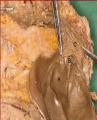
A visible platform Morris water maze was used to determine the visual acuity (Figure 2).

Figure 2: A modified Morris water maze.
This device is based on the principle that rats can instinctively swim and can be trained to escape onto a visible platform. The water maze was a circular galvanized tank with white walls measuring 1.2 m in diameter and 0.6m in height, divided into four equal quadrants. Water at 25°c was used to fill the tank to a depth of 30cm. A circular unfixed platform of 10cm in diameter protruding 1cm above the water surface was used. A bright cloth was used to cover the protruding part of the platform to make it highly visible
All the rats were submitted to pre-training and training session before visual acuity testing was done. During the pre-training session each rat would be placed into the water maze and then allowed to swim, locate and mount the visible platform placed in the center of one of the four quadrats. The rats were given one or more trials of 60 seconds time limit. After succeeding in finding and mounting the platform, the rats were trained for 2 days. In the period of training4 trials were given to each rat on each day. On each trial the platform was moved to the center of a different quadrant, and the rats were released from different starting points and allowed to find and escape onto the platform. After mounting the platform, the rat would be left there for 15 seconds after which it would be removed. The rats would then be put into a holding cage containing pre-warmed towels for 1 minute till the start of the next trial. The rat that would fail to f ind the platform in 60 seconds would be guided to reach it, and this trial would be repeated. The time taken for the rat to find and mount the platform for each trial was recorded as the escape latency (in seconds). Testing was done on day 3during the 1st, 3rd, and 5th week of the experiment. Visual acuity was determined using the mean escape latencies [24].
Data analysis
Using graph pad prisms V6, mean ± SEM were obtained and analysis of variance (ANOVA) was applied to determine group mean differences between the control and test groups. A post-HOC Tuckey test was used to determine which group means differed, considering a p-value of < 0.05 to be statistically significant.
Results
In all the groups the escape latencies obtained during the first week were not significantly different (P > 0.05) revealing no significant change in the visual acuity? Although the escape latencies of all the groups reduced during the 3rd week of the experiment, the differences when compared individually with those in the negative and positive control groups were found to be statistically insignificant (P > 0.05). During the 5th week, the escape latencies of the positive control and the group that received EMB+ 25mg/kg Trifolium Extract (TE) increased (9.65±1.22) and (9.6±0.90) respectively (Table 1).
Table 1: Mean Escape Latency.
|
Experimental Groups
|
Mean time (s) ± SEM
|
|
N
|
Week 1
|
Week 3
|
Week 5
|
|
Negative Control
|
5
|
8.55±0.28
|
6±0.47
|
4.35±0.50##
|
|
Positive Control
|
5
|
8.8±2.02
|
7.9±1.34
|
9.65±1.22**
|
|
EMB + 25mg/kg TE
|
5
|
8.65±1.70
|
7.8±1.15
|
9.6±0.90**
|
|
EMB + 50mg/kg TE
|
5
|
8.7±0.91
|
5.9±0.80
|
4.85±0.65##
|
|
EMB + 100mg/kg TE
|
5
|
8.65±0.48
|
5.75±0.68
|
3.6±0.38###
|
This was statistically significant (P<0.05) when compared against the negative control group (4.35±0.50). There was a further decrease in the escape latencies of remaining groups during the 5th week of the study. When compared against the positive control group, analysis of variance (ANOVA) showed a significant difference(P<0.05) in the escape latencies of the negative control group and the group that received EMB+ 50mg/kg TE, but the difference was more significant in the group that received EMB+ 100mg/kg TE.
Discussion
Lantana trifolium had a dose dependent protection against visual acuity changes induced by Ethambutol (EMB). This was shown by a reduction in the escape latencies using a water maze (Figure 3).

Figure 3: Bar graph showing the escape latency during week 1, 3, and 5. Multiple comparisons; against the negative control **P<0.005; against positive control; ##P < 0.005; ###P <0.0005. (EMB = Ethambutol; TE = trifolium Extract).
There was no significant difference in visual acuity in the 1st week in all the groups probably due to delayed effects of EMB. During the 3rd week, all the groups showed a reduction in the escape latency possibly due to delayed effect of EMB and learning. This observation is similar to earlier findings [25-27], who reported a significant reduction of escape latency in water maze trained rats.
The group differences in the third week were not significant (P > 0.05) when compared individually with those in the negative and positive control groups. This is possibly because the onset of symptoms of EMB induced changes is usually delayed [28]. Previous studies observed that structural injury to the optic nerve occurs earlier than visual impairment [29]. The increase in escape latencies of the positive control and the group that received EMB+ 25mg/kg Trifolium Extract (TE) during the fifth week could be due to a decrease in visual acuity as a result of EMB induced visual impairment. Similar observations have been reported about EMB toxicity [3,30,31], who observed that EMB induces changes in the optic nerve that manifest as a decrease in visual acuity. There was a further decrease in the escape latencies of the group that received EMB+ 50mg/kg TE and the group that received EMB+ 100mg/kg TE during the fifth week, showing that Lantana trifolium ethanolic extract probably prevented EMB induced changes in the visual acuity.
Since the decrease in visual acuity is in part as a result of damage to the central fibers of the optic nerve which are the most vulnerable to EMB [32], we stipulate that Lantana trifolium ethanolic extract significantly prevented EMB induced decrease in visual acuity perhaps by protecting the optic nerve structure against EMB toxicity. This observation could be the reason for its tradition use in providing relief to some eye conditions with unknown causes of loss of visual acuity [33]. In our previous study we observed that Lantana trifolium ethanolic extract significantly reduced EMB induced axonal demyelination and vacuolation [22]. Thus, Lantana trifolium protected the optic nerve structure against EMB toxicity, through its anti-oxidative and anti-inflammatory activity reported in experimental animals [34].
Conclusions and Recommendations
Lantana trifolium ethanolic extract had a dose dependent protective potential against ethambutol (EMB) induced changes in visual acuity. This effect of Lantana trifolium ethanolic extract could be as a result of its ability to protect the optic nerve structure against inflammation and anti-oxidant accumulation. We suggest that studies using more sensitive tests to measure visual acuity in Wistar rats are carried out. Further studies to establish Lantana trifolium exact phytochemical component and mechanism of action responsible for its protective potential should be conducted.
Acknowledgments
The authors would like to appreciate the management of Kampala International University for funding the research work, and the staff of Central Diagnostic Laboratory at the college of veterinary medicine, Makerere University, for the expert technical assistance during histopathology studies
References
1. Deng L, Mikusova K, Robuck KG, Scherman M, Brennan PJ, McNeil MR. Recognition of multiple effects of ethambutol on metabolism of mycobacterial cell envelope. Antimicrobial Agents and Chemotherapy. 1995; 39: 694-701.
2. Gwaza L, Gordon J, Welink J, Potthast H, Leufkens H, Stahl M, et al. Adjusted indirect treatment comparison of the bioavailability of WHO-prequalified f irst-line generic antituberculosis medicines. Clinical Pharmacology and Therapeutics. 2014; 96: 580-588.
3. Talbert Estlin KA, Sadun AA. Risk factors for ethambutol optic toxicity. International Ophthalmology. 2010; 30: 63-72.
4. Forbes M, Kuck NA, Peets EA. Mode of action of ethambutol. Journal of Bacteriology. 1962; 84: 1099-1103.
5. Zelefsky MJ, Kollmeier M, Cox B, Fidaleo A, Sperling D, Pei X, et al. Improved clinical outcomes with high-dose image guided radiotherapy compared with non-IGRT for the treatment of clinically localized prostate cancer. International Journal of Radiation Oncology Biology Physics. 2012; 84: 125-129.
6. Guerrero E, Lemus D, Yzquierdo S, Vilchez G, Munoz M, Montoro E, et al. Association between embB mutations and ethambutol resistance in Mycobacterium tuberculosis isolates from Cuba and the Dominican Republic: reproducible patterns and problems. Revista Argentina de Microbiologia. 2013; 45: 21-26.
7. Zhao Z, Lan Y, Bai S, Shen J, Xiao S, Lv R, et al. Late-onset radiation induced optic neuropathy after radiotherapy for nasopharyngeal carcinoma. Journal of Clinical Neuroscience. 2013; 20: 702-706.
8. Kervinen M, Falck A, Hurskainen M, Hautala N. Bilateral Optic Neuropathy and Permanent Loss of Vision After Treatment With Amiodarone. Journal of Cardiovascular Pharmacology, 2013; 62: 394-396.
9. Yang HK, Park MJ, Lee JH, Lee CT, Park JS, Hwang JM. Incidence of toxic optic neuropathy with low-dose ethambutol. International Journal of Tuberculosis and Lung Disease. 2016; 20: 261-264.
10. Jeanjean L, Dupeyron G. Trouble’s visuels anorganiques [Non-organic visual loss]. Journal Français D’ophtalmologie. 2014; 37: 415-420.
11. Osaguona VB, Sharpe JA, Awaji SA, Farb RI, Sundaram ANE. Optic chiasm involvement on MRI with ethambutol-induced bitemporal hemianopia. Journal of Neuro-Ophthalmology. 2014; 34: 155-158.
12. Pradhan M, Sharp D, Best S, Vincent A, Vaphiades M. Drug-induced optic neuropathy-TB or Not TB. Survey of Ophthalmology. 2010; 55: 378-385.
13. Kirenga BJ, Ssengooba W, Muwonge C, Nakiyingi L, Kyaligonza S, Kasozi S, et al. Tuberculosis risk factors among tuberculosis patients in Kampala, Uganda: implications for tuberculosis control. BMC Public Health. 2015; 15: 13.
14. Mustak H, Rogers G, Cook C. Ethambutol induced toxic optic neuropathy in HIV positive patients. International Journal of Ophthalmology. 2013; 6: 542-545.
15. Julio LS, Leito SG, Lotti C, Picinelli AL, Rastrelli L, Fernandes PD, et al. Flavones and phenylpropanoids from a sedative extract of Lantana trifolia L. Phytochemistry. 2010; 71: 294-300.
16. Nalubega R, Kabasa JD, Olila D, Kateregga J. Antibacterial activity and phytochemical screening of eleven plants used as poultry ethnomedicines in southern Uganda. Agricultural Journal. 2011; 6: 303-309.
17. de Sena Filho JG, Rabbani ARC, dos Santos Silva TR, da Silva AVC, Souza IA, Santos MJBA, et al. Chemical and molecular characterization of fifteen species from the Lantana (Verbenaceae) genus. Biochemical Systematics and Ecology. 2012; 45: 130-137.
18. Imbenzi PS, He Y, Yan Z, Osoro EK, Cheplogoi PK. Chemical Constituents in Extracts from Leaves of Lantana trifolia and Their In Vitro Anti-oxidative Activity. Chinese Herbal Medicines. 2014; 6: 242-246.
19. Uzctegui B, Vila D, Surez-Roca H, Quintero L, Ortega J, Gonzlez B. Anti inflammatory, antinociceptive, and antipyretic effects of Lantana trifolia Linnaeus in experimental animals. Investigacion Clinica. 2004; 45: 317-322.
20. Salabarría IS, Díaz ABV, Morera TG, Turro DG, Pérez THG. Estudio f itoquímico y de actividad alelopática del extracto de n-hexano del follaje de Lantana trifolia L. (Spanish). Revista CENIC Ciencias Quimicas. 2009; 40: 33-37.
21. Perumal PC, Sophia D, Raj CA, Ragavendran P, Starlin T, Gopalakrishnan VK. In vitro antioxidant activities and HPTLC analysis of ethanolic extract of Cayratia trifolia (L.). Asian Pacific Journal of Tropical Disease. 2012; 2: 952-956.
22. Owembabazi E, Izo NH, Fernandez EMD, Isaac E, Monima LA, Ahimbisibwe J, et al. Neuroprotective Potential of Lantana Trifolium Ethanolic Extract against Ethambutol Induced Histological Changes in the Optic Nerve. Anatomy Journal of Africa. 2017; 6: 1071-1079.
23. Izo NH, Claude K, Samuel O. Anticonvulsant and toxicity effects of ethanolic extract of Thevetia Peruviana ( Pers .) leaves. International Journal of Ethnopharmacology. 2016; 2: 7-13.
24. Prusky GT, West PWR, Douglas RM. Reduced visual acuity impairs place but not cued learning in the Morris water task. Behavioural Brain Research. 2000; 116: 135-140.
25. Baldi E, Efoudebe M, Lorenzini CA, Bucherelli C. Spatial navigation in the Morris water maze: Working and long lasting reference memories. Neuroscience Letters. 2005; 378: 176-180.
26. Gulinello M, Gertner M, Mendoza G, Schoenfeld BP, Oddo S, LaFerla F, et al. Validation of a 2-day water maze protocol in mice. Behavioural Brain Research. 2009; 196: 220-227.
27. Dhingra D, Kumar V. Memory-enhancing activity of palmatine in mice using elevated plus maze and Morris water maze. Advances in Pharmacological Sciences. 2012; 7.
28. Garg R, Kumar Verma S. Isoniazid-and ethambutol-induced psychosis. Annals of Thoracic Medicine, 2008; 3: 149.
29. Han J, Byun MK, Lee J, Han SY, Lee JB, Han S-H. Longitudinal analysis of retinal nerve fiber layer and ganglion cell-inner plexiform layer thickness in ethambutol-induced optic neuropathy. Graefe’s Archive for Clinical and Experimental Ophthalmology [Albrecht von Graefes Archiv Fur Klinische Und Experimentelle Ophthalmologie]. 2015; 253: 2293-2299.
30. Heng JE, Vorwerk CK, Lessell E, Zurakowski D, Levin LA, Dreyer EB. Ethambutol is toxic to retinal ganglion cells via an excitotoxic pathway. Investigative Ophthalmology and Visual Science. 1999; 40: 190-196.
31. Kandel H, Adhikari P, Shrestha GS, Ruokonen E-L, Shah DN. Visual function in patients on ethambutol therapy for tuberculosis. Journal of Ocular Pharmacology and Therapeutics : The Official Journal of the Association for Ocular Pharmacology and Therapeutics. 2012; 28: 174-178.
32. Tawse KL, Hedges III TR, Gobuty M, Mendoza-Santiesteban C. Optical coherence tomography shows retinal abnormalities associated with optic nerve disease. Br J Ophthalmol, 2014; 98: 30-33.
33. Eldaly MA, Salama MM, Abu Eleinen KG, Ghalwash D, Youssef M, El-Shiaty AF. Blindness and visual impairment among Egyptian glaucoma patients. Journal of Ophthalmology. 2014: 3.
34. Silva GN, Martins FR, Matheus ME, Leitão SG, Fernandes PD. Investigation of anti-inflammatory and antinociceptive activities of Lantana trifolia. Journal of Ethnopharmacology, 2005; 100: 254-259