Research Article | Volume 1 - Issue 1 | Article DOI :
Download PDF
Paul E Bigeleisen1 *, Jeremy Kaplowitz1 , JooYeon Ha2 , Gerbrand Groen3 and Nizar Moayeri4
1 Department of Anesthesiology, University of Maryland School of Medicine, USA 2 Graphic Design, Sleeping Gorilla Design Studio, USA 3 Department of Anesthesiology, University Medical Center at Gronigen, Netherlands 4 Department of Neurosurgery, University Medical Center at Utrecth, Netherlands
Corresponding Author:
Paul E Bigeleisen, Department of Anesthesiology, University of Maryland School of Medicine, 22 South Greene Street, Baltimore, MD 21201, USA, Tel: 585-944-1087; Email: pbigeleisen@ som.umaryland.edu
Keywords
Cadaver; Micro-anatomy; Ultra-sound
Abstract
We colorized 1860 saggital slices from the brachial plexus of a cadaver including the fascicles, blood vessels and bones. These slices were compiled into a continuous video and correlated with continuous sonography of a live model. The video shows cadaver slices and ultra-sound videos of:
(1) The nerve roots and transverse processes
(2) The intercostal nerves and paravertebral space
(3) The plexus trunks and scalene muscles
(4) The plexus divisions, subclavian artery and pleura
(5) The plexus cords and axillary artery
(6) The formation of peripheral nerves and the brachial artery
Citation
Bigeleisen PE, Kaplowitz J, Ha J, Groen G and Moayeri N. Micro-Anatomy of the Brachial Plexus in a Cadaver with Ultrasound Correlation and Surface Videography in a Live Model. SM J Clin Anat. 2017; 1(1): 1004
Introduction
In order for practitioners to perform ultrasound guided brachial plexus block, they must have a thorough knowledge of the sonographic appearance of the brachial plexus at the interscalene, supraclavicular, infraclavicular and axillary regions [1]. While practice on models and patients and the review of sonographic videos is useful to acquire this skill, it is also useful to understand the microanatomy of the nervous tissues in these regions that produce the relevant sonograpahic images [2]. For this reason, we colorized 1860 slices of the brachial plexus and compiled them in to a continuous video. This video also includes continuous correlative sonography of the brachial plexus and surface anatomy in a live model.
The patient gave informed consent to allow her body to be used for scientific and teaching uses prior to her death. The live model gave informed consent for her videography to be used for scientific and teaching purposes. Approval for the use of donated cadavers for teaching or research is not required at the Medical University of Utrecht where the dissection and photography was performed.
Material and Methods
The authors have shown previously, using cyro-micrometry in cadavers, that the micro anatomy of the brachial plexus varies considerably in the content of neural tissue and stroma as the plexus exits the lateral recess as nerve roots and traverses its path into peripheral nerves in the axilla [2]. Using the same dissection technique from another cadaver, we processed 1860 sagittal slices of the brachial plexus, from the spinal cord to the peripheral nerves in the axilla. Each slice was 78 microns thick. Using PhotoShop, an anatomist and medical illustrator, colorized each of these slices, including the spinal cord, as well as the fascicles in the roots, trunks, divisions, cords and peripheral nerves of the entire brachial plexus. We also colorized surrounding vessels and identified adjacent muscles and bones. Finally, we created sonography and surface videography of the same regions in a healthy adult model. The specimen in Figure 2 was stained with Malory - Cason Trichrome Stain.
Results
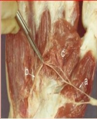
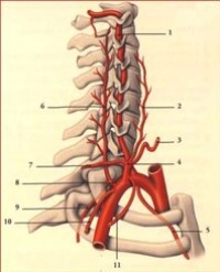
Part 1 of the video (0-9 seconds) shows the spinal cord and surrounding bony canal in saggital section. Part 2 (10-14 seconds) and Figure 1a show the nerve root in the boney canal surrounded by dura mater (green). On ultrasound one also sees the nerve root within the gutter of the 6thtransverse process. Notice also the small intercostal nerves. Part 3 (15-19 seconds) and Figure 1B show the nerve roots between the scalene muscles. Note the dura mater (green) surrounding the nerve roots in Figure 1b.

Figure 1: Sagittal section of the nerve roots with correlative ultrasound. Red bar on the skeletal drawing shows the position of the sagittal section
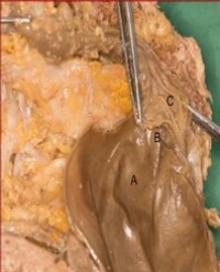
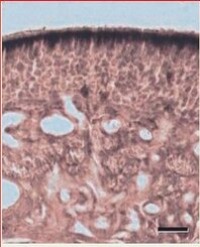
Figure 2

Figure 2: Histological section of the nerve roots
shows a histological preparation of the nerve roots. Note the dura surrounding the C8 and C7 nerve roots and the axons contained by the duramater. Spaces between the axons are filled with Cerebrospinal Fluid (CSF). Also note the formation of fascicles in C6 and C5 and the replacement of the dura mater by epineurium and perineurium. Part 4 and figure 3a (20-30 seconds) show the formation of trunks from the roots. Notice in the cadaver slices (Figures 3a and 3b)

Figure 3: Sagittal section of the plexus trunks and correlative ultrasound. Notice that there are only two trunks in the cadaver. Red bar on the skeletal drawing shows the position of the sagittal section.
that the C5 and C6 roots give rise to a superior trunk, and roots C7 –T1 give rise to a single inferior trunk [3,4]. In most plexuses, C7 gives rise to a middle Trunk and C8 and T1 give rise to the inferior trunk. In the sonography, three separate trunks are seen. In part 5 (31-38 seconds) the trunks have begun to form on sonography and the divsions have begun to form (39-42 seconds). In part 6 (47-66 seconds) the cords have begun to form and these are seen in Figures 4a and 4b.

Figure 4: Sagittal section through the plexus cords with correlative ultrasound. Notice the lipoma surrounding the plexus in the cadaver (figure 4b).
In part 7 (67 80 seconds) the peripheral nerves have begun to form in the cadaver video and in Figures 5a and 5b. Notice the lipoma surrounding the cords in Figure 5a.

Figure 5: Sagittal section through the plexus cords and the origins of the peripheral nerves. Notice the lipoma around the cords in the cadaver (Figure 5a)
Discussion
Although sonography of the spinal cord is possible in some views, the authors have not included sonography of the spine in this manuscript because we wish to focus on the brachial plexus. In parts one and two of the video, the nerve roots and trunks have a hypoechoic image on ultrasound. We believe that this is because these structures are composed largely of axons and CSF within the dura of the roots and within the fascicles of the trunks [2]. In parts 3 -7, the plexus takes on an increasingly hyperechoic appearance, presumably because of the increased presence of stroma around the fascicles
The cadaver demonstrates two anatomic variations. These are
(1) The formation of a single trunk from roots C7, C8 and T1.
(2) The presence of a lipoma within the substance on plexus at the level of the cords.
In summary, using the slices, sonography and videography described above, we have created a video which allows the user to better understand the micro-anatomy of the plexus and a one to one correlation with plexus sonography and surface anatomy. The final composite video, allows the user to better understand the ratio of neural elements (fascicles) to stroma in the plexus and the anatomy of the muscles, bones and vessels adjacent to and surrounding the plexus. This correlation will allow users in anesthesiology to better understand sonographic images of the plexus.
Acknowledgements
Financial Support: Fulbright Foundation, Universities of Maryland, Pittsburgh, Rochester, Utrecht and the Sleeping Gorilla Design Studio.
Figures 2 and 4 reprinted from Ultrasound Guided Regional Anesthesia and Pain Management, Second Edition: Copyright 2015, LWW by permission of the authors and LWW.
Book Chapter: Parts of this manuscript were published in: Foundations of Regional Anesthesia and Acute Pain Medicine, Bentam Science Publishers, 2015, Beijing, China.
Meetings: The work in this manuscript was exhibited at the 2013 meeting of the ASA in San Francisco, CA.