Research Article | Volume 1 - Issue 2 | Article DOI :
Download PDF
Georgios V Georgakis¹, Stephanie Novak², David L Bartlett², Amer H Zureikat², Herbert J Zeh III² and Melissa E Hogg²*
¹Division of Surgical Oncology, SUNY Stony Brook, USA
²Division of Surgical Oncology, University of Pittsburgh Medical Center, USA
Corresponding Author:
Melissa E Hogg, Division of Surgical Oncology, University of Pittsburgh Medical Center, USA, Tel: 412-623-4861; Fax: 412-692-2520; Email: hoggme@upmc.edu
Keywords
Central hepatectomy; Portal lymphadenectomy; Robotic surgery; Cholecystectomy; Hepatobiliary surgery
Abstract
Background: Minimally Invasive Surgery (MIS) is gaining traction within surgical oncology. We aim to evaluate outcomes of patients with gallbladder cancer undergoing MIS surgery compared to open surgery.
Methods: Using the institutional cancer registry and administrative databases, we retrospectively reviewed patients who underwent a central hepatectomy with portal lymphadenectomy for gallbladder cancer from 2011-2014. We excluded gallbladder cancer patients without oncologic resection and those with metastatic disease.
Results: Thirty-four patients underwent surgery: 17 MIS (14 robotic; 3 laparoscopic) and 17 open. There was no statistically significant difference in median operative time (MIS=182 vs open=190 min; p=0.23) or R0 resection (MIS=88.2% vs open=88.2%; p=1.0); however, the MIS cohort had less intraoperative blood loss (median 50 ml vs 400 ml; p=0.006) and placement of peri-hepatic drains (29.4% vs 76.5%; p=0.01) compared to open.MIS cohort went to oral pain medications quicker (2 vs 3 days; p=0.02) and discharged home earlier (4 vs 6 days; p=0.018), than the open cohort. No differences in postoperative 30-day complication rates (52.9% vs 52.9%; p=1.0).
Conclusion: The minimally invasive approach to liver surgery is a safe and equally effective technique for the management of the gallbladder cancer with improvement in blood loss and length of stay.
Citation
Georgakis GV, Novak S, Bartlett DL, Zureikat AH, Zeh HJ and Hogg ME. Revision Posterior Cruciate Ligament Reconstruction or Repair: A Systematic Review. SM Min Inv Surg. 2017; 1(2): 1007.
Introduction
Gallbladder cancer is a rare malignancy [1], for which surgery remains the only curative option. The extent of the surgery has changed throughout the years [2]. Currently a radical cholecystectomy, typically including central hepatectomy with part of segments 4b/5 for a negative margin and portal lymph node dissection, is the recommended procedure for tumors T1b and beyond [3,4]. Minimally Invasive Surgery (MIS) has become the standard of care for routine cholecystectomies for benign disease, since it offers safe outpatient surgery, minimal scarring and comparable surgical outcomes when compared with open surgery [5,6].
In surgical oncology, minimally invasive techniques are gaining tractionin thoracic, esophageal, gastric and colorectal procedures [7-9]. For more complex Hepatobiliary (HPB) procedures, minimally invasive techniques are not yet widespread for several reasons, including the low volume of resectable hepatobiliary cancers coupled with the need for specialized training to perform such surgeries [10]. Because of this, the establishment and standardization of minimally invasive HPB procedures is best achieved in high volume academic centers [11,12].
While technical reports of performing a robotic central hepatectomy have been previously described [13], there are currently no comparative effectiveness studies assessing MIS versus open approaches. Therefore, there is a need for comparison between this newer surgical technique and its open predecessor. The goal of this study is to examine perioperative outcomes between MIS and open surgical approaches to the central hepatectomy and portal lymphadenectomy in patients with known gallbladder cancer.
Patients and Methods
Patient selection
The institutional cancer registry, which populates state and National Databases (NCDB) was queried for all patients within the UPMC healthcare system with the diagnosis of gallbladder cancer from 2011 to 2014. We excluded all patients that did not undergo surgery. The administrative Surginet database was used to find patients undergoing a central hepatectomy during the same time period. These databases were merged to include only patients for whom surgical procedures for gallbladder malignancy were performed. All patients also underwent a central hepatectomy and portal lymphadenectomy; however, many patients had previously undergone a cholecystectomy and were still included. Patients who were found to have metastatic disease, who were simultaneously undergoing chemoperfusion or who did not undergo definitive oncologic surgery were excluded from analysis. Institutional Review Board approval was obtained (PRO15060455).
Data collection and analysis
The cancer registry provides all the data contained within the NCDB data sets, the Surginet database includes basic administrative data such as demographics, operating room time, Estimated Blood Loss (EBL), conversion, length of stay, readmission and death. Additional data was abstracted, including clinical history, prior cholecystectomy status, intraoperative data, pathology, hospital course, postoperative complications, disposition and outcomes. Surgical mortality was defined as death within 30 days of surgery. Clinical staging of disease was assessed according to the American Joint Committee on Cancer (AJCC) staging system (7th Edition).
Robotic resection
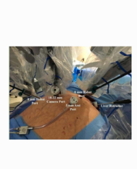
There are two primary portions of the procedure: the portal lymphadenectomy and the liver parenchymal transection. The latter is either done en bloc or after incidental cancer discovered on cholecystectomy. The port placement is depicted in figure 1. Entry into the abdomen is achieved using an optical separator trochar and a zero-degree 5mm scope in the “R1” spot.

Figure 1: Port placement for robotic central hepatectomy. Abbreviations: “R” = robotic 8 mm port, C-12 = 12 mm camera port, A-5 = 5 mm assistant port [optional], and A-12 = 12 mm assistant port.
The camera lines up with porta hepatis - approximately two-fingerbredths above and to the right of the umbilicus. The robotic third arm on the patient’s left side is used primarily to retract the liver. At least 1 assistant 12mm (A-12) port is required. This allows passing of needles, vascular clamp for pringle maneuver and a 10-mm endoscopic bag.
The portal dissection is performed first, allowing for identification of the cystic duct and the Common Bile Duct (CBD). The procedure begins by identifying the hepatic artery lymph node and skeletonizing the Common Hepatic Artery (CHA). The CBD is then identified and cleared off medially by the CHA and then laterally to identify the cystic duct. This margin is sent early for frozen to determine if CBD excision is necessary. This surgery may vary based on whether or not the gallbladder is still in situ. For the video, the gallbladder had previously been removed. The portal vein is then skeletonized laterally and medially. An optional pringle is performed after lymphadenectomy based on the surgeon’s preference.
The liver parenchymal transection can be performed with many different modalities. The robotic options include hook monopolar, scissor monopolar, PK gyrus or bipolar (maryland or fenestrated). A stitch often is placed within the resection bed for retraction, the capsule is cauterized with monopolar and the PK gyrus or bipolar is used for the deeper parenchyma. Suction of smoke or blood in the liver bed from the laparoscopic bed side assistant is critical during this portion.
Statistical analysis
Normality testing was performed, and given the small sample size, non-parametric testing was mostly performed. Means are reported with their Standard Deviation (SD) and medians are reported with the Interquartile Range (IQR). The Mann-Whitney test or Fischer’s exact test were applied for statistical analysis where appropriate. Correlation studies were done by calculating the Pearson product-moment correlation coefficient value r. The p-value was deemed statistically significant when it was ≤ 0.05. Data analysis was performed using Prism Software for Mac, 7th Edition (Graph Pad Software, Inc., La Jolla, CA).
Results
Patient demographics
A total of 34 patients with gallbladder cancer that were treated surgically at our institution: 17 open, 14 robotic, and 3 laparoscopic (Figure 2). Patient demographics are summarized in table 1.

Figure 2: Flowchart of patient exclusion (HIPEC = Heated Intraperitoneal Chemoperfusion).
Table 1: Patient Characteristics.
|
|
Open
|
MIS*
|
P-value
|
|
Number
|
17
|
17
|
|
|
Male (%)
|
1 (5.9%)
|
6 (53.3%)
|
0.085
|
|
Age (years)
|
64 (56.5-69.5)
|
76 (68-81.5)
|
0.043
|
|
BMI (kg/m2)
|
27.17 (23.82-30.99)
|
29.98 (22.21-39.02)
|
0.718
|
|
ASA
|
|
|
0.75
|
|
2
|
2
|
0
|
|
|
3
|
14
|
11
|
|
|
4
|
1
|
3
|
|
|
Stage
|
|
|
1
|
|
I
|
2
|
3
|
|
|
II
|
5
|
6
|
|
|
≥ΑΙΙΙ
|
10
|
8
|
|
|
Neoadjuvant
|
0/17
|
17-Jan
|
1
|
|
Prior GB surgery
|
17-Oct
|
17-Dec
|
0.5
|
Abbreviations: MIS = Minimally Invasive Surgery; BMI = Body Mass Index; ASA = American Society of Anesthesia; GB = Gallbladder; *MIS = 14 robotic + 3 laparoscopic; Bold is statistically significant P-value.
There were no statistically significant differences in sex, body mass index, ASA or stage between the 2 groups. The MIS cohort was younger (median 64years) compared to the open cohort (median 76years p=0.043). In the MIS group, 5 were primary surgeries and 12 were completion surgeries for an incidentally found cancer, and in the open group 7 were primary surgeries and 10 completion surgeries (p=0.5). Only 1 patient received neoadjuvant chemotherapy and he was in the MIS group (p=1.0). No patient required a roux-en-Y hepaticojejunostomy with CBD excision.
Intraoperative data
Median operative time was 182 minutes for the MIS group (IQR 126-228) and 190 minutes for the open group (IQR 146-235), with a p=0.23. The MIS group had significantly less EBL (median 50mL, IQR 30-275) compared to the open group (median 400mL, IQR 50-575), with a p=0.006, (Figure 3A). EBL strongly correlated with the length of surgery in open cohort: MIS group (r =0.59; 95% CI 0.15-0.83; p=0.01) and open (r = 0.73; 95% CI 0.38-0.0.9, p=0.0009) (Figure 3B). Peri-hepatic drains were placed less frequently in MIS (29.4%) compared to open cohorts (76.5%; p=0.01). Ten surgeons overall performed central hepatectomies: two of them performed only open cases and always placed drains and four performed only robotic and did not routinely drain. There were no conversions in the MIS cohort.

Figure 3A: Comparison of EBL between robotic and open cohorts.

Figure 3B: Correlation of EBL with operative time in each cohort.
Postoperative outcomes
Postoperatively, there was no statistically significant difference in the ICU admission rates for the MIS and open cohorts (5/17 vs 5/17, p=1.0). Oral diet initiation was considered full liquids and above (progression beyond clear liquids). As shown in table 2, no statistically significant difference in diet initiation was noted between the MIS (median=3 days) and the open (median=3 days; p=0.24) cohorts. However, conversion to oral pain medications (2 vs 3 days) and length of stay (4 vs 6 days) were decreased in the MIS compared to open cohorts (Table 2). Additionally, after discharge, no difference was noted between the MIS and open group for post-discharge (e.g. visiting nursing) services (50% vs 70.6%; p=0.29).
Postoperative complications
The complication rate was 52.9% in both cohorts (p=1.0) using the Clavien-Dindograding (Table 2). In the open cohort there was one bile leak within surgical drain and one fascial dehiscence requiring re-operation. There was no postoperative 30-day mortality in either group.
Table 2: Patient outcomes.
|
|
Open
|
MIS*
|
P-value
|
|
Recovery
|
|
|
|
|
Advanced Diet (days)
|
3 (2-6)
|
3 (2-4)
|
0.24
|
|
Oral Pain Medicine (days)
|
3 (3-6)
|
2 (1-4)
|
0.023
|
|
LOS (days)
|
6 (4.5-8)
|
4 (3-6)
|
0.018
|
|
Home Nursing
|
12 (70.6%)
|
8 (47.06%)
|
0.296
|
|
Morbidity
|
|
|
|
|
Complications
|
9 (52.9%)
|
9 (52.9%)
|
1
|
|
Clavien 0
|
8 (47.1%)
|
8 (47.1%)
|
.
|
|
Clavien 1
|
3 (17.6%)
|
6 (35.3%)
|
.
|
|
Clavien 2
|
5 (29.4%)
|
3 (17.6%)
|
.
|
|
Clavien 3
|
1 (5.9%
|
0 (0%)
|
.
|
|
Clavien 4
|
0 (0%)
|
0 (0%)
|
.
|
|
30-Day Mortality
|
0 (0%)
|
0 (0%)
|
1
|
|
Oncologic
|
|
|
|
|
R0 Resection
|
15 (88.3%)
|
15 (88.3%)
|
1
|
|
Lymph Node
|
1 (0-5.5)
|
2 (1-3)
|
0.57
|
|
Median Survival months
|
19.8
|
Not reached
|
0.0928
|
|
|
(95% CI19.7-19.9)
|
|
|
Abbreviations: MIS = Minimally Invasive Surgery; LOS=Length of Stay; CI = Confidence Interval
*MIS = 14 robotic + 3 laparoscopic; Bold is statistically significant P-value.
Oncologic outcomes
The video depicts a patient with an incidentally discovered gallbladder cancer T2NxMx with a positive cystic duct margin after a cholecystectomy for acute cholecystitis. Post-operative staging work-up revealed no evidence of disease and a Ca19-9 of 46.6 U/ mL. The patient underwent a robotic portal lymphadenectomy and central hepatectomy with pringle maneuver due to cirrhosis. Intra operative frozen section revealed a negative new cystic duct margin. Final pathology was negative for residual disease in the liver, and one out of three lymph nodes were positive for poorly differentiated adenocarcinoma.
Discussion
For gallbladder cancer, the only potential curative approach for tumors T1b and beyond is a hepatectomy with a negative margin [2]. Historically the standard approach has been an open technique;however, the use of MIS has been reported to be safe and feasible. In this study comparing open to MIS central hepatectomy, we find that patients in the MIS group have lower blood loss, earlier conversion to oral pain meds, and a shorter LOS. Otherwise, the approach has a similar morbidity, mortality, and survival as seen in open surgery. This corroborates with previous literature on MIS surgery in other malignancies [7,8].
A frequent criticism of robotic surgery is an increase in OR time; however, in our study, there was no difference in OR time between open and MIS groups (or robotic specifically-data not shown). Additionally, there was more blood loss in the open group, which is consistent with the findings of Shen et al [13] and the findings of our group for other HPB surgeries [11,14-16] in which the minimally invasive technique was used. This is possibly attributed to the magnification which enables easy visualization of small vessels that may not be seen open, the ability to use multiple hemostatic devices at once, the need to keep the operative field clean and the pneumoperitoneum may additionally tampanade small vessels.
In the case of gallbladder cancer, the cornerstones of a successful cancer surgery with favorable long term oncologic outcomes are R0 resection and adequate lymphnode sampling. For both these factors, our data suggest that the minimally invasive technique is no different compared to the open approach. This is the first time that the minimally invasive technique is directly compared to open surgery for gallbladder cancer. This is an important metric since the extension of the tumor to the liver and the subsequent ability to resect it with a negative margin, as well as the resection and histologic evaluation of adequate number of lymphnodes, affects patient survival [17].
The major limitation of this study is its small sample size and use of retrospective data administrative. However, chart abstraction for additional data was used to ascertain information and intention-to treat which is not feasibly in population based studies. Gallbladder cancer has a prevalence of 11,000 cases a year [18], with most cases found incidentally. This makes the study of gallbladder cancer difficult, especially when specifically evaluating MIS techniques and oncologic outcomes. We were not adequately powered for multivariate modeling to control for factors like the difference in age between the two cohorts given this sample size. For this reason, Cox regression was not able to be performed for survival between the groups. Also, the possibility of a selection bias that could potentially confound our results exists. Additionally, given large number of surgeons performing these procedures, it is likely that differing practice patterns explain some statistically significant differences like drain placement.
Our study is unique because we are one of the few centers in the country that routinely approaches HPB surgical cases with a robotic approach [12,14]. Furthermore we surpassed our learning curve for the platform in 2011 which is why this time period was chosen. Our sample size for this rare cancer is one of the biggest, considering that the robotic platform was only integrated in surgical practice within the last decade.
Conclusion
The MIS approach including robotic approach to the surgery for gallbladder cancer with central hepatectomy and portal lymphadenectomy demonstrated an advantage in length of stay,decreased need for pain medication and lower EBL. Given the low incidence of this disease large randomized controlled trials will not be feasible and confirmation of these findings will likely need to be through review of large multi-institutional national databases.
References
1. Randi G, Franceschi S, La Vecchia C. Gallbladder cancer worldwide: geographical distribution and risk factors. Int J Cancer. 2006. 118: 1591-1602.
2. Bartlett DL, Fong Y, Fortner JG, Brennan MF, Blumgart LH. Long-term results after resection for gallbladder cancer. Implications for staging and management. Ann Surg. 1996; 224: 639-646.
3. Dixon E, Vollmer CM, Sahajpal A, Cattral M, Grant D, Doig C, et al. An aggressive surgical approach leads to improved survival in patients with gallbladder cancer: a 12-year study at a North American Center. Ann Surg. 2005; 241: 385-394.
4. Principe A, Del Gaudio M, Ercolani G, Golfieri R, Cucchetti A, Pinna AD. Radical surgery for gallbladder carcinoma: possibilities of survival. Hepatogastroenterology. 2006; 53: 660-664.
5. Harrell AG, Heniford BT. Minimally invasive abdominal surgery: lux et veritas past, present, and future. Am J Surg. 2005; 190: 239-243.
6. Robinson TN, Stiegmann GV. Minimally invasive surgery. Endoscopy. 2004; 36: 48-51.
7. Giugliano DN, Berger AC, Rosato EL, Palazzo F. Total minimally invasive esophagectomy for esophageal cancer: approaches and outcomes. Langenbecks Arch Surg. 2016.
8. Papageorge CM, Zhao Q, Foley EF, Harms BA, Heise CP, Carchman EH, et al. Short-term outcomes of minimally invasive versus open colectomy for colon cancer. J Surg Res. 2016; 204: 83-93.
9. Suda K, Nakauchi M, Inaba K, Ishida Y, Uyama I. Robotic Surgery for Upper GI Cancer: Current Status and Future Perspectives. Dig Endosc. 2016; 28: 701-713.
10. Lidsky ME, Sun Z, Nussbaum DP, Adam MA, Speicher PJ, Blazer DG. Going the Extra Mile: Improved Survival for Pancreatic Cancer Patients Traveling to High-volume Centers. Ann Surg. 2016; 266: 333-338.
11. Boone BA, Zenati M, Hogg ME, Steve J, Moser AJ, Bartlett DL, et al. Assessment of quality outcomes for robotic pancreaticoduodenectomy: identification of the learning curve. JAMA Surg. 2015; 150: 416-422.
12. King JC, Zeh HJ, Zureikat AH, Celebrezze J, Holtzman MP, Stang ML, et al. Safety in Numbers: Progressive Implementation of a Robotics Program in an Academic Surgical Oncology Practice. Surg Innov. 2016; 23: 407-414.
13. Shen BY, Zhan Q, Deng XX, Bo H, Liu Q, Peng CH, et al. Radical resection of gallbladder cancer: could it be robotic? Surg Endosc. 2012; 26: 3245-3250.
14. Tsung A, Geller DA, Sukato DC, Sabbaghian S, Tohme S, Steel J, et al. Robotic versus laparoscopic hepatectomy: a matched comparison. Ann Surg. 2014; 259: 549-555.
15. Zureikat AH, Moser AJ, Boone BA, Bartlett DL, Zenati M, Zeh HJ. 250 robotic pancreatic resections: safety and feasibility. Ann Surg. 2013; 258: 554-559.
16. Zureikat AH, Postlewait LM, Liu Y, Gillespie TW, Weber SM, Abbott DE, et al. A Multi-institutional Comparison of Perioperative Outcomes of Robotic and Open Pancreaticoduodenectomy. Ann Surg. 2016; 264: 640-649.
17. Ito H, Ito K, D’Angelica M, Gonen M, Klimstra D, Allen P, et al. Accurate staging for gallbladder cancer: implications for surgical therapy and pathological assessment. Ann Surg. 2011; 254: 320-325.
18. Siegel RL, Miller KD, Jemal A. Cancer statistics. CA Cancer J Clin. 2016; 6: 7-30.