Case Report | Volume 6 - Issue 1 | Article DOI :
Download PDF
Kurtova Kalina MD, MSc, Argyriadou Vasiliki – Theodora MD, MSc, Dr Charalampos Triantafyllidis MD, MSc, PhD, ACCP
Corresponding Author:
Kalina Kurtova, 1st Pulmonary Department & Clinic, Kavala General Hospital, Greece
Keywords
Crohn’s Disease; Infliximab; Respiratory Failure; Pneumonia
Abstract
Lung infections are a common complication among immunocompromised patients and they have high morbidity and mortality. The continuous use of Tumor Necrosis Factor-alpha (TNF-α) inhibitors in these patients can further worsen the infections, causing lifethreatening pneumonias. We present a 20 year old male with Crohn’s disease administered infliximab while having an active infection of the respiratory system. The infection quickly evolved to bilateral pneumonia. Empirical antimicrobial chemotherapy was initiated with colistin and cefixime and after a 19 day hospitalization the patient was symptomatically better and was discharged. At one-month’s follow up the patient reported total resolution of symptoms and a chest x-ray revealed normal findings in both lung fields. Immunocompromised patients with Crohn’s disease who are administered Tumor Necrosis Factor-alpha inhibitors are more susceptible to infections. In those patients a simple upper respiratory tract infection can quickly evolve to pneumonia, which can present with severe symptomatology and even acute respiratory failure. Clinicians must be aware of and should monitor vigilantly for such potential complications. We recommend the discontinuation of treatment with TNF-a inhibitors in patients who have an active lung infection.
Citation
Kalina K, Theodora AV, Triantafyllidis C (2023) A case of pneu monia and Acute Respiratory Failure in a patient with Crohn’s disease treated with Infliximab. SM J Pulm Med 6: 4.
Abbreviations
IBD: Inflammatory Bowel Disease; CD: Crohn’s Disease; TNF-α: Tumor Necrosis Factor-alpha
Introduction
Inflammatory bowel disease (IBD) is characterized by chronic inflammation of the colon and small intestine. The most common disorders in this group are Crohn’s disease (CD) and Ulcerative Colitis (UC). While a specific cause has not yet been discovered, the basis of the inflammatory process seems to be a dysregulation of the immune response against different intestinal microorganisms. Table 1 shows the various suspected mechanisms in the inflammatory response. One of them is the initiation of inflammation and the genetic susceptibility of the individual. Genetic predisposition and variations can make individuals more susceptible to CD. Mutations in genes related to the immune system (such as NOD2) have been linked with an increased risk for the disease. Therefore, it is not surprising that CD shows a tendency to cluster in families, with as many as 15% of patients with CD reporting a first-degree family member with the disorder [1]. Moreover, environmental factors, including diet and smoking, have been associated with triggering inflammation in CD. Another important part of the inflammatory process seems to be the changes in the gut microbiome and dysbiosis that have been shown to activate the immune system. Autoimmunity plays a potential role as well since it’s likely for the immune system to mistakenly attack healthy cells of the intestinal lining. Additionally, it is important to mention the release of pro inflammatory cytokines from T-cells and macrophages that have invaded the intestinal mucosa. Among these cytokines are TNF-α, interleukin-12, interleukin-17, which have been associated with chronic intestinal inflammation [2]. Their release can further amplify the inflammation. Last but not least, if the intestinal epithelial barrier is damaged or compromised, bacteria and other pathogens can pass through it and trigger an immune response and further inflammation [3].
The understanding and further research of these mechanisms is important because it can help with the development of novel therapies for CD. Of the mechanisms mentioned, proinflammatory cytokines play a crucial role in the development of CD by activating various cascades and immune cells, eventually leading to tissue damage and inflammation. As mentioned above, one of the most important cytokines in this process is the Tumor Necrosis Factor-alpha (TNF-α) [4]. Novel drugs that bind to the TNF-α have shown to disrupt pro-inflammatory responses as they inhibit the cytokine from interacting with its receptors [5]. However, the long-term use of such drugs can lead to immunosuppression and an increased risk of infections. There are different studies suggestive of worsening infections in patients treated with Infliximab, a monoclonal TNF-α antibody [6]. Upper respiratory tract infections are common not only in immunocompromised patients but in the general population as well, posing a tremendous burden on the healthcare system. According to an analysis based on the Global Burden of Disease, Injuries and Risk Factors Study, the incident cases of upper respiratory tract infections reached 17.2 billion in 2019 [4]. The study was conducted over a period of 39 years, spanned more than 204 countries and territories, and it is important to note that the frequency of upper respiratory tract infections remained stable throughout the analyzed period. Our case report highlights the dangers of continuing treatment with Infliximab in an immunocompromised patient with CD and an active respiratory infection, especially the potential for developing life-threatening pneumonia.
|
Table 1: Different mechanisms responsible for the inflammation associated with Crohn’s disease.
|
|
|
Involved mechanisms
|
|
Initiation of Inflammation
|
-genetic predisposition
-mutations in genes related to the immune system
-environmental factors
|
|
Gut microbiome
|
-changes in the gut microbiome and dysbiosis
|
|
Autoimmunity
|
-immune system that mistakenly attack healthy cells of the intestinal lining
|
|
Cytokine release
|
-release of pro-inflammatory cytokines from T-cells and macrophages (TNF-α, interleukin-12, interleukin-17)
|
|
Epithelial barrier dysfunction
|
-a compromised epithelial barrier can allow the entrance of bacteria and antigens and the activation of the immune response
|
Case Report
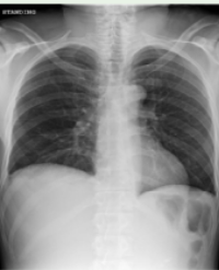
A 20-year-old Caucasian male with a medical history of Crohn’s disease referred to our Emergency Department in August 2022 from a peripheral hospital where he was being treated for sinusitis (Figure 1). The patient had been administered the biologic agent infliximab without consulting his physician, despite the presence of fever and an active infection of the upper respiratory tract for the previous seven days. Despite his treatment with piperacillin-tazobactam and metronidazole, the infection quickly evolved to bilateral pneumonia and Acute Respiratory Failure.

Figure 1: Chest and Paranasal sinus X-Ray on 21/08/2023, before admission, revealing sinusitis.
Upon referral to our Emergency Department, the patient was alert but hemodynamically unstable (blood pressure was 95/55mmHg, heart rate - 100/min, SpO2: 92%, N/C 3lt/min). The patient was alert but lethargic and febrile with a temperature of 38°C. Auscultation revealed bilateral coarse crackles. Relevant laboratory investigations are summarized in Table 2. A Chest X-Ray (CXR) was performed on admission as well, revealing lobar consolidation in the Right Lung suggestive of pneumonia (Figure 2). Subsequently, a CT lung scan was performed, revealing tree-in-bud opacities and areas of consolidation, in addition to enlarged lymph nodes (Figure 3).
Table 2: Relevant initial laboratory findings.
|
Investigation
|
Result
|
Normal range
|
|
WBC count
|
7,59
|
4,0-10,80 K/μl
|
|
Platelet count
|
189,00
|
150-350 K/μl
|
|
Hb
|
10,4
|
13,5-17,5 g/dL
|
|
Sodium
|
137
|
136-145 mmol/l
|
|
Potassium
|
4,1
|
3,5-5,1 mmol/l
|
|
Creatinine
|
1,5
|
0,7-1,3 mg/dl
|
|
C-reactive protein
|
6,8
|
<0,3 mg/dL
|
|
Lactate dehydrogenase
|
1051
|
85-227 U/I
|
|
C.P.K.
|
5052
|
39-308 U/I
|
|
C.P.K. MB
|
3,7
|
0-3,6 ng/mL
|
|
D-dimer
|
2203
|
<500 mg/L
|
|
SGOT
|
532
|
15-37 U/I
|
|
SGPT
|
210
|
16-63 U/I
|
|
Blood cultures
|
No growth
|
-
|
|
SARS-Cov
|
Negative
|
-
|
|
Sputum smear
|
Negative
|
-
|
|
Pneumococcal and Legionella urinary antigen tests
|
Negative
|
-
|
|
Common viruses panel
|
Adenovirus
|
-
|

Figure 2: CXR on admission revealing lobar consolidation in the Right Lung.

Figure 3: Computed Tomography findings on admission (25/08) show the quick progression of the infection, revealing tree-in bud opacities, areas of consolidation with Air-Bronchogram in the Right Lower Lobe, as well as in the Right Upper and Middle Lobe.
Polymerase Chain Reaction (PCR) Testing of sputum samples was performed and detected Adenovirus. The patient was initially treated with linezolid, but subsequent laboratory tests demonstrated leukopenia, anemia and thrombocytopenia which led to the withdrawal of the drug. Due to the worsening lung opacities (Figure 4A) and the deterioration of respiratory function, empirical antimicrobial therapy was initiated with colistin combined with cefixime. The patient became afebrile and showed clinical improvement. After a 19 days hospitalization, the patient was discharged afebrile and hemodynamically stable. The CXR at discharge revealed significant improvement of the lung opacities (Figure 4B). A follow up CXR was performed again in one month’s time after discharge and revealed normal findings in both lung fields. The patient reported total resolution of his symptoms.

Figure 4 Chest X-Rays during hospitalization.
(A) Worsening lung opacities despite treatment.
(B) CXR upon discharge shows improvement of lung opacities after treatment with colistin and cefixime.
Discussion
Immunocompromised patients are more prone to infections. Different studies suggest that patients with CD may be more susceptible to urinary tract infections and gastroenteritis [8]. Additionally, existing infections may progress more rapidly in these patient groups. While our patient first presented and was treated for an upper respiratory tract infection and sinusitis, the administration of the biologic agent infliximab likely contributed to the worsening of his infection. The patient performed no improvement of the clinical symptoms despite treatment with meropenem and linezolid. What is more, after only three days of this initial antimicrobial chemotherapy the patient’s laboratory test results revealed pancytopenia - a rare, but severe adverse reaction of linezolid [9]. Linezolid was immediately discontinued and in the following days leukocyte and platelet count normalized. This confirms findings that linezolid induced myelosuppression is reversible after appropriate treatment and discontinuation of the drug. Additionally, since no microorganism was identified from the blood cultures and tests, empirical antimicrobial therapy with colistin and cefixime was initiated. The patient became afebrile and his symptoms alleviated.
Conclusion
The management of this case aims to highlight the fact that the administration of TNF-α inhibitors to immunocompromised patients with an active infection can lead to life-threatening complications and pneumonias. Clinicians must be vigilant for early signs of infections and worsening of symptoms in patients undergoing treatment with TNF-α inhibitors for underlying inflammatory conditions such as CD. We recommend the discontinuation of therapy with biologic agents until the active infection is treated successfully.
References
1. Crohn’s disease: MedlinePlus Genetics [Internet]. medlineplus.gov. Available from: https://medlineplus.gov/genetics/condition/crohns-disease/#inheritance
2. de Mattos BRR, Garcia MPG, Nogueira JB, Paiatto LN, Albuquerque CG, Souza CL, et al. Inflammatory Bowel Disease: An Overview of Immune Mechanisms and Biological Treatments. Mediators of Inflammation [Internet]. 2015 [cited 2019 Oct 16];2015:1–11. Available from: https://www.ncbi.nlm.nih.gov/pmc/articles/PMC4539174/
3. Plevy S. The immunology of inflammatory bowel disease. Gastroenterology Clinics of North America. 2002 Mar;31(1):77–92.
4. Saez A, Herrero-Fernandez B, Gomez-Bris R, Sánchez-Martinez H, Gonzalez-Granado JM. Pathophysiology of Inflammatory Bowel Disease: Innate Immune System. International Journal of Molecular Sciences. 2023; 24(2):1526. https://doi.org/10.3390/ijms24021526
5. Infliximab (2005) Drugbank. Available at: https://go.drugbank.com/ drugs/DB00065 (Accessed: 17 September 2023).
6. Colombel JF, Loftus EV, Tremaine WJ, et al. The safety profile of infliximab in patients with Crohn’s disease: The Mayo Clinic experience in 500 patients. Gastroenterology 2004; 126:19–31.
7. Jin X, Ren J, Li R, Gao Y, Zhang H, Li J, et al. Global burden of upper respiratory infections in 204 countries and territories, from 1990 to 2019. EClinicalMedicine. 2021 Jul;37:100986.
8. Porter CK, et al. Infectious gastroenteritis and risk of developing inflammatory bowel disease. Gastroenterology. 2008;135(3):781–786.
9. Li MM, Shen WC, Li YJ, Teng J. Linezolid-Induced Pancytopenia in Patients Using Dapagliflozin: A Case Series. Infect Drug Resist. 2022;15:5509-5517. https://doi.org/10.2147/IDR.S375694
10. Plevy S. The immunology of inflammatory bowel disease. Gastroenterology Clinics of North America. 2002 Mar;31(1):77–92.
11. Crohn’s disease: MedlinePlus Genetics [Internet]. medlineplus.gov. Available from: https://medlineplus.gov/genetics/condition/crohns-disease/#inheritance
12. de Mattos BRR, Garcia MPG, Nogueira JB, Paiatto LN, Albuquerque CG, Souza CL, et al. Inflammatory Bowel Disease: An Overview of Immune Mechanisms and Biological Treatments. Mediators of Inflammation [Internet]. 2015 [cited 2019 Oct 16];2015:1–11. Available from: https://www.ncbi.nlm.nih.gov/pmc/articles/PMC4539174/
Citation
Kalina K, Theodora AV, Triantafyllidis C (2023) A case of pneumonia and Acute Respiratory Failure in a patient with Crohn’s disease treated with Infliximab. SM J Pulm Med 6: 4