Back to Journal

Annals of Applied Microbiology & Biotechnology Journal

Approaches to Method Development, Suitability and Validation for Alternative Sterility Test Methods for Complex Parenterals

[ ISSN : 2576-5426 ]

Abstract Background Challenges with Traditional Sterility Testing Sterility by ATP Bioluminescence Investigational Tool Conclusions References
Details

Received: 15-Apr-2024

Accepted: 02-May-2024

Published: 04-May-2024

Miriam Guest* 

Senior Principal Scientific Advisor, Microbial Solutions, Charles River Laboratories, USA

Corresponding Author:

Miriam Guest, Senior Principal Scientific Advisor, Microbial Solutions, Charles River Laboratories, USA

Keywords

Sterility testing; Complex parenterals; Advanced drug delivery systems; Quality control; Pharmaceutical microbiology; Sterility assurance

Abstract

Advancements in pharmaceutical development have led to increased diversity in drug and biological products. From simple aqueous formulations to advanced drug delivery systems, including sustained release depots, nanospheres and microsphere suspensions, as well as the rise of cell and gene therapy products. With this progress, increased complexity in the manufacture, testing and quality control release of these products has been observed. For injectable products, sterility testing at release is a mandatory step in the product release process. While there are limitations in traditional sterility testing, the final sterility test provides additional sterility assurance for the safety of the product being delivered to the patient. Traditional sterility tests are determined by the visual inspection of nutrient media to assess for the presence of microbial proliferation; product formulations can interfere with visual inspection and ultimately will always lead to a subjective result. Typically, following the initial incubation period, samples of the incubated broth are transferred to fresh nutrient media, and a further incubation step is applied prior to visual inspection.

By Adopting Adenosine Triphosphate (ATP) bioluminescence as the detection platform, the subjective assessment can be avoided, with an unequivocal result determining the presence or absence of microbial proliferation. This technique also avoids the subsequent incubation step on sampling from the sterility test containers.

All living organisms contain the ATP and the enzyme Adenylate Kinase (AK) as part of their biochemical processes, during the ATP Bioluminescence test, an enzyme cascade amplifies the presence of microbial ATP and detect the presence of contamination in the test sample

Background

For the purposes of this review, the phrase “complex parenterals” refers to formulations that are not aqueous formulations. Complex refers to formulations which typically render the nutrient media used in sterility testing turbid, making visual determination of the presence or absence or microbial growth challenging. This encompassing a broad spectrum of products and therapeutic modalities,including advance drugs delivery systems, cellular therapies, biologics, nanosuspensions, vaccines, and products containing challenging adjuvants.Sterility testing a critical release criterion for injectable products, classical microbiological methods are utilised [1,2],whereby samples undergo either membrane filtration, rinsing and the addition of nutrient media or direct inoculation into nutrient media,followed by a 14-day incubation period.Visual inspection of the media is then performed, where the test article is inspected for the presence of microbial growth (e.g., turbidity, pellicle formation, precipitation,flocculent growth or other differences between the test media and the un-inoculated media).Whilst training of personnel executing the sterility test is performed, the results can only ever be interpreted in a subjective manner.

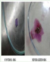
The image (Figure 1) highlights the challenge of visual inspection for a complex formulation and is perhaps one of the worst-case challenges in terms of sample matrix.

Figure 1 Example of Sample Matrix which renders sterility test media turbid.

During traditional sterility testing, a sub-culture step is incorporated at the end of the incubation period, where samples are transferred to fresh broth. During that transfer step, it is possible that product residue could be transferred over to the fresh broth and again make visual inspection for the presence of growth challenging.

The application of ATP bioluminescence requires no additional sub-culture step. The amplified ATP reaction is not impacted by visual artefacts and can provide an unequivocal result of the presence of absence of microbial contamination.

Challenges with Traditional Sterility Testing

Complex Parenterals Membrane filtration is the preferred approach to sterility testing [1,2], which has some benefits over direct inoculation; product residues are removed during the filtration process, whilst potential contamination is captured on the filter membrane, meaning there is less likely to be product related interference impacting microbial proliferation. The nature of the test article should be understood prior to commencing into method development. In the case of complex parenterals, direct inoculation methods are often applied, where samples of the drug product, are placed directly into nutrient media prior to incubation.

During the 14-day incubation period, the broth is visually inspected to assess for the presence of turbidity, pellicle formation, precipitation, flocculant growth and other differences between the test broth and the un-inoculated media. The presence of the drug product itself can cause additional challenges (when compared to simple aqueous formulations undergoing membrane filtrations). The compendia state, that if visual determination cannot be readily assessed, sub-cultures into fresh nutrient media should be conducted. It is prudent to perform both the test set up and the sub-culture steps in a dedicated isolator system, this reduces the risk of cross contamination occurring during the testing itself.

Should growth be suspected, additional sub-culture steps may be applied to fresh nutrient broth and onto plated media. It is recommended that the sub-cultured media is incubated for as long as it took to observe the change in the original test method. Again, this is challenging in products that render the nutrient media turbid, as it may not be obvious when visual growth was originally observed. Sub-culturing is not always reliable [3], and the heavy reliance on these techniques can add additional risk to patient safety.

Sterility by ATP Bioluminescence

The determination of microbial growth by ATP Bioluminescence is a well-established technique, bioluminescence can be observed in nature in glow-worms, marine organisms, and fireflies. The reaction was first described in 1947 [4] and subsequent publications have highlighted the applicability of ATP bioluminescence for the determination of microbial growth [5,6]. All living cells contain ATP and it is a key marker for viable life. The use of ATP bioluminescence in the microbiology laboratory is reliant on the use of a luminometer to determine the light generated and thus, the presence of viable microorganisms in a test article. The technique has been applied for sterility testing, within the pharmaceutical industry with increasing adoption in recent years [7].

The ATP Bioluminescence method amplifies the ATP, and is, therefore, not constrained by the finite amount of metabolic ATP available. Instruments provide automated addition of enzymes, extractants, and buffers, with a luminometer reading the levels of ATP. The amplification technology catalyses and compounds the production of an additional amount of ATP, enabling it to be readily detected. The reaction is essentially linear in nature, a 25-minute reaction time can amplify the amount of ATP by 1000 times more than was originally present. This enables a shortened incubation time, when compared to traditional sterility test methods. Results are reported in Relative Light Units (RLU) and compared to untested broth as a calibrator. Levels of RLU to determine a positive result are established during the method validation process; typically, a three fold increase over the broth calibrator is applied (the broth calibrators will generate low levels of background ATP).

Samples from the broth containers are extracted and transferred in duplicates to cuvettes for testing. Inadvertent cross-contamination at the end of the incubation period will not result in a false positive result, as no additional incubation period takes place, there would be insufficient ATP present to generate an RLU value significantly higher than the broth calibrator.

An overview of traditional sterility testing of complex parenterals is provided in Figure 2.

Figure 2 Traditional Sterility Testing of Complex Parenterals.

Sterility testing involves several manual steps that require technician intervention, each of which increases the risk of cross-contamination during testing. In the event of a sterility test failure or even a suspect positive sterility test result, a complex deviation process ensues.

Sterility test investigations are complex, and often involve a multi disciplinary team. During investigations of suspect-positive sterility test units, production of the drug product may be halted to ensure that root causes are identified and addressed before manufacturing resumes.This can lead to drug shortages and extended downtimes. As with all microbial data deviations, investigations can be prolonged due to the waiting periods required for incubation.

PDA Technical Report 88 [8] provides an overview of the steps required to investigate microbial data deviations. Such deviations include a laboratory review and if no laboratory error is identified, a manufacturing investigation takes place. Some companies may prefer to initiate the manufacturing investigation in parallel, prior to the conclusion of the laboratory investigation.

During the laboratory investigation, a team will consider multiple aspects of the testing:

Equipment:

• Sterility test isolator or laminar flow hood

• Glove integrity testing

• Environmental monitoring data

• Cleanroom certification

• Room conditions acceptable

• Sanitisation and decontamination

Materials:

• Media, QC checks, expiry, storage, integrity

• Could undissolved media cause ambiguous results?

• Could product interference cause ambiguous results?

• Materials transfer

• Glassware management (cleaning & sterilization)

Test Procedure:

• Unusual events documented during testing?

• Complexity of the test

• Sufficient environmental monitoring to detect low levels of background contamination?

• Method suitability conducted appropriately.

Personnel:

• Personnel setting up equipment for the test?

• Personnel performing the test?

• Personnel performing subcultures (if used)?

• Personnel interpreting the test results?

This detailed investigation will require appropriate quality oversight and review.

Determining the presence of absence of microbial growth,by utilizing ATP Bioluminescence can significantly simplify both the testing and investigation process.By reducing manual steps prior to subsequent incubation steps,the risk of extraneous cross-contamination during testing is minimized,thereby reducing the risk of false positives, and leading to simplified investigations.Refer to Figure 3 for an overview of sterility testing by ATP bioluminescence.

Figure 3 Overview of Sterility Testing with ATP Bioluminescence.

Investigational Tool

The ATP Bioluminescence test could be used as an investigative tool, where indeterminate results may stop production until fully investigated. In such cases, the investigation should consider a holistic data set, with a result from an ATP readout being a powerful tool.

Before adopting any change in the QC Microbiology laboratory, a risk assessment should be conducted to understand the proposed future state. In the circumstance of sterility testing, assuming the current method is demonstrated as being suitable for use, as per USP, additional testing could be performed to provide assurance that the ATP bioluminescence response is not impacted by the test article.

In such circumstances, it is anticipated that the sample has undergone a 14-day incubation period, during which period, microbial growth would have generated large quantities of microbial cells.

Adopting an alternative method for investigation purposes may be justified with sound scientific rationale, during investigations a holistic data set should be reviewed. By utilising a non-subjective arbiter of the presence of microbial proliferation, investigations can be expedited. Faster investigations can have broader benefits, by accelerating understanding of the issues faced, the root cause can be more easily identified (by reducing the time to result, the likelihood of detecting the root cause is increased), furthermore, an unequivocal answer on the presence of microbial growth can reduce the manufacturing facility downtime, which may be incurred if a suspect-positive sterility test is observed.

Approach to Method Validation

Should an end user wish to routinely adopt the ATP bioluminescence test, the validation of an alternative microbial method is required. Considerations for the regions where the product sold should be made, but broadly speaking, the application of USP , Ph Eur 5.1.6 and PDA TR 33 should be considered.

Alternative method validation typically directly compares the classical method with the proposed alternative method with the aim to demonstrate non-inferiority of the new method. For qualitative tests, such as sterility, Specificity, limit of detection, ruggedness and robustness should be addressed. It is also important to demonstrate that the test is fit for the intended use; in the case of drug product sample matrices, demonstrating that the test sample matrix does not adversely impact the final result.

Conclusions

Advances in the production of medicinal products has impacts across the manufacturing, testing, and release phases. For advanced drug delivery mechanisms, additional layers of complexity can be observed in the traditional sterility test. By adopting alternative microbiological test methods, such as ATP bioluminescence for complex parenteral formulation, some of these challenges can be overcome, removing subjectivity in test interpretation, and reducing manual activities during the test. Reducing risks of cross contamination during testing can ultimately simplify investigations and can result in reduction of manufacturing downtime and expedite the process of releasing drug products to patients.

References

1. United States Pharmacopeia Sterility Tests.

2.European Pharmacopeia 2.6.1 Sterility Tests.

3.Guest M, Pickard B, Smith B, Drinkwater S. The use of amplified ATP bioluminescence for rapid sterility testing of drug product formulations. PDA J Pharm Sci Technol. 2023; 77: 402-411.

4.McElroy WD. The energy source for bioluminescence in an isolated system. Proc Natl Acad Sci U S A. 1947; 33: 342-345.

5.Stanley PE. A review of bioluminescent ATP techniques in rapid microbiology. J Biolumin Chemilumin. 1989; 4: 375-380.

6.Rapid microbiological methods in the pharmaceutical industry. Martin C Easter Editor. CRC Press. 2003: 288.

7.Deutschmann S, Paul M, Claassen-Willemse M, van den Berg J, IJzerman-Boon P, Grunert da Fonseca V, et al. Rapid sterility test systems in the pharmaceutical industry: Applying a structured approach to their evaluation, validation and global implementation. PDA J Pharm Sci Technol. 2023; 77: 211-235.

8.PDA Technical Report No. 88. Microbial Data Deviations.

9.United States Pharmacopeia Validation of Alternative Microbiology Methods.

10. European Pharmacopoeia 5.1.6. Alternative Methods for Control of Microbiological Quality.

11. PDA Technical Report No. 33 (Revised 2013) Evaluation, Validation and Implementation of Alternative Rapid Microbiological Methods.

Citation

Guest M. (2024) Approaches to Method Development, Suitability and Validation for Alternative Sterility Test Methods for Complex Parenter als. Ann Appl Microbiol Biotechnol J 5: 4.

Other Articles

Article Image 1

Efficacy of a Combination of Plumbago Capensis and Aloe Secundiflora Aqueous and Methanolic Plant Extracts in the Treatment of Leishmania Major in Balb\C Mice

Background & Objectives: Monotherapy and combination therapy studies were to determine antileishmanial activities of the methanolic and water leaf extracts from Aloe secundiflora and Plumbago capensis.

Methods: The plants were analyzed by determining the Minimum Inhibition Concentration (MIC), Nitric Oxide (NO) production stimulation, Infection Rates (IR) and Multiplication Index (MI). Cytotoxicity of these plant extracts was also assessed.

Results: The MIC levels of water and methanolic plant extracts, amphotericin B and Pentostam were 2000μg/ml, 1000μg/ml, 125μg/ml and 250μg/ml respectively against Leishmania major promastigotes. Study revealed that water and methanolic plant extracts significantly inhibited the growth of Leishmania parasites (P≤0.05) as compared to amphotericin B with respect to the parasite infection rates and MIC levels. The IC50 for the water and methanolic plant extracts was 279.488μg/ml and 42.824μg/ml respectively. A similar extraction procedure was done for P.capensis. When administered orally, a combination of P.capensis and A.secundiflora proved to be more effective than that of the methanolic extract with LDU units of 4,800 and 15,300 respectively. Water extracts of P.capensis when used alone gave LDU of 5,200 units, methanolic extract gave 11,000 units. Aloe secundiflora water and methanolic extracts gave the same LDU of 10,700. Methanolic extract combinations of the two plants were the least effective, yielding the highest LDU of 21,000 units.

Interpretation & Conclusion: The results suggest that a combination oral therapy of extracts of the two plants and monotherapy using P.capensis can be effective methods of partially treating leishmaniasis.

Paul K Ndungu1, Johnstone M Ingonga1, Michael Gicheru2 and Christopher O
Anjili1


Article Image 1

Gene Therapy for Treatment of Melanoma Cancer

Melanoma occurs when the pigment-producing cells (melanocytes) that give colour to the skin become cancerous. Malignant melanoma has become one major problem for its higher rate of occurrence among patients than any other type of cancer throughout the world. Hence, it has become very crucial to treat melanoma skin cancers by early detection and prevention. It is remarkable how an advanced therapy like gene therapy is leading towards a better solution for melanoma skin cancer. Not only can gene therapy cure an individual, it also prevents same set of diseases throughout a blood line. With supported evidences from research and clinical trials, gene therapy also provides clue for a new generation treatment for different diseases including cancer.

Oisorjo Chakraborty, Aishwarya Mukherjee and Pranab Roy*


Article Image 1

Mycoplasma leachii Causes Bovine Mastitis: Evidence from Clinical Symptoms, Histopathology and Immunohistochemistry

Twelve quarters of six lactating cows were inoculated with Mycoplasma leachii strain GN407 through intramammary ductal infusion, and another twelve quarters were inoculated with Mycoplasma culture medium as controls. One lactating cow was used as negative control, in which two quarters were inoculated with Mycoplasma culture medium, and another two quarters were not inoculated with any medium. Clinical observations, histopathology and Immunohistochemistry (IHC) detection were performed on Post Inoculation Days (PIDs) 3, 6 and 9 to elucidate the pathogenicity of M. leachii in bovine mastitis. From PIDs 3 to 9, twelve inoculated quarters developed mild to severe clinical mastitis and mammary tissue histopathological changes, including inflammatory cell infiltration and architectural destruction of mammary gland ducts; on PID 9, the control quarters also developed mild mastitis and histopathological changes. Throughout the experimental period, the quarters of the negative control cow were clinically and pathologically normal. The M. leachii antigen was detected by IHC in the mammary tissues of the inoculated quarters as a weak signal on PID 6 and as a strong signal on PID 9; on PID 9, the M. leachii IHC signal was also detected in mammary gland epithelial cells of the control quarters of the inoculated cattle. The M. leachii antigen was not detected in the mammary tissues from the quarters of the negative control cow on PID 9. In conclusion, direct histological and immunohistochemical evidence confirmed that M. leachii causes clinical bovine mastitis through histopathological lesions induced by the invasion of the pathogen into mammary gland cells and inflammatory cell infiltration

Jitao Chang1, Debin Yu2, Jianbin Liang2, Jia Chen2, Fang Wang1, Zhigang Jiang1,
Xijun He3, Rui Wu2* and Li Yu1*


Article Image 1

Phylogenetic and Biochemical Analysis of the Reishi Mushroom (Ganoderma lucidum) Populations from Altai

G. lucidum is a typical representative of wood-rotting polypores of the Ganodermataceae family (Basidiomycetes). In Russia, G. lucidum is predominantly found in southern regions: in Stavropol and Krasnodar krais, Northern Caucasus, as well as in Altai taiga in logging areas. In this study we investigated the phylogeny of G. lucidum specimens from Altai based on the ITS1 ribosomal spacer, and compared them to reishi from other regions of the world. We also studied the phytochemical content of reishi fruit bodies. Results of the screening suggest that ethanol fractions contain mostly flavonoids, phenols, and coumarins; water fractions are dominated by tannins, carbohydrates, and coumarins; and hexane and ethyl acetate extracts, by terpenoids. The main fatty acids were palmitic, oleic, linoleic, and linolenic acids. We found that fruit bodies of Altai G. lucidum contained 32.4 mg of phenols per 1 g of extract (in pyrocatechol equivalent), while flavonoids made up 11.1 mg per g (in quercetin equivalent). Polysaccharide content was 10.72% of the absolutely dry substance.

Slynko NM, Blinov AG, Babenko VN, Mihailova SV, Bannikova SV, Shekhovtsov
SV, Nechiporenko NN, Goryachkovskaya TN, Veprev SG and Peltek SE*


Article Image 1

A Study on Mineralisation of Poly cis 1,4 isoprene (NR) and Synthetic Rubber Gloves (SRG) by the Bacterial Consortium

Objective: The aim of the study wasto isolate bacterial consortium from the effluent contaminated site that is used in the mineralizing both natural rubber and synthetic rubber, to optimise the growth conditions for efficient mineralisation and to biochemically characterise and to use 16s RNA sequencing for identifying the bacterial strains.

Materials and Methods: Natural rubber mineralizing bacterial consortium was isolated from effluent contaminated soil. The mineralisation study was performed for five days at every 24 h interval. Optimization studies were performed with different parameters such as varying concentrations of latex, pH, carbon sources, nitrogen sources, mixed carbon and nitrogen source and different temperature. The bacterial consortium mineralizing nr latex was used to mineralize Synthetic Rubber Gloves (SRG) using the same medium for 40 days at every 5 days interval. Effect of pre-treatment was studied by pre-treating the SRG with acetone and exposing it to sunlight. Mineralisation of the Rubber was confirmed by spectrophotometric and Fourier Transform Infra-Red
(FTIR) studies.

Results: Isolated organism was identified as Enterobater cloacae, Microbacterium laevaniformans and Methylobacterium rhodesianum. Maximum mineralisation of (1.66x10⁻⁴) was shown on the 4th day of incubation. Conformation of NR degradation was done by FTIR analysis that shows the presence of aldehyde and ketone produced due to bacterial degradation. The parameters giving optimum results were concentration of latex -1%, pH- 8.5, carbon source- Xylose, nitrogen source - Ammonium Nitrate, temperature- 37°C. Maximum mineralisation of synthetic rubber was shown on the 20th day (1.3x10⁻⁴). Among the pre-treated and the untreated samples most prominent distortions were visible on the surface of the sunlight sample when visualized under
scanning electron microscopy.

Conclusion: From the present investigation, it can be concluded that the isolated bacterial consortium containing the strains Enterobater cloacae, Microbacterium laevaniformans and Methylobacterium rhodesianum were able to mineralize natural rubber as well as synthetic rubber. This could be applied in the removal of waste rubber products present in the environment.

Veenagayathri Krishnaswamy* and Nikita Ahongsangbam


Article Image 1

Application of Chitosan in the Control of Fungal Infections by Dermatophytes

Dermatophytes are a group of fungi that can invade keratinized tissues of humans and other animals and produce an infection called Dermatophytosis. As chitosan possesses antimicrobial activity, it can potentially be used to treat dermatophytic infections. The main objective of this work was therefore, to evaluate the antifungal activity of chitosan upon some dermatophytes, namely Microsporum canis and Trychophyton rubrum. In view of this, Minimum Inhibitory (MICs) and Minimum Fungicidal Concentrations (MFCs) of chitosans upon the fungi were determined. Moreover, in order to understand the effect of chitosan on fungal activity, hair was infected with these fungi in the presence and absence of chitosan and Scanning Electron Microscopy (SEM) images were obtained and analyzed. Lastly, keratin-azure was used as substrate to evaluate the effect of chitosan on keratin degradation by M. canis and T. rubrum. The results showed that chitosan possesses antifungal activity against T. rubrum and M. canis, presenting MICs and MFCs ranging from 1.1 to 2.2 mg/mL. The antifungal activity of chitosan is concentration dependent. The analysis of SEM images of hair infected with these dermatophytes revealed that chitosan seems to have a protective effect on the hair, reducing the extent of damage when compared to the control. Chitosan also displayed important activity in preventing proteases’ action and in preventing hair damage. Based on the obtained results, it’s possible to conclude that chitosan showed relevant antifungal activity against dermatophytes, which opens good prospects to the use of chitosan as an alternative for the conventional fungal treatments.

Ana I Lopes, Freni K Tavaria* and Manuela E Pintado


Article Image 1

Characterization of Endophyte Microbiome Diversity in Chia Plant (Salvia hispanica L.)

A total of 9347 fungal and bacterial endophytes were isolated from the roots, stem and leaves of chia plant. Roots harbored more number of fungal endophytes than either stem or leaves whereas stem supported more number of bacterial endophytes than either roots or leaves. The nutritious plant supported more of gram negative compared to gram positive bacterial endophytes. The most common bacteria isolated were Pseudomonas Bacillus, and Cocci. The fungal endophytes isolated from root, stem and leaves of the chia plant showed the presence of Penincillium, Aspergillus, Fusarium, and Macrophomina spps. Dominant fungal endophyte was Aspergillus spp. which was found in all the plant parts instigated. Roots of the plant possessed maximum nitrogen fixers followed by stem and leaves. A proportion of 55% for the bacterial endophytes isolated from the plant chia plant were able to fix nitrogen whereas 25% were able to solubilize phosphorous. The phosphate solubilization efficiency was found to be highest for the Aspergillus spp at 83%.

Jasira Jzar1 , Mary Simiyu2 , Joseph Mafurah2*, Joshua Ogendo2 and Anne Osano3


Article Image 1

Isolation and Screening of Novel Isolates of Bifidobacteria from Human Milk as Potential Probiotic with Antidiarrheal Activity

Aims:
The objectives of this research work were isolation of Bifidobacteria from the human milk and its Probiotic characterization such as low pH, bile and in-vitro antimicrobial activity against diarrhea causing pathogen.

Methodology and Results:
In this research work, 47 bifidobacterial isolates were isolated from the human milk of the 50 lactating women and identified by using phenotypic methods. The isolates were examined in-vitro for their tolerance to unfavorable condition at low pH of 2 and 4 and at different concentrations of bile 0.3%, 0.5% and 1%. Further the isolates were tested for the antimicrobial activities by using diarrhea causing indicator stains such as E. coli, Salmonella enterica and Shigella boydii. Antibiotic susceptibility test was performed for the isolates which showed zone of inhibition in antimicrobial testing. Based on the result of in-vitro Probiotic test, the best four isolates Dbs18, Smk9, Smk4 and Smk5 were selected for further evaluation of tolerance test of phenol (0.1%, 0.2%, 0.4%), NaCl (5%, 8%, 12%). Auto aggregation and hydrophobicity assay were also done for the four selected isolates. In in-vitro test of low pH, out of 47 isolates only 14 isolates were able to grow whereas in bile tolerance assay most of the isolates grew well at 0.3% bile concentration but variability of growth of isolates were observed at 0.5% and 1% bile. In antimicrobial assay, 15 isolates out of 47 isolates showed antimicrobial activity after ruling out the inhibitory activity of low pH. In NaCl and phenol tolerance test all the four selected isolates were able to survive the different concentration of phenol and NaCl. The percentage of hydrophobicity and auto aggregation was highest in Dbs18 followed by Smk9 among the four isolates.

Conclusion, significance and impact of study:
Among the four isolates Dbs18 and Smk9 showed good hydrophobicity and auto aggregation ability. These bifidobacterial isolates Dbs18 and Smk9 are found to possess desirable Probiotic properties and will be selected for the in-vivo test and molecular identification will be done for the selected isolates. These bifidobacterial strains may act as a potential candidate of novel Probiotic strain isolated from human milk for the treatment of bacterial gastrointestinal diarrhea.

Sangeeta Huidrom* and Narotam Sharma


Article Image 1

Improving Bioelectricity Generation of Microbial Fuel Cell (MFC) With Mediators Using Kitchen Waste as Substrate

The enhancement of bioelectricity generation in the Microbial Fuel Cell (MFC) necessitated the introduction of exogenous compound (s) (i.e. mediators). The effect of 1ml of various synthetic exogenous mediators including dyes and metallorganics such as Ethylene Diamine Tetra Acid [EDTA], potassium ferricyanide [K3 Fe(CN)6 ], methylene blue [MB], neutral red [NR] and potassium permanganate [KMnO4 ] was investigated in a 21day study during electricity generation in an MFC. The maximum Power Density (PD) obtained without the addition of any mediator was 84.58mW/m2, while those MFCs which utilized mediators recorded higher energy yield. The highest power density and percentage energy contribution of 924.79mW/m2 (993.39%) was obtained using K3 Fe(CN)6, while values obtained with EDTA [803.71mW/m2 (850.24%)]; MB [340.45mW/m2 (302.52%)] and KMnO4 [192.14mW/m2 (121.17%)] as mediators were appreciably higher. Further study on the use of these mediators showed inhibitory effects with the % reduction of microbial load in the following trend as MB (4.96%) < EDTA (6.13%) < NR (11.67%) < Ferricyanide (19.16%) < KMnO4 (21.89%) when compared to the control. Although the application of mediators improved energy production, minimum inhibitory concentration of the mediators should be ascertained to prevent the eradication of electrogens during electricity production.

Adebule AP*, Aderiye BI and Adebayo AA 


Article Image 1

Bacillus Cereus Bacterium: A Human Pathogen

Bacteria belonging to genus Bacillus are endospore-forming bacteria Gram-positive and aerobic that are distinguished by the rod-designed cell morphology. Besides, they are found in varied environments. Bacillus sp., is known to have an economic interest. In fact, various strains or species are employed in animal and human food manufacture. Among Bacillus sp., Bacillus cereus is particularly dangerous for humans. This bacterium is a source of food toxin and involves severe infections.

Karim Ennouri