Research Article | Volume 2 - Issue 1 | Article DOI :
Download PDF
Vishal Dhundale¹, Vijayshree Hemke², Dhananjay Desai¹, Gayatri Aher¹, Pinky Khemchandani¹, Parthsarathi Dikonda¹, and Bharati Thosare¹
¹Department of Microbiology, New Arts, Commerce and Science College, Ahmednagar, India
²Jijamata Mahavidyalay, Buldhana, India
Corresponding Author:
Vishal Dhundale, Department of Microbiology, New Arts Commerce and Science College, Ahmednagar, Maharashtra, India, Tel: +91 9881881000; Email: vrdvishal@gmail.com
Keywords
Microbial fuel cells; Lonar
soda crater; Electricity; Oceanobacillus
Abstract
Microbial fuel cells (MFCs), which can be use bacterial cultures as biocatalyst for the conversion of chemical energy into the electricity from the biomass. The bacteria that can be able to synthesis the electron from biodegradation of organic content and transfer the electron are known as exoelectrogen. The objective of this study was to investigate the electricity generation from the extremophilic bacterium isolated from Lonar Lake (India). Oceanobacillus iheyensis BS1(2), an alkalophilic, Gram-positive, spore forming, was isolated from the anode of a lonar lake sediment MFC that was continuously operated under pH 10.0. Before the culture transfer in anode, strain Oceanobacillus iheyensis BS12 was aerobically cultivated in medium Horikoshi II medium. A totally fifty seven bacterial culture were isolated, from which BS12 was selected for the further investigation of MFC. Phylogenetic analysis based on 16S rRNA gene sequences indicated that strain BS1(2) was affiliated with the genus Oceanobacillus. The experimental results performed that the strain BS1(2) was capable of utilizing organic acids and sugars as electron donors to generate electricity. The MFC was constructed and the electricity generation was measured after various intervals using various parameters, 644mV electricity was generated after 1h, but after 48h the electricity generation dramatically decreases 420mV. The effect of pH on MFC was also studied, pH enhanced electricity (644mV), indicating requirement of pH for bacterium BS12. The present studies, thus serve in finding the proximate values of these course features which basic consequence in optimum bioelectricity generation.
Introduction
Microbial fuel cells (MFCs), which can be use bacterial cultures as biocatalyst for the conversion of chemical energy into the electricity from the biomass. The bacteria that can be able to synthesis [1,2] the electron from biodegradation of organic content and transfer the electron are known as exoelectrogen [3]. Now a days the extensive work is performed on the many exoelectrogen in MFCs which were Gram-negative and most exoelectrogens are cultivated in MFCs under 7 pH such as Klebsiella pneumoniae, Desulfobulbus propionicus, Geobacter sulfurreducens, Rhodoferax ferrireducens, Aeromonas hydrophila and Shewanella putrefaciens [4-9]. The extensive bacterial cultures that have been examined as biocatalyst for the bioelectricity generation in MFC comprise the pure culture of aerobic and anaerobic bacteria and consortial diverse cultures from sea floor sediments [10] and wastewater [11-12]. For direct bio electricity generation in MFC, the ideal bacterial culture must be able to grow aerobically and be electrochemically active, utilizing an anode as an alternative electron acceptor while oxidizing metabolites of various carbon sources. But very less studies have been reported using Gram-positive bacteria as biocatalyst in MFCs like Corynebacterium sp. strain MFC03 [13] Thermincola ferriacetica Z-0001 [14] and Bacillus subtilis [15], Clostridium butyricum EG3 [16], were performed to be enable producing bioelectricity. Electrochemical mechanisms have been used in different fields of biotechnology including biosensors, bioelectrochemical synthesis and biofuel cells [17]. Electricity can be generated directly from sewage sludge with microbial fuel cells (MFCs), combining degradation of organic matter and MFC for the generation of bioelectricity and the degradation of sewage sludgeorganic matter under the alkaline condition studied by Yuan et al. A group of bacteria from the extremophiles that has been tested only to a lower range in MFCs. Extremes such as in pH, salinity, temperature and alkalinity, when combined with materials that perform best under such circumstances would able to result in more graceful MFCs [18]. The search for bacteria that function optimally at higher pH and thereby would have higher catalytic rates was the aim of the present study. Electricity generation by anaerobic bacteria and anoxic sediments from hypersaline soda lakes was studied by Miller and Oremland. The objectives of this study were to examine the possibility of generating electricity in an MFC with alkaliphilic microorganisms as biocatalysts and analyze the composition of the bacterial communities enriched in the MFCs. This study demonstrates that alkaliphilic microbial culture can be used as biocatalysts to generate electricity. Phylogenetic analysis of the enriched bacterial culture in the MFCs showed the evolutionary position in phylogenetic tree. This is the first attempt of exploiting alkaline Lonar soda lake microbial communities for bio electrical energy generation in MFC.
Materials and Methods
Sediment sampling
Sediment samples were collected from alkaliphilic Lonar crater,which is located in the Maharashtra state of India.The sediment samples were collected with the help of a scooper in sterile bottles.They were labeled, transported and stored at 4°C prior to analysis and experiment until analysis [19].
Enrichment and isolation of microorganisms
Enrichment of water samples and sediment samples were carried out in various enrichment media.All flasks were incubated at 37°C on a rotary shaker (100 rpm) for 48h.After enrichment,the organisms were isolated on respective media agar plates and incubated at 37°C for 24h.
Identification of the bacterial culture
Bacterial cultures were examined for their cultural, morphological character, and standard biochemical test were performed according to Bergey’s Manual of systematic bacteriology.
16S rDNA sequences and Phylogenetic analysis:
DNA was extracted from bacilli culture using standard phenol chloroform protocol.The partial sequence of the 16S rRNA gene was amplified by using polymerase chain reaction and universal primer Eubacteria specific primers,
16F 27 (5'CCAGAATTGATCMTGGCTCAG-3') and 16R 1525(5'TTCTGCAGTCTAGAAGGTGWTCCAGCC-3').The PCR condition used were an initial denaturation at 94° C for two minutes,followed by 35 cycles of denaturation at 95°C for one minutes and extension at 72°C for one minutes and final extension at 72°C for 10 minutes.The amplified 16S rRNA gene PCR products from these isolates were directly sequenced after purification by precipitation with polyethylene glycol and NaCl procedure and directly sequenced on the Applied Biosystems Model 3730 DNA sequence (Foster,California USA).The 16S rRNA sequence were analyzed using BLAST program Multiple Sequence Alignment of approximately 900 bp sequences were performed using CLUSTAL W, version 1.8. A phylogenetic tree was constructed from evolutionary distances using the neighbor-joining method of MEGA 4 program package.
Cultural condition
The culture was retrieved by streaking on Horikoshii II (soluble starch 10.0, peptone 5.0, yeast extract 5.0, KH2PO4 1.0, MgSO4.7H2O 0.2, Na2CO3 10.0, agar 20.0.) agar plates and incubated at 37°C. For MFC operation 2-3 isolated colonies were inoculated in 100 ml of Horikoshii II Broth and incubated at 37oC at 160 rpm in 48 hrs in shaking conditions.
Enrichment of culture: The 100 ml Horikoshii II media prepared and inoculated with culture and incubated for 48h at 37°C. All the flasks were incubated at RT on a rotary shaker (120 rpm) for 48h. After enrichment, these cultures serve as a MFC for the electricity generation.
MFC assembly design and component Electrode: Carbon electrode (aluminum) of dimension 15cm×0.2cm were used at both the ends of cathode and anode and tightly fixed with the containers containing medium, culture and distilled water (Figure 1)..

Figure 1: Circuit Assembly.
Cathodic chamber: The cathode chamber of the MFC was made up of 100ml plastic bottles filled with distilled water as catholyte.
Anodic Chamber: 100ml plastic bottles were used for this purpose.The bottles were surface sterilized by washing with 70% ethyl alcohol and followed by UV exposure for 15 minutes.Then 100 ml of previously enriched culture of bacteria was added in sterile soil.
Salt bridge: The salt bridge was prepared by dissolving 1% agarose in distilled water.The mixture was boiled for 2 minutes and poured in the PVC pipes (dimension 10 × 3 cm) under aseptic condition.The salt bridge was properly sealed and kept in refrigerator for proper settling. Two holes were made in the lower side of bottles for the insertion of salt bridge.
Circuit Assembly: Two chambers were internally connected by salt bridge and externally the circuit was connected with aluminium wires which were joined to the two electrodes at its two ends and to the multimeter by another two ends.
Measurement of potential difference and current: The potential difference generated by the Fuel Cell was measured by using multimeter from HAOYUE -DT830D.
MFC operations
All the components of MFC are connected i.e. via salt bridge internally and with externally with wires to the multimeter. The pure colony were aseptically transferred in 100 ml specific broth and incubated at 37°C at 160 rpm for 48 hours. The bottles were surface sterilized prior to operation of MFCs by 70% alcohol and exposed to UV radiation for 15 minutes.The salt bridge was sealed inside the holes under aseptic conditions. Sterile soil was poured in the anodic chamber and BS1(2) culture broth was mixed in sterile soil and prepared liquid suspension. Then in cathodic chamber sterile distilled water was poured. The MFCs was operated at room temperature. The MFC set up was kept at static conditions. The varied carbon source, pH, organic acids and salts concentration was one by one tested on isolates for their ability to generate potential difference. The MFCs was run up to 48 hrs and the voltage was recorded at every 2 hours interval in all the cases.
Results and discussion
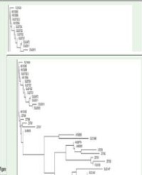
Oceanobacillus is part of a phylogenetic cluster of halophilic genera in the Bacillus sensu lato group, also involving the various genera.The Oceanobacillus,firstly explain with the species O. iheyensis by Lu et al.[20], has been extended recently with two species O. oncorhynchi [21] and Oceanobacillus picturae comb. nov. [22]. Phylogenetic analysis based on 16S rRNA gene sequences indicated that strain BS1(2) was affiliated with the genus Oceanobacillus (Figure 2).

Figure 2: Phylogenetic tree based on a comparison of the 16S ribosomal DNA sequences and some of their closest phylogenetic relatives.The numbers on the tree indicates the percentages of bootstrap sampling derived from 1,000 replications.
According to 16S rRNA gene sequences, strain BS1(2) shown a high level of similarity with the type strain of Oceanobacillus and shown a substantial degree of relatedness to references 16S rRNA sequences of Oceanobacillus in the database. The highest similarity values with the sequences of obligately halophilic and alkaliphilic microorganisms that previously isolated from the bottom of the deep-sea.
Biological processes offer great promising-g alternatives due to their environmental friendliness. Microbial fuel cells (MFCs) are a newly developed and environmentally friendly biotechnology. In the present study total fifty seven bacterial cultures were isolated by using different enrichment media. Out of them five bacterial strained BW43, BW41, BS11, BS12 and BW42 were selected for the electricity generation and screened from the Horikoshi II enrichment medium. In the present study, the 24h alkaline pH (10) culture was aerobically prepared and aseptically pours into anodic chamber. After the preparation, MFC was constructed and measure the electricity generation after various intervals such as 24h to 64h. the optimum bioelectricity generate 113 and 424 mV by BW43 and BW41 after 24h respectively, but 316mV was generated by BS11 at 24h but after 24h the bioelectricity generation was dramatically decrease for these three bacterial strain. While BW42 was generate the bioelectricity 203mV at 24h and increase the electricity generation 230mV after 30h. In present investigation, the BS12 was found prominent for the bioelectricity generation. At 24h the 111mV electricity generated and continuously increase the power generation upto 614mV at 38h, after these interval the generation was goes down to decrease (Figure 2). Oceanobacillus iheyensis BS12 strain was screened and selected for further bioelectricity generation and characterization. Velasquez-Orta et al., [23] was evaluated the performance of MFCs using two different types of algae as substrates. Chlorella vulgaris (a microalgae) and Ulva lactuca (a macroalgae). One of the first pure cultures to be studied as an oxidation catalyst in MFC systems was Shewanella oneidensis [24]. An MFC can be a very robust device when it is subjected to short-term changes of operating parameters such as sugar, NaCl, Acetate, lactate and pH.
Table 1:
| Gram character |
+ |
Growth at pH 12 |
+ |
| Shape of Bacteria |
Long Rod |
Growth at 1% NaCl |
+ |
| Size of Bacteria (Legnth (um)) |
3 |
Growth at 2% NaCl |
+ |
| Size of Bacteria (Width (um)) |
0.6 |
Growth at 3% NaCl |
+ |
| Arrangements of Cell |
Chain |
Growth at 4% NaCl |
+ |
| Spore bearing |
+ |
Growth at 5% NaCl |
+ |
| Position of Spore |
Central |
Growth at 6% NaCl |
+ |
| Shape of Spore |
Cylindrical |
Growth at 7% NaCl |
+ |
| Swollen Sporangia |
- |
Catalase |
+ |
| Capsule |
+ |
Oxidase |
+ |
| Motility |
Motile |
Indol |
- |
| Type of motility |
Highly Motile |
MR |
- |
| Flagella |
+ |
VP |
- |
| Enrichment medium |
Horikoshi II |
Citrate |
Not Utilized |
| Size of Colony |
1mm |
Urease |
- |
| Pigment |
White |
Nitrate |
- |
| Colony Shape |
Circular |
Glucose |
+ |
| Colony Elevation |
Effuse |
Arabinose |
- |
| Colony Edge |
Entire |
Mannitol |
- |
| Internal structure of Colony |
Wavy Interlaced |
Xylose |
- |
| Colony on slant |
Eschinulate |
Lactose |
- |
| Growth at 37º C |
+ |
Trehalose |
- |
| Growth at 45º C |
+ |
Sucrose |
- |
| Growth at 50º C |
+ |
Galactose |
- |
| Growth at 55º C |
- |
Maltose |
- |
| Growth at pH 7 |
+ |
Fructose |
- |
| Growth at pH 8 |
+ |
Salicin |
- |
| Growth at pH 9 |
+ |
Sorbitol |
- |
| Growth at pH 10 |
+ |
Raffinose |
- |
Effect of Sugar on Bioelectricity generation
The most extensive studies has been reported on the bioelectricity generation by using carbohydrate-rich wastes such as food processing wastewater, starch processing wastewater and chocolate based wastewater [25]. Electricity generation by direct oxidation of glucose in mediator less microbial fuel cells was reported by Chaudhuri and Lovley [7].In the present studies, anodic chamber is supplement with various sugars for the bioelectricity generation. Tarte et al., [26] were studies on effect of NaCl and Glucose on generation of electricity MFC prepared from waste water with the different combinations of glucose and NaCl.The bacterium BS12 also supplemented with the various sugar, after the addition of glucose, the electricity generation was found to be increases up to 254mV and also supplement with sucrose (223mV),lactose(209mV),maltose (126mV),manitol (119mV), but the optimum electricity generation was found after addition of starch 445mV (Fig4).Microbial fuel cells are (MFCs) fascinating bioelectrochemical devices that use living catalysts to produce electric energy from organic matter present naturally in the environment or in waste. Kumar et al., [27] investigate bioelectricity generation and treatment of sugar mill effluent using a microbial fuel cell (Figure 3 and Figure 4).

Figure 3: Effect of Sugar on Bioelectricity generation.

Figure 4: Effect of Sugar.
Responses of Voltage Output to pH variations
In recent years, this technology has been studied by many researchers. Generally, different parameters including both operational and designing factors affect the MFC performance. An MFC can be a very robust device when it is subjected to short-term changes of operating parameters such as pH to probe the response of MFC performance to these parameters; an MFC inoculated with a BS12 was constructed. Gonzalez del Campo et al. [28] revealed that In the case of effects of medium pH on the power generation of an MFC, it was found that acidification of the anode affected. In the present investigation, 169mV at 48h old culture generate the bioelectricity at neutral pH while the bioelectricity generation was increase after the pH was 10 (594mV) but at 50h incubation the electricity was optimum generate 644mV (Figure 5).

Figure 5: Responses of Voltage Output to pH variations.
Effect of NaCl on Bioelectricity generation
Salinity effect on the microbial fuel cell performance was investigated. The 1.5% and 2% of Nacl was added into the MFC, after the addition of NaCl, the electricity generation was found to be increase while as the incubation period was increase the electricity was also increases (1h,19mV), after 48h the electricity was 265mV and 120mV generated after addition of 1% and 2% respectively. After 50h incubation the electricity generation was found optimum for 1% NaCl 352 mV while 52 h Incubation bioelectricity generation was optimum for 2% NaCl (520 mV) and The similar result was observed by Luo et al., [29] after the addition of NaCl on electricity generation was decreases. Muralidharan et al., [30] was studied on the impact of salt concentration on electricity production in microbial hydrogen based salt bridge fuel cells. The best of our knowledge, these MFC was most efficient for electricity generation. Parkash et al., [31] studied the impact of various salt and concentration on the electricity generation based on dual chambered MFC. They revealed that the KCl salt bridge was efficient electricity generating that the NaCl (Figure 6).

Figure 6: Effect of NaCl on Bioelectricity generation.
Effect of lactate and acetate on Bioelectricity generation
Biochemical routes that lead to acetate produce more hydrogen than those that lead to butyrate production [32]. However, there are no previous studies on electricity production from lactate, while bioelectricity generation from acetate in two-chambered MFCs is well known, systems. Here we demonstrate that electricity can be generated from lactate and acetate in a double-chambered MFC, and we compare power densities obtained from acetate and lactate with those previously obtained in the same system using sugar. Here in the present investigation the 231mV and 167mV bioelectricity were generated in presence of lactate and acetate respectively (Figure 7).

Figure 7: Effect of lactate and acetate on Bioelectricity generation.
Conclusion
A total twenty eight bacterial isolates obtained in the isolation exercise, cultural, morphological characteristics and standard biochemical test were performed for of all the isolates. A totally fifty seven bacterial culture were isolated, from which BS12 was selected for the further investigation of MFC. Phylogenetic analysis based on 16S rRNA gene sequences indicated that strain BS1(2) was affiliated with the genus Oceanobacillus. In this double chamber MFC using bacterium BS1(2) was used as biocatalyst. Anode chamber was kept up in batch condition for the addition of sugar, lactate and acetate while in cathode chamber salt was added and maintained at continuous condition. The MFC was constructed and measure the electricity generation after various intervals, 644mV was electricity was generated after 1h, but after 48h the electricity generation was dramatically decreases up to 420mV. The effect of salt on MFC, NaCl was enhance electricity generation indicating a bacterium BS1(2) was required NaCl for the MFC and salt, sugar had an even greater effect on execute the electricity generation by MFCs. However, further improvements need to be made to increase power generation.
References
1. Bullen RA, Arnot TC, Lakeman JB, Walsh FC. Biofuel cells and their development. Biosens. Bioelectron. 2006; 21: 2015-2045.
2. Davis F, Higson SP. Biofuel cells - recent advances and applications. Biosens. Bioelectron. 2007; 22: 1224-1235.
3. Logan BE, Murano C, Scott K, Gray ND, Head IM. Electricity generation from cysteine in a microbial fuel cell. Water Res. 2005; 39: 942-952.
4. Zhang LX, Zhou SG, Zhuang L, Weishan L, Jintao Z, Lifang D, et al. Microbial fuel cell based on Klebsiella pneumoniae biofilm. Electrochem. Commun. 2008; 10: 1641-1643.
5. Holmes DE, Bond DR, Lovley DR. Electron transfer by Desulfobulbus propionicus to Fe(III) and graphite electrodes. Appl. Environ. Microbiol. 2004; 70: 1234-1237.
6. Bond DR, Lovley DR. Electricity production by Geobacter sulfurreducens attached to electrodes. Appl. Environ. Microbiol. 2003; 69: 1548-1555.
7. Chaudhuri SK, Lovley DR. Electricity generation by direct oxidation of glucose in mediatorless microbial fuel cells. Nat. Biotechnol. 2003; 21: 1229-1232.
8. Pham CA, Jung SJ, Phung NT, Lee J, Chang IS, Kim BH, et al. A novel electrochemically active and Fe(III)-reducing bacterium phylogenetically related to Aeromonas hydrophila, isolated from a microbial fuel cell. FEMS Microbiol. Ecol. 2003; 223: 129-134.
9. Kim HJ, Park HS, Hyun MS, Chang IS, Kim M, Kim BH. A mediatorless microbial fuel cell using a metal reducing bacterium, Shewanella putrefaciens. Enz Microb Technol. 2002; 30:145-152.
10. Bond DR, Holmes DE, Tender LM, Lovley DR. Electrode-reducing microorganisms that harvest energy from marine sediments. Science. 2002; 295: 483-485.
11. Min B, Logan BE. Continuous electricity generation from domestic wastewater and organic substrates in a flat plate microbial fuel cell. Environ Sci Technol. 2004; 38: 5809-5814.
12. Rabaey K, Boon N, Siciliano SD, Verhaege M, Verstraete W. Biofuel cells select for microbial consortia that self-mediate electron transfer. Appl Environ Microbiol. 2004; 70: 5373-5382.
13. Liu M, Yuan Y, Zhang L, Zhuang L, Zhou S, Jin-ren Ni. Bioelectricity generation by a Gram-positive Corynebacterium sp. strain MFC03 under alkaline condition in microbial fuel cells. Bioresource Technology. 2010; 101: 1807-1811.
14. Marshall CW, May HD. Electrochemical evidence of direct electrode reduction by a thermophilic Gram-positive bacterium, Thermincola ferriacetica. Energy Environ. Sci. 2009; 6: 699-705.
15. Nimje VR, Chen CY, Chen CC, Jean JS, Reddy AS, Fan CW, et al. J. Power Sources. 2009; 190: 258-263.
16. Park HS, Kim BH, Kim HS, Kim HJ, Kim GT, Kim M, et al. A novel electrochemically active and Fe(III)-reducingbacterium phylogenetically related to Clostridium butyricum isolated from a microbial fuel cell. Anaerobe. 2001; 7: 297-306.
17. Dhundale V, Hemke V, Chaudhari A, Darade R, Charude S. Evaluation of Electricity generation by Microbial Fuel Cell from hypersaline Indian soda lake. Int J Pharm Bio Sci. 2017; 8: 363-369.
18. Lee J, Phung NT, Chang IS, Kim BH, Sung HC. Use of acetate for enrichment of electrochemically active microorganisms and their 16S rDNA analyses, FEMS Microbiology Letters. 2003; 223: 185-191.
19. Song T, Wei-min Tan, Xia-yuanWu , Charles CZ. Effect of graphite felt and activated carbon fiber felt on performance of freshwater sediment microbial fuel cell. J Chem Technol Biotechnol. 2012; 87: 1436-1440.
20. Lu J, Nogi Y, Takami Y. Oceanobacillus iheyensis gen. nov., sp. nov., a deep sea extremely halotolerant and alkaliphilic species isolated from a depth of 1050 m on the Iheya Ridge. FEMS Microbiol Lett. 2001; 205: 291-297.
21. Yumoto I, Hirota K, Nodasaka Y, Nakajima K. Oceanobacillus oncorhynchi sp. nov., a halotolerant obligate alkaliphile isolated from the skin of a rainbow trout (Oncorhynchus mykiss), and emended description of the genus Oceanobacillus. Int J Syst Evol Microbiol. 2005; 55: 1521-1524.
22. Lee SY, Oh TK, Kim W, Yoon JH. Oceanobacillus locisalsi sp. nov., isolated from a marine solar saltern of the Yellow Sea, Korea. Int J Syst Evol Microbiol. 2010; 60: 2758-2762.
23. Velasquez-Orta SB, Curtis TP, Logan BE. Energy from Algae using Microbial Fuel Cells. Biotechnol. Bioengineering. 2009; 103: 1068-1076.
24. Bretschger O, Obraztsova A, Sturm CA, Chang IS, Gorby YA, Reed SB, et al. Current production and metal oxide reduction by Shewanella oneidensis MR 1wild type andmutants. Appl Environ Microbiol. 2007; 73: 7003-7012.
25. Chaturvedi V, Verma P. Microbial fuel cell: a green approach for the utilization of waste for the generation of bioelectricity. Bioresour. Bioprocess. 2016; 3: 38.
26. Tarte S, Pawar V, Kadam A, Chandre M. Studies on effect of NaCl and Glucose on generation of electricity from waste water. Int J Curr Biotechnol. 2015; 3: 12-15.
27. Kumar R, Singh L, Zularisam AW. Bioelectricity Generat ion and Treatment of Sugar Mill Effluent Using a Microbial Fuel Cell. Journal of Clean Energy Technologies. 2016; 4: 249-252.
28. Gonzalez del Campo A, Cañizares P, Lobato J, Rodrigo M, Fernandez Morales FJ. Effects of External Resistance on Microbial Fuel Cell’s Performance. In: Lefebvre G, Jimenez E, Cabanas B. (eds) Environment, Energy and Climate Change II. The Handbook of Environmental Chemistry. 2014; 34: 175-197.
29. Luo Y, Heng-yun W, Juan-juan J, Xiu-feng L, Qi-ming Z, Ming-ping ZC, et al. Salinity effect on the microbial fuel cell performance. Appl. Mechanics Materials. 2014; 651-653: 1365-1369.
30. Muralidharan A, Babu A, Nirmalraman K, Ramya M. Impact of salt concentration on electricity production in microbial hydrogen based salt bridge fuel cells. Indian Journal of Fundamental Appl Life Sci. 2011; 1: 178 184.
31. Parkash A, Aziz S, Soomro SAJ. Impact of salt concentrations on electricity generation using hostel sludge based dual chambered Microbial Fuel Cell. Bioprocess Biotech. 2015; 5: 252.
32. Liu H, Cheng S, Logan BE. Production of Electricity from Acetate or Butyrate Using a Single-Chamber Microbial Fuel Cell. Environ. Sci. Technol. 2005; 39: 658-662.
Citation
Dhundale V, Hemke V, Desai D, Aher G, Khemchandani P, Dikonda P, et al. Improvement and Stable High Bioelectricity Generation Using Alkaliphilic Oceanobacillus iheyensis BS1(2) in Microbial Fuel Cells and Effect of Different Anodic Operating Conditions. Ann Appl Microbiol Biotechnol J. 2018; 2(1): 1010.