Case Report | Volume 1 - Issue 1 | Article DOI :
Download PDF
Rae Leonor Gumayan¹ and John A Sandoval²,³*
¹Southern Illinois University School of Medicine, USA
²HSHS St. John’s Children’s Hospital, USA
³Department of Pediatric Surgery, Baptist Children’s Hospital, USA
Corresponding Author:
John A Sandoval, Department of Pediatric Surgery, HSHS St. John’s Children’s Hospital, USA, Tel: 217-757-6535; Email: john.sandoval@hshs.org
Keywords
Recurrent hypertrophic pyloric stenosis; Minimally invasive surgery; Atropine sulfate; Pediatric anesthesia and neurodevelopment effects
Abstract
Recurrent pyloric stenosis is a rare occurrence that presents weeks after initial operative management and a history of complete cessation of symptoms. We report on a case managed with a repeat laparoscopic pyloromyotomy with a successful outcome. Brief commentary is provided on the emerging significance of administration of general anesthesia and the possible long-lasting deleterious neurocognitive effects in the pediatric population
Citation
Gumayan RL and Sandoval JA. Operative Management of Recurrent Hypertrophic Pyloric Stenosis: A Case Report and Review of the Literature. SM Min Inv Surg. 2017; 1(1): 1004.
Introduction
Idiopathic Hypertrophic Pyloric Stenosis (IHPS) is a common disease of infancy that requires surgical management. The gold standard of treatment is a pyloromyotomy that results in high curative rates with low morbidity to the patient. An incomplete pyloromyotomy is a known but infrequent post-operative complication that usually presents within five days and necessitate immediate evaluation. Even more rare are cases of recurrent IHP Srequiring re-operation.
Case Report
A 2.5 kg previously healthy female infant born at term presented at three weeks of age with non-bilious projectile emesis for several days, constipation, decreased urine output, and weight loss from her birth weight of 2.9 kg. On physical exam, the patient was malnourished and dehydrated. Laboratory findings indicated mild hypokalemia, but severe hypochloremic metabolic alkalosis. An abdominal ultrasound revealed hypertrophic pyloric stenosis with a wall thickness of 4.3 mm and a 22 mm channel length (Figure 1).

Figure 1: The initial ultrasound showed hypertrophic pyloric stenosis. The pyloric channel had a length of 25 mm and a thickness of 4.3 mm.
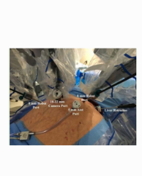
The patient was resuscitated with intravenous fluids over a 48 hour interval and prepared for a laparoscopic pyloromyotomy. An open technique was used to insert a 5 mm step trocar through the umbilicus with the verification of intraperitoneal placement prior to the pneumoperitoneum pressure of 10 mmHg. Two stab incisions were placed in the right upper quadrant and the left upper quadrant. The liver was retracted and the proximal duodenum was grasped to allow for stabilization of the pylorus, which was obviously hypertrophied. A longitudinal incision was made with electrocautery along the anterior aspect of the pylorus from the white line onto the proximal stomach and antrum. Muscle was split with blunt dissection until the mucosa was visualized. The stomach was distended with air and there was no evidence of leak. The 2 portions of the pyloric muscle were grasped and moved independently of each other. She tolerated the procedure well and had an unremarkable postoperative course. She was started on immediate oral feeding and was discharged on Postoperative Day (POD) 1. At 2 weeks follow up, she was tolerating feeds well and had complete resolution of emesis.
On POD 29, she was re-admitted for choking apneic spells, worsening reflux, and occasional projectile emesis for one week. Her weight on this admission was 3.0 kg. A repeat abdominal ultrasound demonstrated 3.6 and 4.2 mm width with 2 cm length but a patent pyloric channel consistent with pyloric stenosis. She was feeding 3 oz every 3 hours with moderate reflux and one episode of projectile emesis. Within two days, she progressively had emesis at almost all her feeds and she was placed on maintenance IV fluids. A non-barium contrast enema suggested possible functional immaturity of the colon. Upper gastrointestinal contrast study done on POD 32 showed pyloric beaking with complete gastric outlet obstruction and prominent shouldering on the gastric antrum, highly suggestive of recurrent hypertrophic pyloric stenosis (Figure 2).

Figure 2: Upper GI study performed on your patient on POD 32 showed complete gastric outlet obstruction. The bulge of the hypertrophied muscle is shouldering (arrow) into the antrum (A) of the stomach and a beak-like taper of the pylorus channel is observed (arrow head).
The patient underwent a second redo laparoscopic pyloromyotomy (Figures 3a-c). The previous site of the pyloromyotomy was visualized and 2 portions of the pyloric muscle were clearly identified and there was concern for inadequate pyloromyotomy length (Figure 3a). The previous pyloromyotomy site was extended onto the stomach antrum using a 3 mm right angle instrument and electrocautery such that the muscle was split with blunt dissection until the mucosa was clearly visualized (Figures 3b & 3c). The infant was allowed to feed ad libitum postoperatively. She had an uneventful recovery and was discharged on POD 2 tolerating full feeds without emesis.

Figure 3: Intraoperative findings at the time of redo laparoscopic pyloromyotomy (Figures 3a-c).
Figure 3a shows the previous site of the pyloromyotomy and concern for an inadeaquate pyloromyotomy length.
Figures 3b and 3c demonstrate extension of the previous pyloromyotomy site towards the stomach antrum.
Discussion
A 2008 review of 30 years of data from the Hospital of Sick Children in Toronto yielded a mean of 100 cases of IHPS annually with only two recorded cases of recurrent pyloric stenosisin total (<0.07%) [1]. In review of the English literature, a total of only nine cases of true recurrent IHPS have been reported requiring surgical intervention [2-9]. True recurrent pyloric stenosis has been defined under the criteria that after a successful pyloromyotomy, the patient has absence of emesis for ≥3 weeks accompanied by weight gain before re-emergence of symptoms and confirmed by an upper GI study or an operative view of the hypertrophied pylorus [2]. Our patient’s second abdominal ultrasound revealed an open pyloric channel, however, an upper GI study performed three days later demonstrated a complete gastric outlet obstruction. Her clinical presentation corroborated with a rapid progression of intolerance of feeds and she underwent a laparoscopic repyloromyotomy 33 days after a first successful pyloromyotomy. An incomplete gastric pyloromyotomy was attributed to the etiology of her failed initial pyloromyotomy. This observation has been previously reported for incomplete pyloromyotomies by Khoshoo and colleagues [10].
The etiology of IHPS remains unclear, but some authors speculated that IHPS may be an acquired disease that continues to progress despitea primary surgical intervention [9] or that it is simply the nature of the disease [3]. While both open and minimally invasive approaches are considered comparable surgical options, we prefer the laparoscopic approach because of the shorter operative time without the risk of higher complications or cost to the patient and better cosmetic advantage in comparison to an open operative technique [11-13]. Nonsurgical methods for IHPS include medical and fluoroscopic mechanical balloon dilation. Atropine and for historical purposes, methyl scopolamine nitrate have been described for medical management [14,15]. The present use of atropine is not currently accepted in Western countries as a primary therapy for HPS, as opposed to Japan, where medical management using IV atropine is used as first-line therapy [16-19]. A recent meta-analysis by Wu and coauthors showed the efficacy of atropine in the management of IHPS medical treatment with either oral or IV atropine was an appropriate alternative to pyloromyotomy for IHPS patients, particularly in the infant subset population with major concurrent disease or when parents were unwilling to let their infants undergo surgery [20]. Enthusiasm for oral atropine as a rescue therapy for incomplete pyloromyotomy or persistent emesis following laparoscopic pyloromyotomy has attracted attention for these challenging postoperative senarios [21,22]. Moreover, limited data supports the use of fluoroscopic pyloric balloon dilation for treatment of recurrent IHPS, the technique requires a skilled and experienced interventional radiology department [1]. If balloon dilation fails to relieve the obstruction, a redo pyloromyotomy is generally felt to be required. Regardless of the above management options, none of the reported cases resolved spontaneously, ultimately requiring surgical intervention. The most appropriate study to confirm recurrent pyloric stenosis is an upper GI study that can demonstrate partial or complete gastric outlet obstruction as seen in our patient. A repeat abdominal ultrasound is unreliable as it can appear thickened and will take up to 8 months to decrease in width [7]. Lastly, given the concerns linking anesthesia exposure in infants with poor neurocognitive outcome [23] and the possibility that longer duration, or repetitive, anesthetic exposures may cause neurocognitive impairment in children [24], strategies to either limit the exposure of these agents oridentify viable alternative anesthetics should be taken into consideration, including the use of oral atropine in the context of recurrent pyloric stenosis.
References
1. Nasr A, Ein SH, Connolly B. Recurrent pyloric stenosis: to dilate or operate? A preliminary report. J Pediatr Surg. 2008; 43: 17-20.
2. Al-Ansari A, Altokhais TI. Recurrent pyloric stenosis. Pediatr Int. 2016; 58: 619-621.
3. Ankermann T, Engler S, Partsch CJ. Repyloromyotomy for recurrent infantile hypertrophic pyloric stenosis after successful first pyloromyotomy. J Pediatr Surg. 2002; 37: 1-2.
4. Cappiello C, Strauch E. A rare case of recurrent hypertrophic pyloric stenosis. Journal of Pediatric Surgery Case Reports. 2014; 2: 519-521.
5. Granström A, Felder S, Frykman P. Laparoscopic repyloromyotomy following open pyloromyotomy for recurrent pyloric stenosis: a case report and review of the literature. Eur J Pediatr Surg. 2013; 23: 499-501.
6. Hiller DJ, Petty JK, Sieren LM. Recurrent Pyloric Stenosis: A Rare Entity. Am Surg. 2015; 81: 330-331.
7. Iranmanesh AM, Pawley BK, Rice MM, Richer EJ. Imaging findings in infants with recurrent vomiting after pyloromyotomy: a pictorial review. EmergRadiol. 2015; 22: 691-695.
8. Louati H, Zitouni H, Belhajmansour M, Ben Dhaou M, Jallouli M, Mhiri R, et al. A Difficult Recurrent Hypertrophic Pyloric Stenosis. Journal of Neonatal Surgery. 2017; 6: 48.
9. vanHeurn LW, Vos P, Sie G. Recurrent vomiting after successful pyloromyotomy. Pediatr Surg Int. 1999; 15: 385-386.
10. Khoshoo V, Noel RA, LaGarde D. Endoscopic balloon dilatation of failed pyloromyotomy in young infants. J Pediatr Gas troenterol Nutr. 1996; 23: 447-451.
11. Kim SS, Lau ST, Lee SL, Schaller R Jr, Healey PJ, Ledbetter DJ, et al. Pyloromyotomy: a comparison of laparoscopic, circumumbilical, and right upper quadrant operative techniques. J Am Coll Surg. 2005; 201: 66-70.
12. Greason KL, Thompson WR, Downey EC, Lo Sasso B. Laparoscopic pyloromyotomy for infantile hypertrophic pyloric stenosis: report of 11 cases. J Pediatr Surg. 1995; 30: 1571-1574.
13. Sathya C, Wayne C, Gotsch A, Vincent J, Sullivan KJ, Nasr A. Laparoscopic versus open pyloromyotomy in infants: a systematic review and meta-analysis. Pediatr Surg Int. 2017; 33: 325-333.
14. Nagita A, Yamaguchi J, Amemoto K, Yoden A, Yamazaki T, Mino M. Management and ultrasonographic appearance of infantile hypertrophic pyloric stenosis with intravenous atropine sulfate. J Pediatr Gastroenterol Nutr. 1996; 23:172-177.
15. Corner, B.D. Hypertrophic pyloric stenosis in infancy treated with methyl scopolamine nitrate. Arch Dis Child. 1955; 30: 377-386.
16. Yamataka A, Tsukada K, Yokoyama-Laws Y, Murata M, Lane GJ, Osawa M, et al. Pyloromyotomy versus atropine sulfate for infantile hypertrophic pyloric stenosis. J Pediatr Surg. 2000; 35: 338-341.
17. Singh UK, Kumar R, Prasad R. Oral atropine sulfate for infantile hypertrophic pyloric stenosis. Indian Pediatr. 2005; 42: 473-476.
18. Kawahara H, Takama Y, Yoshida H, Nakai H, Okuyama H, Kubota A, et al. Medical treatment of infantile hypertrophic pyloric stenosis: should we always slice the “olive”? J Pediatr Surg. 2005; 40: 1848-1851.
19. Takeuchi M, Yasunaga H, Horiguchi H, Hashimoto H, Matsuda S. Pyloromyotomy versus i.v. atropine therapy for the treatment of infantile pyloric stenosis: nationwide hospital discharge database analysis. Pediatr Int. 2013; 55: 488-491.
20. Wu SF, Lin HY, Huang FK, Chen AC, Su BH, Li CI, Lin HC. Efficacy of Medical Treatment for Infantile Hypertrophic Pyloric Stenosis: A Meta-analysis. PediatrNeonatol. 2016; 57: 515-521.
21. Owen RP, Almond SL, Humphrey GM. Atropine sulphate: rescue therapy for pyloric stenosis. BMJ Case Rep. 2012; 2: 2012.
22. Cubas RF, Longshore S, Rodriguez S, Tagge E, Baerg J, Moores D. Atropine: A Cure for Persistent Post Laparoscopic Pyloromyotomy Emesis?. J Neonatal Surg. 2017; 6: 2.
23. Creeley CE. From Drug-Induced Developmental Neuroapoptosis to Pediatric Anesthetic Neurotoxicity-Where Are We Now?. Brain Sci. 2016; 6: 32.
24. Montana MC, Evers AS. Anesthetic Neurotoxicity: New Findings and Future Directions. J Pediatr. 2017; 181: 279-285.