Case Report | Volume 6 - Issue 1 | Article DOI : https://dx.doi. org/10.36876/smdj790598
Download PDF
Claire Barber*, Emily Wichelt, Lindsey Braden, Racquel Kaizer, Luna Emogene, Adam El-Newihi, Amber Boudreaux, and Mohamed Aziz
Department of Pathology, American University of the Caribbean School of Medicine, 1 University Drive at Jordan Road Cupecoy, St.Maarten
Corresponding Author:
Claire Barber, Department of Pathology,
American University of the Caribbean School of Medicine, 1 University Drive at Jordan Road Cupecoy, St. Maarten, Tel: 512-919-6276;
Keywords
Eccrine; Skin; Adnexal; Recurrence; Metastasis
Abstract
Eccrine porocarcinoma (EPC) is a rare type of skin cancer arising from the intraepidermal portion of eccrine sweat glands or acrosyringium that represents 0.005-0.01% of all cutaneous tumors. EPC can present as a primary tumor or, more commonly, as a malignant transformation of an eccrine poroma (EP). Eccrine and apocrine neoplasms present a bewildering array of morphologies, which often defy precise classification. This case describes a 62-year-old female with a soft tissue ulcerated mass at the front aspect of right thigh that reoccurred two years after a wide local excision was performed. EPC represents a problematic diagnosis as it is a locally aggressive neoplasm, with high recurrence rates, metastases to lymph nodes and to distant solid organs. While initial surgical treatment is curative for most of the cases, no standard treatment protocols exist for metastatic EPC. Clinicians, therefore, need to be aware of this rare entity, as porocarcinomas often show a marked morphologic variability, which has led to misdiagnosis and delayed treatment with disastrous consequences.
Citation
Barber C, Wichelt E, Braden L, Kaizer R, Emogene L, et al. (2020) Eccrine Porocarcinoma, a Rare Skin Adnexal Tumor. Report of a Case and Review of the Literature. SM Dermatol J 7: 5. doi: https://dx.doi. org/10.36876/smdj790598
ABBREVIATIONS
EPC: Eccrine Porocarcinoma, MMS: Mohs micrographic surgery, WLE: Wide Local Excision, IHC: Immunohistochemistry
INTRODUCTION
Eccrine porocarcinoma (EPC) is a rare type of skin cancer arising from the intraepidermal portion of eccrine sweat glands or acrosyringium [1]. Although EPC is relatively rare, representing only 0.005% of epithelial cutaneous tumors, the literature widely recognizes it as the most common of the sweat gland adenocarcinoma types [2, 14]. Eccrine sweat glands consist of secretory coils in the lower dermis or subcutis that secrete electrolytes and water into a thin duct terminating in the acrosyringium, the intraepidermal ductal portion opening directly to the surface of the skin [3]. The greatest distribution of sweat glands and eccrine poromas is on the palms of the hands and soles of the feet. However, the distribution of EPC does not correlate well with the concentration of sweat glands [4]. Eccrine porocarcinoma classically presents as a verrucous plaque or nodule of the distal extremities, particularly the lower extremities, the head and neck or trunk of an individual typically in the sixth to ninth decades of life [5]. Less than 300 cases have been described in the entire world medical literature [6]. Porocarcinoma affects both sexes equally and all races may be affected [11]. This tumor can appear de novo or as a progression of a benign poroma [6]. The time to malignant transformation of a benign poroma is highly variable and can range from months to decades [6]. Transition from a benign eccrine poroma to a malignant tumor may be indicated by ulceration or bleeding, sudden growth, or a new multinodular morphology [4]. It usually manifests clinically as a solitary lesion with a highly non-characteristic macroscopic appearance [7]. Often EPC presents as an ulcerated nodule or as a plaque, polypoid, or verrucous lesion [7].
Porocarcinoma is a dangerous disease because of high rate of recurrence after resection and metastasis to vital organs [8]. Metastasis occurs in about 20% of cases with a very poor outlook and high mortality [9]. When lymph node metastases occur, the mortality rises up to approximately 65% [10]. Most commonly, multiple cutaneous metastases develop however metastases have also been reported in lung, retro-peritoneum, bone, liver, breast, bladder, peritoneum, and ovary [6]. Because of EPC’s rarity and lack of distinct clinical features, its diagnosis is often challenging [12]. The low degree of clinical suspicion of EPC could be due to the low incidence rate, non-specific macroscopic appearance, and vague dermoscopic pattern of neovascularization. As a result, the diagnosis and appropriate treatment are often delayed, thereby providing time for the cancer to spread throughout the body, with lethal consequences [6]. Studies have been limited due to its rarity, and controversies regarding tumor behavior and treatment are present in the literature [14]. Clinicians, therefore, need to be aware of this rare entity, as porocarcinomas often show a marked morphologic variability, which has led to misdiagnosis and delayed treatment with disastrous consequences in previously reported cases [10].
EPC is classified into three eccrine porocarcinoma subtypes: “infiltrative,” “pushing” and “pagetoid” eccrine porocarcinoma [15, 16]. It can be intraepidermal, an in-situ variation, growing horizontally, and produces an extensive pagetoid infiltrate. This epidermotropic or intraepidermal type has been reported to have local recurrences. The invasive dermal type usually presents as nodular aggregates associated with a broad deep margin pushing down into the dermis (pushing type) or with an infiltrative growth pattern (infiltrative type) [6]. Dividing tumors into those with a “pushing” or “infiltrating” advancing margin was also predictive of outcome with the latter having an increased risk of local recurrence [16]. The pagetoid variant seems to have an aggressive local potential, although this could not be statistically investigated in studies because of the limited number of cases [15].
CASE PRESENTATION
A 62-old-woman presented with 3.8 cm soft tissue ulcerated mass at the front aspect of the right thigh. She first observed the mass two years earlier, but only recently noticed an increased in size with ulceration at presentation. Biopsy of the mass was diagnostic of Eccrine Porocarcinoma (Malignant Eccrine Poroma- Adnexal skin carcinoma). The mass’s depth of invasion was 1.2 cm into the subcutaneous tissue and was excised with a 1.5 cm save margin. Inguinal sentinel lymph node sampling was performed and was negative, no vascular or neural invasion were noted, and there was no lymph nodes dissection.
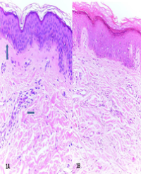
Pathologic microscopic examination revealed an infiltrative tumor border with areas of horizontal growth pattern. The tumor displayed small cuboidal cells manifesting intercellular bridges and eosinophilic cytoplasm with sharply defined cell borders. The tumor showed nuclear atypia, increased mitotic activity 8 mitosis/10 high power field, and moderate necrosis (Figure 1a-d). Immunohistochemistry (IHC) studies were performed, and the tumor cells were positive for EMA, CEA, CAM 5.2, CK 7, while negative for CK 20, CD 10, and CD34. Only basal layers were stained by S-100. Final diagnosis was consistent with the biopsy diagnosis of Eccrine Porocarcinoma. No post-operative treatment was given.

Figure 1: Microscopic description of original tumor,
Figure A & B: Infiltrative tumor border with areas of horizontal growth pattern (H&E stain X20)
Figure C & D: Small cuboidal cells manifesting intercellular bridges and eosinophilic cytoplasm with sharply defined cell borders (H&E stain X40 and X60)
Two years later, a 2.4 cm irregular soft mass developed at the site of prior excision. The mass showed surface hemorrhagic ulceration. Biopsy confirmed recurrent Eccrine Porocarcinoma (Figure 2a-d). The mass was excised with a 2 cm safe margin and additional treatment with regional chemotherapy combined with immunotherapy were given.

Figure 2: Microscopic description of the recurrent tumor.
Figure-2 A & B: The mass showed surface hemorrhagic ulceration
Figure-2 C & D: Small cuboidal cells manifesting intercellular bridges and eosinophilic cytoplasm with sharply defined cell borders. Increased mitosis and necrosis (H&E stain X40)
Three years later, patient developed metastasis to the inguinal regional lymph nodes followed by disseminated metastasis including lung, liver and brain. Patient expired as a result of widespread metastasis.
DISCUSSION
Porocarcinomas are commonly overlooked or misinterpreted as squamous or basal cell carcinomas as well as other common malignant and even benign skin tumors [10]. The differential diagnosis of EPC clinically may include squamous cell carcinoma, basal cell carcinoma, verucca vulgaris, and metastatic adenocarcinoma [4]. Eccrine porocarcinomas can also mimic many other cutaneous malignancies, including Bowen’s disease, amelanotic melanoma, cutaneous metastasis of a visceral carcinoma, or common benign lesions such as seborrheic keratosis, verruca vulgaris or pyogenic granuloma [10].
EPC like EP is composed of small cuboidal cells manifesting intercellular bridges and eosinophilic cytoplasms with sharply defined cell borders. The cells and their nuclei are both larger than those seen in EP but are, nonetheless, smaller than the tumor cells of squamous cell carcinoma [5]. Tumor borders may be “infiltrative,” “pagetoid,” or “pushing.” Infiltrative EPCs are characterized by a poorly defined lower margin with malignant cell clusters infiltrating the dermis; pagetoid EPCs have intraepidermal spread of tumor cells mimicking Paget disease; pushing EPCs have a distinct dermal lining around the polypoid tumor [3]. While analyzing 69 EPC cases, Robson et al. found a variety of histological patterns including a clear, squamous, and spindle cell differentiation, mucus cell metaplasia, and colonization by melanocytes [16]. Given EP’s morphologic range, it is not surprising that the histologic differential diagnosis is long [2]. The pathology of these lesions may resemble inflamed poroma, Bowen’s disease, or Paget’s disease. There is also histologic overlap with malignant acrospiromas, basal cell carcinomas and squamous cell carcinomas [4]. For tumors with a nodular growth pattern, the differential diagnosis reportedly includes eccrine acrospiroma, sebaceous carcinoma, proliferating trichilemmal tumor, balloon cell melanoma, squamous cell carcinoma and metastatic adenocarcinoma (e.g., renal cell carcinoma) [2]. For tumors with a horizontal growth pattern, the list includes poroma, clonal seborrheic keratosis, Paget disease and Bowen’s disease [2].
EPCs are histologically diverse however, common characteristics on hematoxylin & eosin (H&E) staining include nuclear atypia, increased mitotic activity, and necrosis [3]. Histological appearance alone seems less useful because the tumor cells can be confused with squamous, clear, spindle, or Paget cells or melanocytes [3]. Immunohistochemical stains used for the diagnosis of EPC include carcinoembryonic antigen, cytokeratin (CK) (pan-CK and CK5/6), epithelial membrane antigen, p53, and p63 [6]. CD117 has been reported to be highly effective in identifying EPC and recent evidence has indicated that CD117 may be helpful in distinguishing EPC from squamous cell carcinoma [3].
It has been noted that up to 18% to 50% of porocarcinomas degenerate from initially benign poromas. Factors hypothesized to play a role in this transformation include chronic light exposure and immunosuppression [3]. YAP1 and WWTR1 fusions were exclusive to poromas and porocarcinomas among skin tumors, and thus the detection of these fusion transcripts or protein products would have diagnostic applications [12]. While examining other skin tumors Sekine et al. found out of 24 squamous cell carcinomas, 32 basal cell carcinomas, 5 cutaneous adenocarcinomas, 9 Merkel cell carcinomas, and 27 seborrheic keratoses, there were no recurrent YAP1 fusions. These observations indicate that YAP1 fusions are highly recurrent and specific to poromas and porocarcinomas among skin neoplasms [12]. The common presence of YAP1 fusions in poromas and porocarcinomas supports their histogenetic relationship, which is consistent with the idea that a significant proportion of porocarcinomas develop through the malignant transformation of preexisting poromas [12]. In our case, molecular studies were not performed due to medical insurance issue.
From others perspective, chronic light exposure, exposure to chemical agents and immunosuppression could be the predisposing factors for de novo eccrine porocarcinoma [8]. Porocarcinomas tended to harbor a larger number of mutations than poromas (average 3.0 vs. 0.96 mutations/ lesion). However, only 3 genes were recurrently mutated in porocarcinomas: KRAS, SETD2, and TP53 [12]. Their findings implicate multiple tumor suppressors in the tumorigenesis of porocarcinoma, including TP53 and RB1 [13].
Because of the rare incidence of EPC, there have been few large studies to investigate its management. As a result, there is no protocol to dictate the optimum method of excision, the utility of radiation therapy, or the best method of chemotherapy if needed [4]. Its early identification and complete excision gives the best chance of a cure. Neither chemotherapy nor radiation therapy has been proven to be of clinical benefit in treating metastatic disease [1]. Diagnosis and treatment of EPC is extremely important because of its aggressive nature [4]. Approximately 20% of patients experience local recurrence and 20% experience metastatic disease after treatment. In patients with lymph node involvement, mortality rates are approximately 67%. The survival period for patients with distant metastases is reported to be 5 to 24 months [3]. In a review of 453 cases of porocarcinoma, it was found that 31% of the cases had concomitant metastases at the time of presentation and the most common organs are lymph nodes (57.7%), also evidenced in our case, followed by visceral metastases to lung (13%), liver (9.9%), brain (9.9%), skin (5.8%), bone (3.2%) and stomach (0.6%) [17]. Most of the patients present with mass without symptoms (poroma) long time before converting to malignant type (porocarcinoma). This finding may suggest that aggressive management of benign subcutaneous mass is advised to prevent malignant conversion especially when the diagnosis is not confirmed to be benign [8]. Our findings add to the growing body of evidence that early diagnosis and surgical treatment are the main good prognostic factors [14].
Surgery was the main mode of therapy with adjuvant chemoradiotherapy if metastasis and/or recurrence occurred. [8]. Although WLE (Wide Local Excision) was reportedly the most common treatment modality, more recently, there is increasing evidence for MMS (Mohs micrographic surgery) and complete margin assessment as the preferred treatment of the primary tumor [3]. MMS becomes most important for “infiltrative” and “pagetoid” eccrine porocarcinomas as the literature as showed that wide surgical margins (> 2 cm) associated with conventional histology did not seem to reduce the risk of recurrence [15]. Practically it is recommended that a clinically suspected malignant squamous lesion should be removed by an excisional complete resection first with 3-mm limited margins; when the pathology report is diagnostic for eccrine porocarcinoma, the histological subtype should be clearly mentioned as “infiltrative,” “pagetoid” or “pushing.” If it is a “pushing” variant, no additional surgery is warranted, while additional surgery is required for “infiltrative” and “pagetoid” variants, with a further 5-mm margin using a modified micrographic Mohs technique [15]. There are no data to support the use of adjuvant chemotherapy or radiotherapy, and there are currently no agreed criteria to define patients at high risk of relapse [6].
Sentinel lymph node biopsy should also be considered if patients possess high-risk features but do not have palpable lymphadenopathy, in which case FNA would be performed [3]. The location of the primary tumor should be considered in the decision to use these modalities as well because it seems that primary lesions on the upper and lower limbs, trunk, and genitalia/ buttocks have high risk of lymph node metastasis, whereas head and neck tumors seem to have a comparatively lower risk [3]. In addition, a mitotic index of more than 14 mitotic cells per 10 high-power fields, lymphovascular invasion, and a tumor depth exceeding 7 mm and poorly differentiated tumor may suggest the need for diagnostic imaging and often predict a worse prognosis. [6]. According to Robson et al., sentinel lymph node sampling may be justified in tumors with > 7 mm depth or high mitotic counts because they are more likely associated with lymph node involvement or death [4, 16]. Performance of prophylactic lymphadenectomy in EP is a controversial issue, despite the fact that the incidence of lymph node involvement is significant [7]. Prophylactic lymphadenectomy seems to have no impact on disease-free survival [17]. Literature does agree however that radical lymphadenectomy of the affected territory be performed if the sentinel lymph node biopsy is positive [6].
Sentinel lymph node biopsy should also be considered if patients possess high-risk features but do not have palpable lymphadenopathy, in which case FNA would be performed [3]. The location of the primary tumor should be considered in the decision to use these modalities as well because it seems that primary lesions on the upper and lower limbs, trunk, and genitalia/ buttocks have high risk of lymph node metastasis, whereas head and neck tumors seem to have a comparatively lower risk [3]. In addition, a mitotic index of more than 14 mitotic cells per 10 high-power fields, lymphovascular invasion, and a tumor depth exceeding 7 mm and poorly differentiated tumor may suggest the need for diagnostic imaging and often predict a worse prognosis. [6]. According to Robson et al., sentinel lymph node sampling may be justified in tumors with > 7 mm depth or high mitotic counts because they are more likely associated with lymph node involvement or death [4, 16]. Performance of prophylactic lymphadenectomy in EP is a controversial issue, despite the fact that the incidence of lymph node involvement is significant [7]. Prophylactic lymphadenectomy seems to have no impact on disease-free survival [17]. Literature does agree however that radical lymphadenectomy of the affected territory be performed if the sentinel lymph node biopsy is positive [6].with systemic immunotherapy (interferon alpha) [10]. Recent studies have also shown that isotretinoin in monotherapy may be an effective adjuvant agent in metastatic EP, although this needs to be further studied [7]. Although retinoid’s (such as isotretinoin) mechanism of action remains unknown, beneficial effects have been observed with respect to diverse cancerous and precancerous lesions of the skin in a limited number of cases [7].
This case and literature review seek to discuss the inconstant presentation of Eccrine porocarcinoma, and further examine the challenges in diagnosing and treating this neoplasm. Delay in early detection due to its similarities with many other benign and malignant counterpoints poses a great threat to patients. EPC’s definitive diagnosis is made through biopsies with immunochemistry staining to aid in eliminating similar differentials. While there are no official protocols to treating EPC, Mohs micrographic surgery has been growing in popularity especially for more aggressive lesions. The best chances of successfully treating EPC are early detection and excision before metastasis occur.
ACKNOWLEDGMENT
Special thanks to Fadi Jamaleddin Ahmad, Jessica Jahoda, and Mataline Shine, MD candidates, American University of the Caribbean for their assistance in reviewing the final manuscript.
REFERENCES
1. Marone U, Caracò C, Anniciello AM, Di Monta G, et al. Metastatic eccrine porocarcinoma: report of a case and review of the literature. World J Surg Oncol 2011;9:32.
2. Bonadio J, Armstrong W, Gu M. Eccrine porocarcinoma: report of a case with fine needle aspiration cytology, histopathology and immunohistochemistry. Acta Cytol 2006;50:476–80.
3. Nazemi A, Higgins S, Swift R, In G, Miller K, Wysong A. Eccrine Porocarcinoma: New Insights and a Systematic Review of the Literature. Dermatol Surg. 2018 Oct;44(10):1247-1261. doi: 10.1097/ DSS.0000000000001566. PMID: 29894433.
4. Cowden A, Dans M, Militello G, Junkins-Hopkins J, et al. Eccrine porocarcinoma arising in two African American patients: distinct presentations both treated with Mohs micrographic surgery. Int J Dermatol 2006;45:146–50.
5. Crowson, A. N., Magro, C. M., & Mihm, M. C. (2006). Malignant adnexal neoplasms. Mod Pathology, 19(S2). doi:10.1038/modpathol.3800511
6. Fernández-Ferreira, R., Alvarado-Luna, G., Motola-Kuba, D., Mackinney-Novelo, I., Cervera-Ceballos, E. E., & Segura-Rivera, R. (2020). Intergluteal Cleft Eccrine Porocarcinoma with Metastasis to Inguinal Region and Lung: Case Report and Review of the Literature. Case reports in oncology, 13(3), 1463–1473. https://doi. org/10.1159/000510311
7. González-López MA,Vázquez-LópezF,SolerT,Gómez-Die ́zS,etal. Metastatic eccrine porocarcinoma: a 5.6-Year follow-up study of a patient treated with a combined therapeutic protocol. Dermatol Surg 2003;29:1227–32
8. Salih, A., Kakamad, F., Baba, H., Salih, R., Hawbash, M., Mohammed, S., . . . Abdulkarim, N. (2017, June 20). Porocarcinoma; presentation and management, a meta-analysis of 453 Cases. Retrieved March 05, 2021, from https://www.sciencedirect.com/science/article/pii/ S2049080117302078
9. Aaribi I, Mohtaram A, El Youbi M, Kharmoum J, et al. Successful management of metastatic eccrine porocarcinoma. Case Rep Oncological Med 2013;2013:1–3.
10. Gerber PA, Schulte KW, Ruzicka T, Bruch-Gerharz D. Eccrine porocarcinoma of the head: an important differential diagnosis in the elderly patient. Dermatology 2008;216:229–33.
11. Elston D, Maguire C, Kazlouskaya V, Buchen D, et al. Porocarcinoma with perineural invasion. Indian Dermatol Online J 2015;6:122.
12. Sekine, S., Kiyono, T., Ryo, E., Ogawa, R., Wakai, S., Ichikawa, H., . . . Mori, T. (2019, September 03). Recurrent yap1-maml2 and yap1-nutm1 fusions in poroma and porocarcinoma. Retrieved March 06, 2021, from https://www.jci.org/articles/view/126185
13. Harms PW, Hovelson DH, Cani AK, Omata K, Haller MJ, Wang ML, Arps D, Patel RM, Fullen DR, Wang M, Siddiqui J, Andea A, Tomlins SA. Porocarcinomas harbor recurrent HRAS-activating mutations and tumor suppressor inactivating mutations. Hum Pathol. 2016 May;51:25-31. doi: 10.1016/j.humpath.2015.12.015. Epub 2016 Jan 7. PMID: 27067779.
14. Behbahani S, Malerba S, Karanfilian KM, Warren CJ, Alhatem A, Samie FH. Demographics and outcomes of eccrine porocarcinoma: results from the National Cancer Database. Br J Dermatol. 2020 Jul;183(1):161-163. doi: 10.1111/bjd.18874. Epub 2020 Feb 23. PMID: 31954060.
15. Belin E, Ezzedine K, Stanislas S, Lalanne N, et al. Factors in the surgical management of primary eccrine porocarcinoma: prognostic histological factors can guide the surgical procedure. Br J Dermatol 2011;165:985–9.
16. Robson A, Greene J, Ansari N, et al. Eccrine porocarcinoma (malignant eccrine poroma): a clinicopathological study of 69 cases. Am J Surg Pathol 2001; 25: 710 – 720.
17. Lehmann, C., Rodriguez Ossa, P., Vargas Manrique, M., Acosta, A. E., & Quintero Pérez, Y. (2020). Eccrine Porocarcinoma with Zosteriform Metastasis. Cureus, 12(2), e6873. https://doi.org/10.7759/ cureus.6873
18. Günhan O, Karslioğlu Y, Alömeroğlu M, Berberoğlu U. Eccrine porocarcinoma: a case with an obscure primary tumor diagnosed from lymph node metastasis. Am J Dermatopathol. 2007 Apr;29(2):176-9. doi: 10.1097/DAD.0b013e31802e490f. PMID: 17414442.