Case Report | Volume 7 - Issue 1 | Article DOI :
Download PDF
Viviana Crespo1, Alan Bencosme2, Clifford Davis1, Jodie Simelda1, Haden Morton1, Phillip Pearson3 Racquel Kaizer4 , Jessica Jahoda2,5 and Mohamed Aziz5*
1American University of the Caribbean School of Medicine USA
2Florida Atlantic University, Wilkes Honors College, Florida, USA
3Philadelphia College of Osteopathic Medicine, Georgia, Dept. of Biomedical Sciences, USA
4Adventhealth Redmond, Internal Medicine Dep. Rome, Georgia, USA
5Research Writing & Publication (RWP), LLC, NY, USA
Corresponding Author:
Mohamed Aziz*, Research Writing & Publication
(RWP) LLC, 2475 Lefferts Place, Bellmore, NY 11710, USA
Keywords
Neurothekeoma, Myxoid, Mixed, Cutaneous, Benign, Differential diagnosis
Abstract
Neurothekeomas are uncommon benign superficial cutaneous tumors with various histologic patterns, such as myxoid, cellular, or mixed types, depending primarily on the quantity of myxoid matrix present. They generally affect the head and neck, are more common in women than men, and typically start in the second and early third decades of life. Once believed to originate from the nerve sheath, the source is now thought to be fibroblasts that can differentiate into myofibroblasts and entice histiocytes. Neurothekeomas can present a significant challenge for pathologists to diagnose, and strict histomorphologic features and immunohistochemistry studies are essential to establish the diagnosis. We present a recurrent scalp myxoid Neurothekeoma case in a 7-year-old girl and review the pertinent literature.
Citation
Crespo V, Bencosme A, Davis C, Simelda J, Morton H, et al. (2023) Recurrent Scalp Myxoid Neurothekeoma in a 7-year-old girl. Case Report of Uncommon Tumor and Brief Review of the Literature. Case Report and Review of the Literature. SM Dermatol J 8: 5.
Abbreviations
Neurothekeoma (NT), Immunohistochemistry (IHC), Glial fibrillary acidic protein (GFAP),
INTRODUCTION
Neurothekeoma (NT) is an uncommon benign cutaneous tumor with an ambiguous pathophysiology and many diagnostic challenges [1]. It is a rare, unique, benign soft tissue neoplasm that can be cutaneous or superficial, and it typically develops in the head and neck, shoulder, or upper extremities. Although it is likely that the cellular lineage of cellular Neurothekeoma is fibroblastic/myo-fibroblastic or fibro-histiocytic, the histogenesis of the tumor is undetermined. [2]. NT becomes increasingly prevalent in the second decade of life [3]. The median age upon diagnosis is 17 years; 25% of cases involve patients under the age of 10, 59% under the age of 20, and 80% under the age of 30 [3,4, 5]. The 2:1 ratio in females may be explained by identifying local trauma and estrogen use as triggering variables [3,6]. Because of overlapping clinical presentation and histology, nerve sheath myxoma has been inadvertently included within the myxoid variant of Neurothekeoma. However, Neurothekeoma as described by Barnhill and Mihm in 1990, appears to be a separate and distinct entity from true nerve sheath myxoma [7].
CASE PRESENTATION
A 7-year-old girl presented with a recurrent right frontal scalp mass. There were two prior excisions of masses at the same site during the last three years. The first excision was diagnosed as scalp nodular fasciitis, and the first recurrence was diagnosed as scalp myxoma. The parents were concerned about the frequent recurrences in three years and brought their daughter to our cancer center for evaluation. Scalp examination was significant for a pink, brown, tan, well-circumscribed soft to rubbery subcutaneous lobular nodule measuring 1.8 x 1.5 x 0.8 cm. Although the nodule was generally soft, the margins were indurated and appeared to infiltrate the scalp tissue. The nodule was slightly tender. The parents reported no other significant medical history of the patient or other family members. Imaging studies, including CT scans, were inconclusive. A decision was made to excise the entire mass with adequate, safe margins and the use of skin grafts.
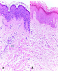
Microscopic examination showed that the lesion is made up of a proliferation of epithelioid and satellite cells that ranges from faintly nodular to solid in appearance in a myxoid background. The tumor cells showed mostly pink eosinophilic cytoplasm and round to oval nuclei with small micronuclei. Scattered pleomorphic and hyperchromatic atypical cells were mixed with more typical tumor cells. Although the tumor showed increased cellularity and focal nuclear atypia, there was no significant morphologic nuclear atypia to support a myxoid variant of sarcoma (Figure 1 A-B). By immunohistochemistry (IHC), the tumor cells were positive for vimentin, CD10, Mit-F, and focal reactivity was noted for smooth muscle actin (SMA) and CD68. They were negative for S100, glial fibrillary acidic protein (GFAP), desmin, CD34, Pan cytokeratin, MUC4, and Melan A (Figures 2 A-B). Slides from prior excisions were examined; although they showed similar features to the current recurrence, the present tumor was much more cellular. Although S-100 and GFAB immunostaining were reported positive in prior excision, the staining was equivocal and insufficient for a definitive determination; the tumor was also focally seen at one of the margins. We received the case in a consultation, and paraffin blocks were unavailable for additional studies. Equivocal staining resulted from an attempt to destain the H&E slides and restain them with S-100. Low-grade fibromyxoid sarcoma was less likely, as they are usually positive for only vimentin and MUC4.In addition, myxoid Dermatofibrosarcoma Protuberans (DFSP) is typically positive for vimentin and CD34. The proliferation study showed only 2% staining with Ki-67. The tumor cells were morphologically inconsistent with other types of sarcomas due to limited nuclear atypia and low Ki-67. The dense induration at the tumor’s margins was explained as prominent scarring from the prior two excisions.

Figure 1: Microscopic examination of the mass
1A: The lesion is made up of a proliferation of epithelioid-like and satellite cells that ranges from faintly nodular to solid in appearance in a myxoid background (H&E Stain X20).
1B: The tumor cells showed mostly pink eosinophilic cytoplasm and round to oval nuclei with small micronuclei (H&E Stain X60).

Figure 2: Immunohistochemistry studies on the mass
2A: Tumor cells positive for Mit-F
2B: Tumor cells positive for CD10
The family requested molecular testing, which produced no significant findings, and the myxoid type of sarcomas was ruled out. Surgical margins were adequate with more than 1 cm safe margin. The final diagnosis rendered was cellular myxoid Neurothekeoma. The patient was followed up for 39 months with no evidence of recurrence.
DISCUSSION
Neurothekeoma is a rare benign skin tumor with Schwann cells and perineural cells in a myxoid matrix [8]. These lesions usually present as a dome-shaped, subcutaneous papule or nodule less than 2 cm in size. They may be flesh-colored, pink tan, or red-brown [4]. Most lesions are asymptomatic, but some may present with tenderness on palpation. Lesions are superficial and often grow slowly [4]. The head and neck are commonly involved, sometimes presenting on the shoulder or upper extremities, but rarely deeper subcutaneous fat, skeletal muscle, or mucosa [5]. The myxoid variant clinically presents as solitary, asymptomatic nodules frequently mistaken for dermal nevi or adnexal neoplasms [9].
Neurothekeomas have been classified into three pathologic variants: cellular, myxoid, and mixed, depending on how much myxoid matrix is present [8]. Generally, neurothekeomas are lobular to plexiform masses containing spindle or stellate shaped cells with an abundant myxoid matrix [10]. The myxoid type is well-circumscribed, has poor cellularity, a high degree of myxomatous alterations, and scattered spindle cells [11]. Tumor cells are often positive for neurogenic cell markers, such as S100, GFAP, and NGFR proteins [9]. However, the myxoid variant is typically positive for NK1/C3 and negative for S100, distinguishing them from melanoma cells [8]. Neurothekeomas have overlapping features with other neural tumors, such as schwannomas, nerve sheath myxomas, and neurofibromas, making them difficult to diagnose [11].
Neurothekeomas arise from fibroblast-like cells composed chiefly of spindle cells with some epithelioid features, which form nests and cords [4,12,13]. The background consists of dense sclerotic collagen bundles and myxoid mucinous matrix, with the latter determining the subtype [12,13]. Tumor cells have abundant pale eosinophilic cytoplasm with round to oval nuclei and small pinpoint nucleoli [13, 14]. Mitotic activity and nuclear pleomorphism are variable [4,5]. Associated inflammatory cells, usually lymphocytes, are present [4]. Osteoclast-like giant and multinucleated cells have also been described [4,13].On low power, multiple closely situated discrete unencapsulated nodules are seen; the lesions form a whorled pattern in the dermis and superficial subcutis but can be infiltrative at the borders [12,14]. On intermediate power, bland and moderately atypical tumor cells in nodules with varying amounts of myxoid background are seen [4]. On high power, abundant pale eosinophilic granular cytoplasm with moderate nuclear atypia is seen [12, 16].
Cellular Neurothekeomas have positive staining for vimentin, NKI-C3 (CD63), CD10, and microphthalmia-associated transcription factor (MITF-1); they are always negative for S100, glial fibrillary acidic protein (GFAP), and Melan A [4, 14,16,17]. Previous studies have also shown positivity for KBA.62 and CD56, focal reactivity for smooth muscle actin (SMA), Factor XIIIa, and CD68, and predominantly negative for NSE, HMB-45, cytokeratin, SOX-10, and EMA [4,16,17]. Many of the stains are nonspecific, and the most useful for confirming cellular neurothekeoma diagnosis is CK1/C3 positivity (14). Maram et al. reported that Cellular neurothekeomas and dermatofibromas share demographic, histopathologic, and immunohistochemical features, including shared expression of MITF and NKI/C3, especially cellular dermatofibroma [18]. Nerve sheath myxoma, which has similar histology and presentation, stain positive for S100, distinguishing it from Neurothekeoma [19]. Cellular neurothekeomas also show positive nuclear staining for TFE3 without gene rearrangement or amplification on FISH, which can help differentiate it from other fibrohistiocytic and malignant granular cell tumors, which also stain positive with CD63 [12]. PRAME (preferentially expressed antigen in melanoma) expression varies between 10% to 75% in all cellular neurothekeomas investigated, making it less reliable in diagnosing malignant melanocytic tumors [15]. The intensity of expression did not correlate with the histologic or clinical presentation [16]. However, its lack of expression in plexiform fibrohistiocytic tumors can help distinguish these two closely related entities [15].
Microarray analysis confirmed the fibro histiocytic origin of neurothekeomas and showed a gene expression profile distinct from nerve sheath myxomas and schwannomas, which have peripheral nerve sheath origin [19]. The molecular profile of neurothekeomas is more closely related to fibrous histiocytomas, which also have similar histological and clinical characteristics [19]. They differentially expressed collagen protein genes, whereas nerve sheath myxomas and schwannomas expressed genes that coded for cell adhesion molecules and neuronal cell intercellular signaling [19]. Point mutations on carcinogenic genes P13K, ALK, SMO, and ERBB3 have been identified using next-generation sequencing [13].
The primary means of treating Neurothekeoma is Surgical Excision [20]. While there is no agreed-upon standard for excision margin, clear microscopic margins and 2-3 mm of grossly negative margins are considered adequate [8]. Advanced techniques can be regarded as if the Neurothekeoma appears in a cosmetically sensitive area such as the face. Mohs surgery is a surgical technique that removes the tissue one layer at a time, examining each layer under a microscope until only healthy tissue remains [20]. This technique can enhance the quality of life in Neurothekeoma patients with tumors in these trouble areas. A majority of Neurothekeomas are small and have minimal extension into surrounding tissue. However, Neurothekeomas have been reported to grow to larger sizes and display infiltration into subcutaneous fat, skeletal muscle, and even the underlying vasculature [21]. With complete excision, reported recurrence rates of Neurothekeoma are low [20] We report an additional case of myxoid Neurothekeoma as we believe it is critical to be aware of these unusual soft tissue lesions as well as the dangers of myxoid and mixed-type Neurothekeomas, which frequently result in difficulties with diagnosis. We hope by this report we assist in preventing malignant mesenchymal tumor misdiagnoses that could have major repercussions, such as prolonged surgical therapy or radiation.
Human subjects: Ethical review and approval were not required for the study on human participants following the local legislation and institutional requirements. The paper has been sufficiently anonymized to keep the patient’s confidentiality.
Conflicts of interest: In compliance with the ICMJE uniform disclosure form, all authors declare the following:
Payment/services info: All authors have declared that no financial support was received from any organization for the submitted work.
Payment/services info: All authors have declared that no financial support was received from any organization for the submitted work
Other relationships: All authors have declared that no other relationships or activities could appear to have influenced the submitted work.
Patient’s consent: Patients was lost to follow-up, and all attempts to reach the patient or family member were unsuccessful. Therefore, the paper has been sufficiently anonymized to maintain patient confidentiality.
Author contributions: Study concept and design: Jessica Jahoda, Viviana Crespo, and Haden Morton. Writing the manuscript: Viviana Crespo, Alan Bencosme, Clifford Davis, Jodie Simelda, and Racquel Kaizer. Data collection: Haden Morton, Alan Bencosme and Viviana Crespo. Analysis and interpretation: Alan Bencosme and Racquel Kaizer. Reviewing and editing the manuscript: Jessica Jahoda and Mohamed Aziz. Editing, critical review, and final approval: Mohamed Aziz
Acknowledgment: Special thanks to Alisa Lu, Fadi Jamaleddin, Tallen Cavenah and Melissa Candela, for their assistance in reviewing the final manuscript.
REFERENCES
1. Kao EY, Kernig ML. Neurothekeoma [Internet]. Treasure Island (FL): StatPearls Publishing; c2020 [citado em 20 Mar 2021]. Disponível em: https://www.ncbi.nlm.nih.gov/books/NBK519525/.
2. Gallager RL, Helwig EB. Neurothekeoma--a benign cutaneous tumor of neural origin. Am J Clin Pathol. 1980 Dec;74(6):759-64. doi: 10.1093/ ajcp/74.6.759. PMID: 7446487
3. Navarrete-Dechent C, Curi-Tuma M, Marín C, González S, Sandoval-Osses M. Cellular neurothekeoma: case report and its (un) relation with nerve sheath myxoma. Anais Brasileiros de dermatologia. 2015 May;90:156-9.
4. Fetsch JF, Laskin WB, Hallman JR, Lupton GP, Miettinen M. Neurothekeoma: an analysis of 178 tumors with detailed immunohistochemical data and long-term patient follow-up information. The American journal of surgical pathology. 2007 Jul 1;31(7):1103-14
5. Hornick JL, Fletcher CD. Cellular Neurothekeoma: detailed characterization in a series of 133 cases. The American journal of surgical pathology. 2007 Mar 1;31(3):329-40.
6. De Giorgi V, Alfaioli B, Franchi A, Gori A, Sestini S, Papi F, Lotti T. Cellular neurothekeoma in a girl: could oestrogens favour the development and growth of this rare tumour?. Journal of the European Academy of Dermatology and Venereology. 2008 Sep;22(9):1149-50.
7. Almeida TFA, Verli FD, Dos Santos CRR, Falci SGM, Almeida LY, Almeida LKY, Mesquita ATM, León JE. Multiple Desmoplastic Cellular Neurothekeomas in Child: Report of the First Oral Case and Review of the Literature. Head Neck Pathol. 2018 Mar;12(1):75-81.
8. Boukovalas S, Rogers H, Boroumand N, Cole EL. Cellular Neurothekeoma: A rare tumor with a common clinical presentation. Plastic and Reconstructive Surgery Global Open. 2016 Aug;4(8).
9. Yun SJ, Park HS, Lee JB, Kim SJ, Lee SC, Won YH. Myxoid cellular Neurothekeoma: A new entity of S100-negative, CD68-positive myxoid Neurothekeoma. Annals of dermatology. 2014 Aug 1;26(4):510-3.
10. Yamashita N, Minami S, Yu M, Miyazaki T, Sagami S. Dermal nerve sheath myxoma. The Journal of Dermatology. 1990 Sep;17(9):564-8.
11. Akhtar K, Zaheer S, Ray PS, Sherwani RK. Myxoid Neurothekeoma: a rare soft tissue tumor of hand in a male toddler. Nigerian Journal of Surgery. 2013 Oct 18;19(1):32-4.
12. Cheng Y, Zhang N, Deng Z, Guan X, Xu J, Qu L, He L. Expression of TFE3 in Cellular and Myxoid Type of Neurothekeoma: Four Cases in Young Children and Adolescents. Fetal Pediatr Pathol. 2023 Aug;42(4):679-684. doi: 10.1080/15513815.2023.2181626. Epub 2023 Feb 21.PMID: 36802993.
13. Ortega M, Sparks J, Nava VE, Smith SDB. Neurothekeoma With PI3K w552*, ALK P1469S, SMO G461S, and ERBB3 L77M Genetic Alterations. Am J Dermatopathol. 2022 Dec 1;44(12):958-960. doi: 10.1097/DAD.0000000000002292. Epub 2022 Sep 7. PMID: 36075574.
14. Murphrey M, Huy Nguyen A, White KP, Krol A, Bernert R, Yarbrough K. Pediatric cellular neurothekeoma: Seven cases and systematic review of the literature. Pediatric dermatology. 2020 Mar;37(2):320-5.
15. Cesinaro AM, Piana S, Paganelli A, Pedroni G, Santandrea G, Maiorana A. PRAME expression in cellular Neurothekeoma: A study of 11 cases. J Cutan Pathol. 2022 Apr;49(4):338-342. doi: 10.1111/cup.14163. Epub 2021 Nov 22. PMID: 34761425.
16. Shankar V. Neurothekeoma. PathologyOutlines.com website. https:// www.pathologyoutlines.com/topic/softtissueneurothekeoma.html. Accessed July 20th, 2023
17. Page RN, King R, Mihm MC Jr, Googe PB. Microphthalmia transcription factor and NKI/C3 expression in cellular Neurothekeoma. Mod Pathol. 2004 Feb;17(2):230-4. doi: 10.1038/modpathol.3800043. PMID:14685254.
18. Abdaljaleel M, North JP. Positive MITF and NKI/C3 Expression in Cellular Neurothekeoma and Dermatofibroma. Applied Immunohistochemistry & Molecular Morphology. 2021 Jul 1;29(6):440-5.
19. Sheth S, Li X, Binder S, Dry SM. Differential gene expression profiles of neurothekeomas and nerve sheath myxomas by microarray analysis. Mod Pathol. 2011 Mar;24(3):343-54. doi: 10.1038/ modpathol.2010.203. Epub 2011 Feb 4. PMID: 21297585.
20. Kao EY, Kernig MN center for biotechnology L. Neurothekeoma [Internet]. StatPearls (updated November 7, 2022); [cited 2023 Aug 1]. Available from: https://www.ncbi.nlm.nih.gov/books/NBK519525/
21. Busam KJ, Mentzel T, Colpaert C, Barnhill RL, Fletcher CD. Atypical or worrisome features in cellular neurothekeoma: a study of 10 cases. Am J Surg Pathol. 1998 Sep;22(9):1067-72.