Research Article | Volume 7 - Issue 1 | Article DOI :
Download PDF
Britta Elkenhans1*, Manuel Stern1, Ingmar Vieregge2, Markus Henningsson3, Rene Botnar4 and Christian Heiss5
1Department of Cardiology, University Hospital, Duesseldorf, Germany
2University of Luebeck, Luebeck, Germany
3Linköping University, SE-581 83 Linköping, Sweden
4Division of Imaging Sciences, King’s College London, Strand, London WC2R LS, UK
5Department of Cardiology, University of Surrey, Guildford, Surrey, GU2 7XH, UK
Corresponding Author:
Britta Elkenhans, Department of Cardiology, University Hospital, Duesseldorf, Germany
Keywords
MRI; T2; Peripheral Artery Disease; Atherosclerosis
Abstract
Aim: The aim of this study was to investigate whether vessel wall imaging using magnetic resonance imaging (MRI) with a T2 preparation pulse with dynamic implementation could accurately predict a proxy for atherosclerosis, carotid artery intima media thickness as measured by ultrasound. Ultrasound is still investigator dependent and a follow up of the femoral artery on its way to the foot and especially of the wall thickness is not possible due to anatomical reasons. Peripheral Arterial Disease (PAD) is a common disease in developed countries and it would be very helpful to have a predictive marker such as vessel wall thickness.
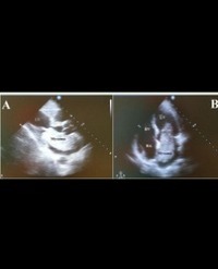
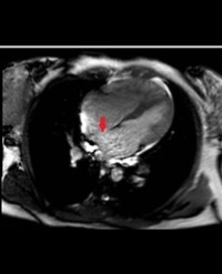
Methods: Five healthy volunteers and seven patients with known PAD underwent MRI of the femoral artery on a 1.5-T scanner with a SENSE-XL-Torso coil (Philips GmbH Market DACH, Hamburg, Germany) using TRANCE and a dynamic T2 preparation pulse afterwards. These measurements were compared with intima media thickness of the carotid artery as assessed by ultrasound.
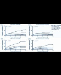
Results: As measured by MRI, patients with PAD had significantly thicker vessel walls than healthy volunteers (p < 0.01). Carotid artery thickness matched femoral artery in patients with known PAD (Pearson r = -0,99, p < 0,014). Interobserver reproducibility was good (bias = -0.0088, 95% limits of agreement = -0.1089 to 0.09123), and ultrasound imaging results correlated closely to those from MRI (Pearson r = -0.99, p < 0.027).
Conclusion: Conducting MRI with a dynamic implementation of a T2 preparation pulse seems to be an adequate, non-invasive method to assess vessel wall thickness as a prognostic factor for atherosclerosis without using contrast agent.
Citation
Elkenhans B, Stern M, Vieregge I, Henningsson M, Botnar R, et al. (2023) Vessel Wall Imaging of the Femoral Artery with a Dynamic T2 Preparation Pulse for Peripheral Arterial Disease Prediction. SM J Cardiol Cardiovasc 7: 4.