SM Journal Clinical and Medical Imaging

Archive Articles

Article Image 1

TIRADS-Based Artificial Intelligence Systems For Ultrasound Imaging of Thyroid Nodules: A Systematic Review

Objective : The Thyroid Imaging Reporting and Data Systems (TI-RADS) is a standard terminology that classifies thyroid nodules according to their potential risk of cancer to reduce unnecessary biopsies, minimize variations in interpreting thyroid nodule images, and improve diagnostic accuracy. This study aims to comprehensively review articles that utilize AI techniques to develop decision support systems for analyzing ultrasound images of thyroid nodules, following different TIRADS guidelines.

Materials and Methods : In this review, we followed a five-step process. This included identifying the key research questions, outlining the literature search strategies, establishing criteria for including and excluding studies, assessing the quality of the studies, and extracting the relevant data. We created a comprehensive search string to gather all relevant English-language studies up to January 2024 from the PubMed, Scopus, and Web of Science databases, and we also followed the PRISMA diagram.

Results: In this review, forty-four papers were included, and the most important properties of these papers, including dataset characteristics, AI technical specifications, results and outcome metrics, metrics, limitations, and contributions, were extracted.

Conclusion: In this review, we evaluated the technical characteristics and various aspects used in the development of artificial intelligence CAD systems based on various TI-RADS. This review demonstrates that AI advancements, especially deep learning methods, have significantly enhanced CAD systems for evaluating thyroid nodules. However, comprehensive datasets, multimodal images, and standard evaluation metrics are needed to further enhance machine learning models. Our study aims to provide researchers and physicians with a summary of the current advancements in this field to guide future investigations.

Yasaman Sharifi1*, Morteza Danay Ashgzari2, Zeinab Naseri3, and Amin Amiri Tehranizadeh3


Article Image 1

Perceptions of Radiographers on Reporting Chest Images at Public Hospitals in Malawi

Introduction : Malawi health facilities’ major challenge is inequalities in access to reporting services due to a shortage of radiologists to report on radiographic images. Radiographers in the developed world acquire formal training in image reporting, however, there is no postgraduate training programme in Malawi. Thus, there is a need to explore radiographers’ perceptions of reporting chest images in Malawi’s hospitals. The study aims to explore radiographers’ perceptions of being tasked with the responsibility of reporting on chest images at public hospitals in Malawi.

Method: The study was conducted through a quantitative cross-sectional design. Data was collected using a self-developed questionnaire. Data collected was analyzed with the assistance of a statistician using the Statistical Package for Social Scientists (SPSS) version 29. The measurement scales and questions included quantitative 10-point Likert. Statistical analysis used the Mann -Whitney U test.

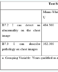
Results: Eighty (71) radiographers representing 81% of the total population from five public hospitals participated in the study. The results from this study found that most radiographers (97.2%), agreed that radiographers needed to report on chest images in the department. Findings demonstrated that the majority of radiographers (80.3%), supported ongoing professional development, and of the radiographers (78.9%), also agreed that reporting on chest images required post-graduate training. Overall confidence in reporting chest images was 81.6% with no difference by years worked as a qualified radiographer (p=0.196), with the less than 10 years were as confident to report chest images as 10 years or more . Confidence in detecting abnormality on chest images (78.8%), demonstrated no variation by years worked as qualified radiographer (p=0. 196) or level in department (junior of senior radiographer (p =0.323). Perceived impact of radiographer’s involvement in chest image reporting to improved cost effective service delivery, work efficiently, and potential impact on patient care and safety.

Conclusion : Based on the findings of this study, there were positive perceptions among the majority of radiographers about the reporting of chest images in Malawi. It was recommended that policymakers change Malawi’s health care policy, especially the radiological sector to allow radiographers to report on chest images

George Simwaba¹*, Lynne Janette Hazell², and Jennifer Motto2


Article Image 1

Comparative Efficacy of Effective Diameter Versus Water-Equivalent Diameter in Size-Specific Dose Estimation for Chest CT: A Retrospective Cohort Study on Patient-Specific Radiation Optimization

Purpose : To investigate the discrepancy between effective diameter (dEFF) and water-equivalent diameter (dW) in size-specific dose estimate (SSDE) calculations for chest CT examinations.

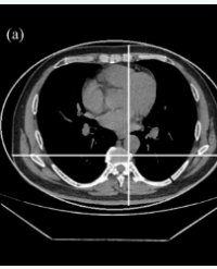
Methods : This retrospective study analyzed 201 patients undergoing chest CT. The volumetric CT dose index (CTDIvol) was recorded for each case. Both dEFF and dW were calculated from the central axial image, with subsequent derivation of size conversion factors (fEFF, fW) and SSDE values (SSDEEFF, SSDEW). Patients were stratified into tertiles based on lateral chest diameter (dLAT): Group A (dLAT<32.955 cm), Group B (32.955-35.190 cm), and Group C (dLAT>35.190 cm). The comparison between different dLAT groups was conducted using the Friedman rank sum test. All statistical analysis was significant as P <0.05.

Results : The mean CTDIvol was 10.42 ± 0.44 mGy. Significant discrepancies were observed between dEFF and dW (t = -16.24, P < 0.001),
corresponding conversion factors (U = 5,030.50, P < 0.001), and SSDE values (U = 12,590.50, P < 0.001). Intergroup analysis revealed statistically significant differences across all parameters (ANOVA F = 134.000-357.249, all P < 0.001).

Conclusion : Water-equivalent diameter demonstrates superior accuracy over geometric measurements for SSDE calculation in chest CT, particularly for patient-specific dose optimization. These findings, while specific to GE scanners, highlight the clinical importance of attenuation-corrected metrics in radiation dose management.

Advances in knowledge : This study provides robust evidence for the feasibility and accuracy of attenuation-corrected dose calculations. The findings redefine personalized dosimetry paradigms, demonstrating that integrating inherent tissue attenuation variability significantly improves treatment planning reliability.

Ai Li1,2 Qiyu Zhang3, Miao Yu2, Xiaohui Zhao2, Lei Xu2, Huijun Ma1, Ziyin He1, Nan Yang2, and Hongtao Niu1*