The Synergistic Effect of Chronic Hyperglycemia and Dehydration on the Progression of Cerebral Infarction
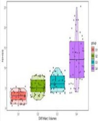
Background : Studies on the role of chronic hyperglycemia and dehydration in the progression of acute cerebral infarction have confirmed that chronic hyperglycemia can promote the progression of acute cerebral infarction, and dehydration can also accelerate the development of cerebral infarction. However, there are few studies on the co-effects of chronic hyperglycemia and dehydration on infarct core volume. Since it is not uncommon for stroke patients to have chronic hyperglycemia and dehydration at the same time, it is of certain clinical significance to study the interaction of the two on stroke. Method: A total of 270 patients with acute cerebral infarction were included and admitted to hospital to improve the examination of glycosylated hemoglobin, blood urea nitrogen and blood creatinine. We would use blood urea nitrogen (BUN)/creatinine (Cr) ratio as a dehydration marker.The dehydrated group was defined as the blood urea nitrogen/creatinine ratio >15, and the non dehydrated group was defined as the blood urea nitrogen/creatinine ratio ≤15. HbA1c >7% was defined as chronic hyperglycemia group, HbA1c ≤7% was defined as non-chronic hyperglycemia group. According to the above definition, they were divided into four groups: dehydration + chronic hyperglycemia group, dehydration + non-chronic hyperglycemia group, non-dehydration + chronic hyperglycemia group, and non-dehydration + non-chronic hyperglycemia group. At the same time, the magnetic resonance DWI examination was improved, the infarct core volume was processed by the software after the acute cerebral infarction magnetic resonance image, and the correlation between the infarct core volume and glycosylated hemoglobin and blood urea/creatinine ratio was evaluated. hyperglycemia group. Results: The core volume of cerebral infarction in chronic hyperglycemia group was larger (infarct volume >43.28ml, HBA1c IQR 6.5%). The core volume of cerebral infarction in dehydrated group was larger (infarct volume >43.28ml, BUN/Cr IQR 17.632). The core volume of cerebral infarction was the largest in dehydration + chronic hyperglycemia group, followed by dehydration + non-chronic hyperglycemia group, non-dehydration + chronic hyperglycemia group, and non-dehydration + non-chronic Conclusion: Chronic hyperglycemia in patients with acute cerebral infarction is likely to lead to dehydration, which is the main reason for the enlargement of the infarct core. Therefore, it is possible for patients with chronic hyperglycemia to avoid the expansion of the infarct core by correcting the key link of critical dehydration.
Huanyin*


