SM Dentistry Journal

Archive Articles

Article Image 1

In-vitro Comparative Evaluation of Cariostatic Potential and Marginal Microleakage of Commonly-used Glass-Ionomer Restorative Materials as Interim Therapeutic Restorations

In-vitro Comparative Evaluation of Cariostatic Potential And Marginal Microleakage Of Commonly-Used Glass-Ionomer Restorative Materials as Interim Therapeutic Restorations.

Introduction: Glass ionomer cements (GIC) can ideally serve many restorative purposes in paediatric dentistry as it has advantages of chemical adhesion to tooth structure, as well as fluoride release and uptake into hard tissues.

Aims: To compare four commercially available conventional GIC based on the amount of fluoride release, marginal integrity and ability to increase microhardness of underlying artificial dentinal caries via remineralization.

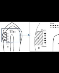
Methods and Material: Standardized cavities were prepared on the buccal surface of 60 extracted sound premolars and artificial caries were induced onto the dentinal floor. Specimens randomized into four groups of fifteen teeth and restored with Fuji VII®, Riva Protect®, Riva Self Cure® and Fuji IX GP® Extra respectively. Measurement of fluoride release was done for 60 days. Subsequently, similar restorations prepared on lingual surface for microleakage test according to standard protocols.Dentin underlying buccal restorations were subjected to microhardness test while lingual restorations were evaluated for marginal microleakage.

Results: One-way ANOVA statistical analysis revealed significant difference was found in the amount of f luoride release between materials on all days of measurement (p<0.05). Riva Protect® released the highest amount of fluoride, followed by Fuji VII®, Riva Self Cure® and Fuji IX GP® Extra. Wilcoxon signed-rank test showed Riva Protect has theability to significantly improve the microhardness of artificial caries to the depth of 100 µm (p<0.05). There was a significant difference in the marginal sealing ability between materials with more microleakage seen with low-viscosity materials, analysed by Pearson’s Chi-Square test.

Conclusions: All materials possess cariostatic potential. However, Riva Protect® is suggested to exert the greatest cariostatic effects among the test materials

Nurulnazra MA, Mahyuddin A and Sockalingam SNMP*


Article Image 1

Effect of Eutectic Mixture of Local Anaesthetics (EMLA) on Pain Perception During Scaling & Root Planing (SRP) Split-Mouth, Controlled, Randomized Clinical Trial

Background: Scaling and Root Planning (SRP) is the most commonly used procedure for treating gingivitis and periodontitis, which may be perceived as painful. The fear of pain during treatment has been identified as a major factor in preventing patients from seeking dental care. Pain control is considered to be an extremely important outcome measure for successful periodontal therapy.

Methods: Local application of Eutectic Mixture of Local Anaesthetic agent (EMLA) having higher concentration of local anaesthetic agent, considered as an effective way to reduce pain.

Results: The mean Visual Analogue Scale (VAS) when EMLA cream was used was lower compared to when EMLA cream was not used. The mean Verbal Rating Scale (VRS) was also lower when EMLA cream was applied.

Conclusion: The results of the present study warrant the use of EMLA in routine practice.

Patel KR*, Bhavsar NV and Brahmbhatt NA


Article Image 1

Dental Implants: the Good, the Bad and the Downright Dangerous

T his article is written by a board certified Oral and Maxillofacial Surgeon with over 12 years of experience in the specialty. The author has been involved in the placement of thousands of dental implants of all types and has particular expertise in managing “salvage” or multiply operated patients who have had failed implants placed by other providers. The author also has expertise in the surgical management of temporomandibular joint disorders and complex infections of the head and neck region. The author has appointments at several well-known academic institutions. T he use of dental implants to replace missing teeth and in fact replace a patient’s entire dentition has become more commonplace over the past several years. One only has to turn on the television to notice that there are multiple facilities offering “teeth-in-a-day” or cheap implants, to replace ones missing dentition. These commercials, although alluring, can be very confusing for prospective patients and may lead to inadequate research being done not only into the procedure, but also into the clinician performing the procedure. Dental implants are safely placed by multiple specialties within the realm of dentistry and good outcomes are not specifically dependent on one’s specialty training but more contingent upon actual clinical experience.

Raza A Hussain*


Article Image 1

Specificities of Canine Replacement by an Implant Supported Crown

In the 1980s, the work of Professors Bräne mark and Schroeder led to the introduction of a new surgical technique: implantology. In the beginning, the position of the implant depended mainly on the available bone volume. Successful treatment was based solely on osteo integration. In a few years, the reliability of this therapy has allowed the treatment from single tooth replacement to full-archrestoration, thus paving the way for an additional requirement: an aesthetic integration. Consequently, the position of the implant had to coincide with the future prosthesis. We moved from an implant ology paradigm guided by bone anatomy to implant ology guided by prosthetic requirements.When it concerns a canine replacement, both fuctional and esthetic require mentsare considered. In fact, the replacement of a canine tooth by an implant will, in its neuro physiologic consequences, exchange a protection system based on the presence of periodontal membrane receptors with one that depends on an implant anky losed in bone that can transmit only feeble information to be filtered by sensitive superior ganglia.A bonded bridge on lateral in cisor and first premolar can be a temporary solution during peri-implant bone healing, it requires a reversible tooth preparation limited to enamel.

Marwa Chakroun1, Anissa Ben Moussa1, Dalenda Hadyaoui1*, Mohamed Ben Khalifa2, Fathi Troudi3, Belhassen Harzallah1 and Mounir Cherif1


Article Image 1

Comprehensive Multidisciplinary Team Approach in the Treatment of Amelogenesis Imperfecta- Case Report

Amelogenesis imperfecta comprises a group of hereditary enamel defects, characterized by enamel hypoplasia, hypomaturation, or hypocalcification of the teeth. We describe the treatment of an 8 year old boy diagnosed with autosomal dominant smooth hypoplastic AI. The patient had hypodontia and teeth with abnormal morphology; missing enamel, and a functional anterior crossbite. A treatment plan was implemented that improved function by restoring posterior teeth, improved esthetics by enhancing teeth appearance, eliminated functional deviation of the jaw, preserved the existing teeth, and promoted low cardiogenic nutrition and good oral hygiene. Treatment of young patients with AI should be oriented toward functional and esthetic rehabilitation.

Anat Baniel1*, Eldad Sharon2, Shahar Menashe3 and Joseph Shapira1


Article Image 1

Dentigerous Cyst Involving Permanent Incisor: A Case Report

Background: This paper aims to describe a case report about dentigerous cysts related to trauma in the primary teeth.

Case report: An eight-year-old boy attended the Pediatric Dental Trauma Clinic. He had a delay in the eruption of the tooth 11 and when he was four years old suffered a trauma in the tooth 51. The radiograph showed a dentigerous cyst involving the tooth 11. The lesion was treated through enucleation and the permanent tooth erupted.

Conclusions: A correlation was observed between the dentigerous cyst, trauma in the deciduous incisor and the successor teeth. Early diagnoses as well as clinical and radiographic follow-ups of traumatized primary teeth are important to minimize possible sequelae in the successor teeth.

Mariana Salazar, Mariana Canano Séllos, Gabriela Caldeira Andrade Americano, Mariana Pires da Costa, Mirian de Waele Souchois de Marsillac, Vera Campos


Article Image 1

Association between Oral Health Status and Cognitive Function among Geriatrics

Background: Oral health disorders such as loss of teeth, periodontitis and root caries are associated with cognitive impairment in a group of elders.

Objective: This study was designed to assess the association between oral health status and cognitive function among a community-dwelling geriatric population in Chennai.

Materials and Methods: This was a cross sectional study involving 150 elderly from two geriatric institutions and two day care centers, who were interviewed and examined for oral health status and screened for cognitive function using Montreal Cognitive Assessment Test (MoCA).

Results: Among 150 participants 53 (35.3%) had mild cognitive impairment (MCI) with a mean cognitive score of 22.17 (SD = 1.7), 73 (48.7%) had severe cognitive impairment (Dementia) with a mean cognitive score of 10.81 (SD = 5.4) and about 24 (16%) had normal cognition with a mean score of 27.08 (SD = 1.4). It was found that of the indices assessed, Russell’s periodontal index had a negative association with cognitive function (R = - 0.212).In the linear regression model, adjusted for age and education, Russell’s periodontal index was a predictive factor for cognitive function among older adults.

Conclusion: Periodontitis may serve as an early risk predictor for cognitive impairment among geriatrics.

Arthi B1*, Madankumar PD1 and Sridhar V2


Article Image 1

Peripheral Ossifying Fibroma: A Case Report

Peripheral ossifying fibroma is a non-neoplastic enlargement of the gingiva that usually arising from the interdental papilla. A 36-year-old female admitted to our clinic for routine care and management of recurring gingival growth. Examination revealed an approximately 1.5 x 1 cm pedunculated, not-tender, firm, pinkish red growth present on the interdental papilla of the maxillary incisors. Excisional biopsy was performed to obtain histopathologic evidence. A confirmatory diagnosis of peripheral ossifying fibroma is made by histopathologic evaluation of biopsy specimen. Although Peripheral ossifying fibroma is a bening and reactive lesion, the recurrence rate is quite high. Therefore, regular follow-up are necessary.

Metin ÇALIŞIR1* and A Cemil TALMAÇ2


Article Image 1

Lichen Planus Lesions in Oral Cavity in Patients with AIDS and Coinfected with Hepatitis C V

Lichen planus is a mucocutaneous, chronic inflammatory and very common dermatological disease. The importance of this disease is related to its frequency in the general population, its multiplicity of aspects, and its possible connection with malignancy. In the skin the lesions consist of reddish or white papules that can present a central depression. The buccal lesions are often multiple, bilateral, striated and appear as whitish plaques in the reticular form and exposing the connective tissue, in the erosive or atrophic form and rarely the bullous. Basal epithelial cells are the main target in lichen planus. The mechanism of basal cell damage is related to a cellular immunization process involving Langerhans cells, T lymphocytes and macrophages. Lichen planus is a middle aged disease that affects men and women in almost the same proportion. Patients with Hepatitis C virus and HIV patients are also more likely to develop oral lichen planus due to adverse effects of their drug therapies, as well as the use of Interferon (IFN) alpha, Ribavirin, and Zidovudine (for patients HIV). Children are rarely affected. Frequently, the severity of the disease is parallel to the patient’s degree of stress, and immunosuppression.

Giovani EM, Souza RS, Camila Correia dos Santos*, Georgevich Neto R and Colloca M


Article Image 1

Recent Advancements in Diagnostic Aids in Orthodontics - A Review

Every medical, dental and surgical procedure is solely based on the diagnosis of the underlying pathology or disorder. In recent times there has been a tremendous advancement in scientific technology which has helped dental practitioners all over the world. The use of computers in cephalometry has made it easier to view and measure various readings. Computer aided design and computer aided manufacturing have increased the accuracy and quality of all dental materials. In the field of Orthodontics, there have been many advances. This article summarizes the recent advancements in diagnostic aids in Orthodontics which has helped revolutionize treatment planning for the Orthodontic fraternity.

Ashish Kakadiya1, Ragni Tandon2, Aftab Azam2, Rohit Kulshrestha3* and Madhvi Bhardwaj4


Article Image 1

Functional Electromyographic Activities of Facial Muscles in Different Growth Patterns

Objective: To evaluate and compare the electromyographic (EMG) activity of masseter, temporalis, mentalis and orbicularis oris muscles during function in patients with different facial growth patterns.

Materials and methods: 75 subjects (38 males and 37 females) with no history of previous orthodontic treatment, jaw surgeries, facial trauma and temporomandibular joint disorder were taken and divided into three groups based on their mandibular plane angle and gonial angle as hypodivergent, normodivergent and hyperdivergent. Lateral cephalograms were traced manually to determine facial type. Electromyographic recordings were done and muscle activities were evaluated at clenching position.

Results: For temporalis hypodivergent group had the highest activity but the difference was not statistically significant (p=0.904). For orbicularis oris hypodivergent group had the highest activity and this difference was statistically significant (p=0.007). For mentalis hypodivergent group had the highest muscle activity and this difference was not statistically significant (p=0.052). Only orbicularis oris muscle showed statistically significant difference in all the three groups during maximum clenching.

Conclusion: Different facial growth patterns had a significant effect on the EMG activity of facial muscles during function. Orbicularis oris muscle activity showed significant difference among the three groups. Muscle activity was higher for the hypodivergent group as compared to the other groups. This indicates that the musculature in hypodivergent patients is strong and in hyperdivergent group is weak. This must be kept in mind while diagnosing and making the treatment plans for both the facial types.

Ankit Gupta1, Kamlesh Singh1, Ragni Tandon1, Ashish Chauhan1, Rohit Kulshrestha2* and Himanshu Trivedi1