SM Dermatology Journal

Archive Articles

Article Image 1

Sclerodermatous Skin: An Immunohistochemical Study

Objective: Systemic Sclerosis (SSc) is primarily characterized by autoimmunity, microangiopathy, and tissue fibrosis. Hypoxia, a powerful stimulator of Vascular Endothelial Growth Factor (VEGF), may be responsible for excessive expression of chronic VEGF in SSc. The oxygen-regulated α-subunit of HypoxiaInducible Transcription Factor-1 (HIF-1α) plays an important role in transcriptional regulation of VEGF. The protein phosphatase and tensin homolog (PTEN) is responsible for dephosphorylation of proteins and thereby promotes tissue repair.

The present study aimed to examine the expressions of HIF-1α, PTEN, and VEGF in patients with scleroderma in an attempt to estimate prognosis and guide therapeutic decisions. We therefore studied the expressions of HIF-1α, VEGF, and PTEN in skin samples exhibiting the features of scleroderma.

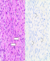
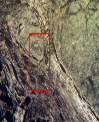
Methods: Skin biopsies of patients were obtained and samples were stained with primary antibodies following PTEN, VEGF and HIF-1α, immunohistochemically.

Results: The SSc and the control group were compared with respect to staining of epidermal keratinocytes, fibroblasts, and the endothelial cells for the antibodies. SSc patients displayed a reduced VEGF expression in endothelial cells. The lesion group showed a significantly lower staining of epidermal cells and endothelial cells for PTEN. SSc group was also characterized by a significantly weaker HIF-1α expression compared to the control group.

Conclusion: In scleroderma skin samples, manipulated expression of these molecules at varying stages of the disease may affect the prognosis of scleroderma.

Rabia Bozdogan Arpaci1*, Tuba Kara1 , Yasemin Yuyucu Karabulut1 , Didar Gursoy1 , Yalçın Polat2 , Gulhan Orekici3 and Umit Tursen4


Article Image 1

Role of Erythroid Differentiation Regulator 1 (Erdr1) On Inflammatory Skin Disorders: A Review

Inflammatory skin diseases remain as one of the most common problems in dermatology. Skin inflammation is a complex process that starts with the introduction of a stimulus such as an allergen or an antigen that triggers the skin to produce inflammatory substances called cytokines and chemokines. At present, the approved drugs for treating inflammatory skin conditions are corticosteroids, yet these can have negative side effects on the skin. Erythroid Differentiation Regulator 1 (Erdr1) is a cytokine previously thought to be connected with hemoglobin synthesis only but was recently found to be involved in inflammation. This review examined the role of Erdr1 as a therapeutic target in the treatment of inflammatory skin disorders. By reviewing previous studies, it was evident that Erdr1 has pro-apoptotic activities; it is able to induce apoptosis in melanoma cells by the regulation of factors such as Bcl-2 and Bax. Treatment with Recombinant Erdr1 (r Erdr1) was also able to inhibit rosacea by inhibiting blood vessel growth or angiogenesis and preventing further infiltration of inflammatory cells. There was also significant reduction of erythema, inflammatory cell infiltration, and microvessel density with Vascular Endothelial Growth Factor (VEGF). Altogether, these studies suggest that Erdr1 has a therapeutic effect on inflammatory skin diseases, particularly psoriasis, rosacea and other skin disorders.

Saad Sami AlSogair*


Article Image 1

Efficacy of Lasers on Vascular Lesions and Rejuvenation

Our purpose is to study the efficacy of the NdYAG Laser (1064 nm) when treating telangiectasias and hemangiomas, and the efficacy of the NdYAG Laser and fractional laser on rejuvenation. We used data from our private office from the past five years (2011-2015). We also studied the side effects of the therapies. We treated 500 patients with telangiectasias and 300 patients with hemangiomas with NdYAG Laser. As far as telangiectasias were concerned, the results were much better when the face was treated. In contrast, the recurrence rates were much higher when the legs were treated. The results were impressive in almost all cases of cherry hemangiomas. The combination of NdYAG Lasers and fractional lasers on a monthly basis had satisfactory results in rejuvenation.

Maria Voyatzi*1 , Julia Grammatikopoulou1 , Eleni Karlafti2 , Androniki Mavroudi1 ,

Foteini Lamprou1 , Genovefa Kypri1 , Theodosia Gkentsidi1 and Maria Tsirmigka1


Article Image 1

Refining Techniques in Eyebrow Transplantation

There has been a growing demand for facial hair restoration including eyebrow hair transplantation. The demand for non-scalp hair restoration is reflective of the cultural trends where full eyebrows are in vogue. Our clinic has seen a large increase in non-scalp hair restoration. Given the large experience allowed by our patient population, we have been able to refine our techniques for optimal results in eyebrow restoration. Presented in this article are our techniques developed from over 1,000 eyebrow transplantations.

Anthony Bared* and Jeffrey S. Epstein


Article Image 1

Safety and Efficacy of a New Regimen in Homogenizing and Brightening Skin Complexion Among Filipino Women

Objectives: To evaluate the safety and efficacy of a new regime of treatment based on the combination of two technologies: Retinsphere® and Fernblock®, topical and oral respectively, in decreasing the visibility of skin blotches and creating a more even complexion. Secondly, the anti-aging effects were also assessed.

Methods: Randomized, double blind, vehicle-controlled study. 80 women aged between 35 and 55 years old were recruited and grouped in 4 arms of 20 subjects: (A) topical single treatment, (B) oral single treatment, (C) combined (oral and topical treatment) and (D) control. Efficacy was objectively evaluated with non-invasive devices (Chromameter®, Mexameter®, Tewameter®, Corneometer®, Triplesense®, SkinSys® and DermaLab®). Baseline values were compared to outcomes obtained at T30 and T90, and also each group (A, B and C) to control group (D). Subjective assessment was performed at the end of the study by the investigator as well as the patient, using a questionnaire especially developed for this purpose. Adverse events were monitored and followed up throughout the 90-day study period.

Results: Active regimens (A, B and C) were significantly better improving skin brightness, lightening and aging signs compared to control. They were also scored positively by the investigator and the patients regarding skin lightening, wrinkle correction and firmness. The overall safety evaluation revealed good tolerance for all regimens. Two patients of combined treatment arm (C) reported mild to moderate erythema and itchiness.

Conclusions: Globally, the outcomes obtained with the combined regime (C) were superior to single treatments separately (A and B). Furthermore, subjects that received combined regimen obtained faster results, reaching better scores within the first month. This could lead to improvement of the patient’s adherence.

Truchuelo MT1*, Gabriel MT2 , Chan HP2 , Chan GP3 and Vitale M4


Article Image 1

Sexual Therapy in Psoriasis? Is It Necessary?

Psoriasis is one of the most common a chronic inflammatory skin disease which could be associated with numerous sexual dysfunctions. According to this, the aim of this study was the analysis of sexual problems in patients with psoriasis. To be included in the review, a study had to: (i) Human data; (ii) concerned patients’ sexual problems in psoriasis; (iii) be published in an English-language journal. Studies were identified through online database searches of PUBMED, MEDLINE, Web of Knowledge (2000-2016), in all analyzed publications, researcher stressed that there was a strong relationship between psoriasis and human sexual functioning. Moreover, sexual impairment in psoriasis patients occurs in all components of the sexual response such as: sexual interest; excitation; orgasm; and erection (only in males case), as well as global sexual satisfaction. Moreover, the prevalence of sexual dysfunction was significant higher than in controls. What is interesting, the large majority of analyzed publications were focused on male patients with psoriasis. All reported publication about sexual problems in psoriasis was focused on adult patients. There were no publications in context of adolescents. The main conclusion of this paper is fact that, in dermatology, doctor needs to pay special attention to the prevention of the sexual dysfunction development in patients with skin disorders. Currently, there is no specific sexual therapy for patients with psoriasis, which would prevent the development of sexual dysfunction. This suggests the need to develop adequate interventions forms which are aimed at supporting the patients’ psychosexual development on the one hand, and prevent the development of sexual dysfunction in psoriasis on the other hand.

Mariusz Jaworski*


Article Image 1

An Unexpected Case of Measles

Outbreaks of measles in London and the South East have been reported recently despite relatively high vaccination rates. We describe the case of a 50 year old female with a delayed diagnosis of atypical measles who presented with respiratory symptoms, fever and a maculopapular eruption, who was treated for a community acquired pneumonia. Steroid and antimicrobial therapy was commenced and blood tests showed raised liver enzymes, inflammatory markers and lymphopenia. She developed a widespread maculopapular eruption which was suggestive of measles and confirmed through the presence of IgM antibodies. She was treated conservatively and made a full recovery. Our case highlights the importance of considering measles in patients presenting with a maculopapular rash and respiratory symptoms, as this can be consistent with a diagnosis of atypical measles.

Faisal Usman Qureshi1 , Sreedhar Kumara Krishna1 , Akhil Sawant2, Alberto Barea1, Saskia Reeken1 and Janakan Natkunarajah1


Article Image 1

Blistering Perioral Rash in an Infant

The correct answer is B: phytophotodermatitis. As suggested by the term, exposure to a light-sensitizing compound from plants (a furocoumarin) and UV light is required to cause this reaction. The acute eruption can range from minimal erythema to marked red edema and vesiculation within 24 hours after initial exposure. The rash is typically nonpruritic but can be painful. As the acute rash fades, hyperpigmentation appears which can last months to years [1].

While the features of the rash are characteristic, the distribution and shape are widely variable. The well-demarcated erythema, edema, and bullae appear in bizarre patterns on sun-exposed skin consistent with the area of plant exposure. Digitate or drip-like patterns may occur from citrus or celery juice that drips during food preparation. Linear patterns may occur from brushing up against outdoor plants such as wild parsnip or hogweed. While classic cases involve obvious sun exposure outdoors, window glass does not block the ultraviolet A waves that cause the phototoxic reactions of furocoumarins [2].

LT Kathryn Barbante1*, LCDR Thomas Barlow2 and LCDR Jennifer Eng-Kulawy2


Article Image 1

Sentinel Lymph Node Melanosis: A Report of Two Cases of Regressed Melanoma Metastases

Tumoral melanosis is found on histopathologic evaluation of a primary melanoma and is typically caused by regression of the tumor. The prognostic value of regression related to primary cutaneous melanoma has been controversial. Very few cases of melanosis in the sentinel lymph node have been described. We present two patients who presented with cutaneous melanoma and were found to have completely regressed metastatic melanoma present within the sentinel lymph node. One patient underwent completion lymph node dissection whereas the other opted for observation. Based on the outcomes of our two cases as well as review of other case reports of this phenomenon we discuss the biologic significance of sentinel lymph node melanosis and how it should be interpreted in relation to adjuvant therapies, staging, and follow up. Based on the unpredictable course associated with this finding we recommend that these patients be treated in a multidisciplinary fashion with treatment decisions influenced by the patient’s goals of care.

Steven H Sun1 , Sara B Peters2 and J Harrison Howard1*


Article Image 1

Non-Healing Lesions in a Patient with Chronic Plaque Psoriasis

Pyogenic granuloma is a bright red benign lesion which can result in bleeding and discomfort. The aetiology is unclear, however drugs, trauma and pathogens are some causes linked to their formation. We describe a case of a 92-year-old female who presented with a rapidly growing nodule on her right second toe with no history of trauma. She had previously been prescribed the retinoid acitretin for plaque psoriasis. After excision of the first lesion the patient presented with another lesion, this time on her left heel. Her lesions resolved on cessation of acitretin therapy. Histological analysis provided a diagnosis of acitretin induced pyogenic granuloma. It is well documented that pyogenic granulomas are rare side effects in acne and psoriasis patients who take retinoid treatment. Paradoxically, retinoids inhibit the formation of vascular endothelial growth factor and would be expected to inhibit rather than stimulate the formation of pyogenic granulomas. It is possible that retinoids favour trauma or share a proangiogenic target; retinoids decrease attachments between keratinocytes and cause nail brittleness. Resolution can occur spontaneously or on cessation of the drug.

Faisal Usman Qureshi1*, Sreedhar Kumara Krishna1 , Akhil Sawant2, Alberto Barea1 , Saskia Reeken1 and Janakan Natkunarajah1


Article Image 1

Biomineralogy of Selected Skin Cancers

Investigation of Carcinoma basocellulare solidum exulcerans, Carcinoma basocellulare superficial multicentricum, and Trichoepithelioma was performed using histology and biomineralogical methods. Obtained data confirmed elevated levels of some elements in altered skin tissues. Moreover, rare micrograins of phosphates were observed. Additionally, examination of biomineralization of human tissues suggests that higher local mineralization (of tissue fluids) may lead to mistakes in DNA code at the moment of cell division. It is possible that cancer tissues are secondarily mineralized by activity of cancer cells. Further research is needed to answer questions that arose.

Maciej Pawlikowski1* and Magdalena Miler2


Article Image 1

The Clinical Picture of Rosacea Depending on Demodex Mites

The article presents the analysis of the clinical picture of patients with rosacea of different degrees of severity complicated with demodicosis. The dermatovenereology life quality index was evaluated for patients with rosacea in the presence and absence of Demodex mites. Presence of Demodex mites reduces the life quality of patients with rosacea. The duration of pathological processes on the facial skin, as well as the number of relapses were assessed. The course of rosacea was analyzed depending on the species identity of Demodex mites (Demodex folliculorum and Demodex folliculorum brevis). In the structure of rosacea diagnoses Demodex folliculorum is significantly more prevalent (n = 20; 66.7%) then Demodex folliculorum brevis (n=8; 26.7).

Kubanov Alexey, Gallyamova Yulia and Kravchenko Anzhela*


Article Image 1

Malassezia spp. beyond The Mycobiota

Malassezia species are part of the normal mycobiota of skin of animals and humans but they can cause skin and blood stream infections as well. These yeasts are all lipid dependent explained by the absence of fatty acid synthase genes in their genome. At the same time, metabolic reconstruction revealed differences in the metabolism of fungal steroids and degradation of CoA-activated long-chain FAs, arachidonic acid, and butanoate metabolism between Malassezia yeasts. In addition, differences in the assimilation of palmitic acid were predicted. Indeed, M. furfur was able to metabolize palmitic acid but M. globosa, M. sympodialis, M. pachydermatis, and an atypical variant of M. furfur were not able to do so. Tools to genetically modify Malassezia have become available recently, which will speed up the process to decipher mechanisms underlying growth and pathogenicity of these yeasts. Here, we will provide an overview about the genus Malassezia and make an assessments to the new insights in this yeast.

Celis AM1,2, Wösten HAB1 , Triana S2 , Restrepo S2 and de Cock H1*


Article Image 1

The Dermatome and Its Thickness Setting: What You Set Is Not What You Get

Background: Skin grafting is a vital part of the treatment of deep skin defects. The preferred thickness of the graft is determined on the parameters of the wound which needs to be covered. Although, it is not known in how far the dial setting of the dermatome corresponds with the actual thickness of the harvested skin graft. To determine the usability of a dermatome setting regarding the resulting grafts thickness.

Methods: During the harvesting of split skin grafts in the operation theatre, the average thickness of harvested skin grafts was compared to the dermatome thickness settings.

Results: A total of 50 grafts were collected from 30 patients, leading to 250 thickness measurements (5 per graft). The measured thickness differed significantly from the dermatome setting: the grafts were up to 50% thinner than the dial setting when the settings of 0.25 mm and 0.30 mm were used. Scalp harvest thickness varied from 50% thinner to 200% thicker than the setting. Thickness was more or less constant across the skin grafts both lengthwise and laterally. We did not detect an effect of the blade becoming blunt regarding its accuracy after repeated use.

Conclusion: These results imply that the average measured skin graft thickness is up to 50% thinner than the thickness setting. Graft shrinkage alone does not seem to pose an adequate explanation for this difference. In scalp harvests, there is more variability in the thickness of the grafts. Furthermore, our results suggest that grafting blades do not have to be changed during the procedure, because we found no signs of bluntness.

Annebeth Meij-de Vries1*, Floris E Aartsen2 , Pieta Krijnen2 , Dorotka T Roodbergen1 , Jos FPM Vloemans1 and RS Breederveld1,2


Article Image 1

NIR Laser Therapy in the Management of Feline Stomatitis

Stomatitis with involvement of the caudal area is a severe inflammation of the oral tissues in cats, which heavily impairs the animal health. The affected gingiva and mucous membrane show different degrees of inflammation, proliferation and ulceration. The only way to manage this serious disease is suppression of inflammation, but often reduction of inflammation at tolerable levels is considered a satisfactory therapeutic result. Many different treatments have been proposed, but currently a therapy of choice and a standardized treatment for stomatitis management have not yet identified. Therefore, new therapeutic strategies are needed to effectively counteract inflammation and improve symptoms.

Laser therapy has been applied with significant results in the management of human patients affected by Oral Mucositis (OM), where it has been effective in reducing inflammation, pain, severity and duration of OM ulcers. The present paper reports the results of a preliminary proof-of-concept clinical study aimed at verifying the hypothesis that NIR laser therapy could be usefully applied to treat cat mucositis.

The enrolled patients were divided into two groups, in order to compare two treatments differing for the energy density released. The effectiveness of laser therapy in reducing inflammation and favoring ulcer healing was assessed by clinical examination of patients, histological analysis and Immunofluorescence microscopy on tissue specimens collected from the animals before and after the cycle of laser therapy.

The outcomes of this preliminary study demonstrate that laser therapy with suitable treatment parameters can be effective in the management of stomatitis.

Even if with different response times in the two groups, laser therapy induced an improvement of the symptoms that was greater and faster in the group treated with lower energy density.

Paolo Squarzoni1, Daniele Bani2, Francesca Cialdai3 and Monica Monici3*


Article Image 1

Childhood Hypopigmented Mycosis Fungoides: The Greatest Imitator

Mycosis Fungoides (MF) is the most common type of cutaneous T-cell lymphomas, especially found in individuals over the age of 50. The incidence of childhood MF is low, only about 0.01-0.03% per year. The clinical manifestations of MF show great variation, with scaly erythematous patch as the most common presentation. In children, hypopigmented patch manifests frequently and can mimic several skin disorders, from leprosy to post inflammatory hypopigmentation. We reported a case of a 5-year-old girl with multiple red patches on her trunk and extremities. Our first differential diagnoses were leprosy, erythema annulare centrifugum, and granuloma annulare. Histopathology and immunochemistry findings correspond to hypopigmented MF. She was treated with mometasone furoate 0.1% cream and Narrow Band Ultraviolet B (NBUVB) for over a year.

Nahla Shihab*


Article Image 1

Intractable Vaginismus - Management by Incision of Spasmodic Perivaginal Muscles and Resurfacing with Labia Minoraflaps - New Appraoch

Introduction: Vaginismus is defined as recurrent or persistent involuntary spasm of the musculature of the lower third of the vagina, which interferes with coitus resulting in matrimonial disharmony. There are many methods described for the management of the condition. But most of times all methods fail to treat such patients. We have developed a newer approach for management of this condition in which spasmodic muscles are incised and resultant defect is resurfaced with Labia Minora Flaps.

Material and method: We have treated fourteen females who were married more than 5 years before and had undergone all types of treatment without success.

Results: All of the females could initiate sexual intercourse within 3 weeks of surgery

Conclusion: Incision of spasmodic muscles and resurfacing with labia minora flaps is one of good option if other treatment had failed, for management of vaginismus.

Vishwa Prakash1* and Neeta Garg2


Article Image 1

An Atypical Case of Leishmaniasis with HIV Co-Infection

An article describes an atypical case of rural type of cutaneous leishmaniasis at the HIV-infected patient, clinical stage 4. There is no case, described in literature until this day, when the clinical presentation of rural type of cutaneous leishmaniasis extended for year and half. According to authors, the length and severity of disease’s progression is caused with HIV-co-infection and a secondary bacterial infection-accompanying pathological process.

Azizov BS1*, Nabiev FH3 and Karimova MK2