SM Journal Clinical and Medical Imaging

Archive Articles

Article Image 1

Chronic Cholecystitis which Mimics Gallbladder Cancer: a Case Report

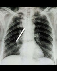
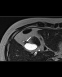
A 50-year-old male visited our hospital because he was experiencing epigastralgia. On Computed Tomography (CT), a bile duct stone was detected as a high-density material at the level of the lower biliary tract. After admission, biliary drainage by endoscopic procedures, endoscopic sphincterotomy, and stone extraction were performed. Coincidently, fundal-type adenomyomatosis of the gallbladder was suspected on Magnetic Resonance Imaging (MRI) with Magnetic Resonance Cholangiopancreatography (MRCP). Two months after hospital discharge, the biliary stone was not detected on follow-up MRI with MRCP, but focal thickening of the gall bladder had progressed in comparison to the thickness observed on previous CT and MRI with MRCP. The possibility of gallbladder cancer could not be denied; therefore, an extended cholecystectomy was performed. Histopathological examination revealed chronic cholecystitis without malignancy and no active inflammation. There was no active inflammation in the mucosa, muscularis propria, and subserosa. Quantitative visual assessment using diffusion-weighted imaging in addition to dynamic CT was useful for the diagnosis of chronic cholecystitis.

Kenji Motohashi1*, Takao Igarashi1, Hirokazu Ashida1, Kunihiko Fukuda1 and Satoru Chiba2


Article Image 1

Tracheobronchomegaly

A 48 year old man occasional smoker was symptomatic since 4 years with cough with copious amount of expectoration and exertional dyspnoea and recurrent infective exacerbations. There was past history of tuberculosis 15 years back treated with empirical antituberculosis therapy for a period of 1 year. There was no significant family history. Physical examination revealed presence of post exercise desaturation, grade III clubbing and coarse crackles in bilateral lung fields. Chest Radiograph (CXR) showed the presence of bilateral cystic opacities. High Resolution Computerised Tomography (HRCT) of the chest (Figure) (1a) (1b) revealed bilateral cystic bronchiectasis and tracheobronchomegaly with tracheal, right and left main bronchus dimensions being 30 mm, 27.2 mm, 22 mm respectively. Spirometry showed Forced Vital Capacity (FVC) of 1.55 L (of predicted), Forced Expiratory Volume in 1 second (FEV1) of 1.02 L (32% of predicted), and FEV1/FVC of 66%. A diagnosis of tracheobronchomegaly-Mounier Kuhn Syndrome (MKS) was made.

Suhas HS, Ketaki Utpat, Unnati Desai and Jyotsna M Joshi*


Article Image 1

Radiologic Guide to Surgical Treatment of Kienbock

Kienbock’s disease, or avascular necrosis of the lunate, is a progressive disease ultimately resulting in end-stage arthrosis. Various surgical treatments are available for different Lichtman stages of disease. This article reviews surgical treatments of Kienbock’s disease from a radiologic perspective. The reader will learn preoperative indications and imaging, normal and abnormal post-surgical appearance and potential complications of operative treatments of Kienbock’s disease.

Erika M Nealey1, Jonelle M Petscavage-Thomas2*, Felix S Chew1, Christopher Allan3 and Alice S Ha1


Article Image 1

Lumbar Nerve Root Anomalies are Still a Diagnostic and Therapeutic Challenge. Case Report and Review of Literature

Even in the MRI era lumbar nerve root anomalies in patients with lumbar radiculopathies are still frequently un- or misdiagnosed and usually discovered during spine surgery only. The existing literature suggests that patients with lumbar nerve root anomalies are especially prone to develop radiculopathies and suffering operative complications. Therefore, preoperative diagnosis is imperative to enable appropriate surgery and avoid complications. We describe a case of a patient with unilateral lumbar radiculopathy misdiagnosed with a lumbosacral disc herniation on magnetic resonance imaging. During surgery a Neidre/MacNab type 2b lumbar nerve root anomaly rather than a disc herniation was verified, which required a wide decompression of the involved neural structures. The literature is reviewed and the clinical and radiological features of lumbar nerve root anomalies are discussed from a neurosurgical perspective.

Fabio Freguia*, Katharina Koeck*, Kirsten Schmieder and Stefan Grossauer*


Article Image 1

Microwave-Assisted Synthesis of Small and Water-Dispersible Lanthanide Doped NaGdF4 Nanocrystals for PL/CT/ MR Multimodal Imaging

Multimodal imaging is advantageous in overcoming the deficiencies of individual imaging modalities. Herein, small, monodisperse and water-dispersible NaGdF4 : Ln (Ln = Eu, Tb, Dy) Nano Crystals (NCs) were successfully prepared in a facile manner by one-pot microwave-assisted solvothermal reaction in ethylene glycol. The morphology, size distribution, zeta potential, and crystal structure of the resulting NCs were well characterised by transmission electron microscopy, Nano-ZS90 Zetasizer, and X-ray diffraction analyses. These as-synthesised NaGdF4 : Ln (Ln = Eu, Tb, Dy) NCs exhibited a low cytotoxicity for Hela and C9H2 cells. Moreover, the doping concentrations of Eu3+, Tb3+, and Dy3+ ions were optimised for efficient Photoluminescence (PL) under the excitation of 273 nm in water. Furthermore, these NCs exhibited excellent relaxivity parameters (r1 ) in Magnetic Resonance (MR) imaging and Hounsfield units in X-ray Computed Tomography (CT) imaging in vitro. Therefore, these NCs have great potential to construct a unique platform for PL/CT/MR multimodal imaging simultaneously.

SongxiaYu1, Zhiqiang Wang2, Ruijun Cao1 and Lingjie Meng1*


Article Image 1

Artificial Joints: Chief Directions in Contemporary Orthopedics (Review)

Modern trends in joints investigation are presented, particularly the tribological aspects of natural and artificial joints. The extent of the achievements in this field at the turn of the 21st century is discussed. This period is distinguished by the discovery of liquid-crystalline and quasi-electret states in synovial liquids; ascertainment by AFM of new boundary lubrication mechanisms in joints, which are realized at the nanolevel; development of methods for in vitro study of friction in joints using electromagnetic fields able to simulate the natural articular biofield. A relationship is shown between biotribology and the chief directions in contemporary orthopedics including: intraarticular chondroprotection exercised via local therapeutic methods based on tribological monitoring of pharmaceutic substances; injection of drugs based on blood serum; development of a new generation of articular endoprostheses able to simulate the biophysical properties of synovial joints. It is shown that progress in modern biotribology has provided scientific substantiation of orthopedic treatment procedures.

Victor A Goldade1* and Yuliya M Chernyakova2


Article Image 1

Interest of Diffusion MRI in Hypoglycemic Coma

Hypoglycemia is a frequent and severe cause of coma, especially of diabetic patients. Prognosis is related to irreversible neuronal lesions and the most important predictive factors are the severity and duration of hypoglycemia. Cerebral cortex, hippocampus and basal ganglia are the most vulnerable sites. We report a case of hypoglycemic coma with cortical and basal ganglia hyper-intensities on diffusion weighted MRI suggesting cerebral complication, also we focused on the interest of Diffusion weighted MRI sequences in this issue through a literature review.

Jalal H*, Analy I, Bouroumane MR, Berghalout L, Moulattaf MounaEL and Cherif Idrissi Gannouni N


Article Image 1

Air in the Kidney

A 55-year-old woman admitted to our emergency department with three days history of abdominal pain, fever and vomiting. She was a known diabetic patient for last 25 years. On admission, she had fever (390C), heart rate of 100BPM and a, normal blood pressure and respiratory rate. The physical examination revealed an ill-looking patient with distended abdomen. There was extremely tender right renal angle with a diffusely tender right loin. The laboratory support showed leukocytosis, creatinine 2.06mg/dl and random serum glucose 512mg/dl. Urinalysis revealed numerous pus cell an RBC but ketonuria was absent. The cultures from the blood and urine sample showed Escherichia coli organism. Abdomen CT showed gas into the renal parenchyma and into the perirenal space on the right (Figures 1,2). The patient was diagnosed Emphysematous Pyelonephritis and successfully treated with antibiotic and supportive therapy. EPN is a uniformly fatal illness, if left untreated [1]. Treatment should be aggressive, starts with vigorous fluid resuscitation, antibiotic therapy, and control of blood sugar and electrolytes [2,3]. Before the advent of interventional radiology, early surgery and nephrectomy was a mandatory procedure.

 Rohat Ak*, Fatih Doğanay and Özge Ecmel Onur


Article Image 1

Strangulated Right Diaphragmatic Hernia: A Diagnostic Trap to Avoid

An 78 year old woman with Chronic Obstructive Pulmonary Disease (COPD) post tobacco (30 packs annualy) and Cardiac Arrhythmia was admitted to our hospital. It was noted a medical history of laparotomy sub costal for for cholecystectomy 20 years ago. She present with worsening of respiratory symptoms such as dyspnoea of rest for more than one week. Physical examination showed the patient was afebrile with acute respiratory distress (oxygen saturation at 87%) associated with confusion (Glasgow score : 10) and a high blood pressure situation (200/100 mm Hg) accompagnied by tachycardia (heart rate approximately 125 BPM) and abdominal distension comes with vomiting alimentary. Laboratory investigations indicated : respiratory acidosis (pH: 7.07) with hypercapnia at 91 mm Hg, a PaO2 at 135 mm Hg and alcaline reserve at 36 mmol/l. The rest of biological examinations did not show any other specific abnormalities.

Razafimanjato NNM1*, Ravoatrarilandy M2, Hunald Fa2, Rakotovao HJL2 and Castier Y3


Article Image 1

Mondor

A 75-year-old woman presented with a six-months history of recurrent painful lumps on the palmar aspect of different fingers on both hands (Figure 1). They resolved spontaneous in few days. Patient didn’t remember any local trauma. She was anticoagulated with vitamin K-antagonists because of previous pulmonary embolism and prosthetic mechanical mitral valve. She had no neoplasms, infections or systemic diseases in her medical history. Physical examination revealed bluish nodules on the volar side of the proximal interphalangeal joints of the index and middle f ingers (Figure 2). They were painful,of hard fibrous consistency, subcutaneous and 3-5 mm in size. Laboratory tests were normal. The clinical picture was not typical for paroxysmal finger haematomas, infective endocarditis(Osler’s nodes, Janeway lesions) or vasculitis. A diagnosis of recurrent spontaneous thrombosis of palmar digital veins was confirmed by echography. An antiphospholipid syndrome was rouled out and ibuprofen was locally applied. Mondor’s Disease was first described in 1939 and it is a rare condition which involves thrombophlebitis of the superficial veins of the breast and anterior chest wall. It sometimes occurs on the penis or on the fingers. The diagnosis is made by the typical clinical aspect. Radiological or Histological examinations are usually not necessary. Mondor’s Phlebitis is a self-limiting and generally benign disease. The specific aetiology remains uncertain. Trivial local traumas (for example handwork) are presumed. Surgery, infection, malignancy or hypercoagulable state are responsible for the disease only in rare cases.

Brenno Balestra* and Carlo Pusterla


Article Image 1

Isolated Hypoglossal Nerve Palsy in a Patient with Non-Small Cell Lung Carcinoma: Paraneoplastic Neurological Syndrome?

Paraneoplastic neurologic syndrome is a group of disorders resulting from damage to the nervous system in carcinoma, remote from primary site and not due to metastasis, infection, or metabolic complication associated with cancer. Patient with small cell lung cancer sometimes shows various paraneoplastic neurological syndromes, but patient with non-small cell lung cancer rarely shows paraneoplastic neurologic syndromes. Here we wish to report a case of 65-year-old female presented to our clinic with isolated right sided hypoglossal nerve palsy and poorly differentiated adenocarcinoma of lung.

Himanshu Juneja1*, Surekha Dabla2 and Manoj Yadav2