SM Journal Clinical and Medical Imaging

Archive Articles

Article Image 1

Fluorescence Spectroscopy, Phosphorescence Spectroscopy and Luminescence Spectroscopy Comparative Study on Malignant and Benign Human Cancer Cells and Tissues under Synchrotron Radiation with the Passage of Time

In the current study, we have experimentally and comparatively investigated and compared malignant human cancer cells and tissues before and after irradiating of synchrotron radiation using fluorescence spectroscopy, phosphorescence spectroscopy and luminescence spectroscopy. It is clear that malignant human cancer cells and tissues have gradually transformed to benign human cancer cells and tissues under synchrotron radiation with the passage of time (Figures 1–3) [1–119]. It can be concluded that malignant human cancer cells and tissues have gradually transformed to benign human cancer cells and tissues under synchrotron radiation with the passage of time.

Alireza Heidari*


Article Image 1

Emergence of Radiomics: Novel Methodology Identifying Imaging Biomarkers of Disease in Diagnosis, Response, and Progression

Radiomics is an emerging area within clinical radiology research. It seeks to take full advantage of all the information contained in multiple medical imaging modalities. With a radiomics approach, medical images are not limited to providing only a qualitative assessment but can also provide quantitative data by parameterizing image features. These parameters can be used to identify regions and volumes of interest and discriminate normal healthy tissue from abnormal or diseased tissue. Radiomics is an interlinked sequence of processes of vital importance that begins with the acquisition and selection of medical images that involve standardization of acquisition protocols and inter-equipment normalization. This is followed by the identification and segmentation of regions or volumes of interest by expert radiologists through the use of computational tools that offer speed while reducing variability and bias. The segmentation process is the most critical stage in radiomics. This sometimes requires the incorporation of a pre-processing stage consisting of advanced techniques (reconstruction processes, filtering, etc.). Thereafter, representative characteristics of the region or volume of interest are extracted by approaches based on statistics, morphological features, and transform-based variables. Next, a statistical selection of the parameters that provide a high association and correlation with the clinical condition of interest is performed. Finally, processes such as data integration, standardization, classification, and mining processes can be applied as needed for particular applications. Ongoing research in radiomics aims to reduce the time and costs involved in interpreting medical images while simultaneously increasing the quality of diagnoses and monitoring of as well as the selection of treatment strategies. The results of many studies combining radiomics with standard medical techniques are highly encouraging, and these new approaches are increasingly used. This review article details the components of radiomics and discusses its applications, challenges, and future directions for this exciting new field of study.

Edward Florez1, Ali Fatemi1,2, Pier Paolo Claudio2,3 and Candace M Howard1*


Article Image 1

Subcutaneous Emphysema in Critically Ill Children

Aim: To analyse the aetiology, pathophysiology and prognosis in a series of patients who developed clinically significant subcutaneous emphysema in a pediatric intensive care unit.

Method: Retrospective investigational study, recording and diagnosing all cases of clinically significant subcutaneous emphysema observed in the pediatric intensive care unit of a Spanish hospital during the period from January 2007 to December 2017.

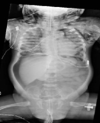
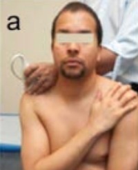
Results: 19 patients (7 girls and 12 boys), aged from 4 months to 14 years, were diagnosed with clinically significant subcutaneous emphysema during the study period. Regarding the aetiology of the condition, the origin of the emphysema was traumatic in 31, 5 % of patients (6 cases); associated with an invasive procedure or technique in 37% of patients (7 cases); and emphysema occurring in the context of a medical illness in 31, 5% of patients (6 cases). The emphysema occurred associated with pneumothorax in the 42% of patients but when the emphysema was due to traumatic event the association appeared in the 83% of cases. Three patients required specific treatment for emphysema (15, 8%) and one died due to its severity (5%).

Conclusions: Small subcutaneous emphysemas are asymptomatic and occur very frequently, associated with medical illnesses, traumas or technical/surgical procedures that are performed daily in intensive care units. Clinically significant, symptomatic emphysemas, however, are exceptional and can aggravate the clinical state of a patient who is already in a critical condition and may even be life threatening. In our serial, emphysema occurred most frequently associated to pneumothorax and it appeared isolated in the 16% of cases. Unfortunately we have not found any published data to compare our results. Despite the low mortality in our serial, emphysemas should be diagnosed as early as possible, and their evolution and clinical impact closely monitored.

Abril Molina Ana* and Ocete Hita Esther


Article Image 1

Modified

We report a novel ultrasonography technique of demonstrating partial tears of supraspinatus tendon using modified scarf technique. Correct diagnosis of the partial tears assist in appropriate management of these injuries.

Rajesh Botchu1* and Winston J Rennie2


Article Image 1

Isolated Vastus Intermedius Rupture- Case Report and Review of Literature

Quadriceps ruptures are common but isolated tears of the vastus intermedius are uncommon. We present a case of a 45-year-old man presenting with swelling and pain in the thigh after trauma who had an isolated tear of the vastus intermedius.

Azzopardi C1, Vidoni A1*, Goswami V2 and Botchu R2


Article Image 1

Developing a Texture Analysis Technique using Fluid-Attenuated Inversion Recovery (FLAIR) to Differentiate Tumor from Edema for Contouring Primary Intracranial Tumors

Purpose: Treatment protocols for malignant tumors generally call for surgical removal followed by tumor-bed irradiation. Irradiation ideally affects the tumor volume while limiting damage to surrounding normal tissues; this requires accurate determination of 3-D treatment volumes. Glioblastoma multiforme (GBM) and meningioma are common primary intracranial tumors. Edema surrounding meningioma is vasogenic, while edema surrounding GBM may also have tumor cell infiltration, difficult to differentiate by Fluid-Attenuated Inversion Recovery (FLAIR). Hypothetically, the FLAIR signals of edema and GBM should differ. We used T2W-FLAIR spin-echo to determine edema type and differentiate tumors through their textural properties.

Methods: Enrollment was 20 patients with GBM and 10 with meningioma. Patients were scanned using a 3-D multiecho GRE sequence, measuring the FLAIR signal of edema around the tumor. Segmentation identified two regions of interest (ROI, edema and tumor); texture analysis was applied to each ROI with- and without normalization. LASSO analysis of texture parameters selected the best parameters for separating ROIs; using them, we performed a Receiver Operating Characteristic (ROC) analysis.

Results: Two intracranial tumor types with- and without normalization provided four scenarios per ROI. First-order statistics using 1%-percentile feature was chosen in all scenarios and had the best discriminant ability for meningioma. Second-order statistics using correlation features was also selected across scenarios, although angle and magnitude varied. Higher-order statistics using short-run emphasis features and gray level non-uniformity provided the best discrimination for GBM images with- and without normalization, respectively. ROC curves display the results of both the single best discriminator and the discriminant ability of the model using all features selected by LASSO. All univariate models had good discriminant ability (AUC>0.83), and all multivariate models had excellent discriminant ability (AUC>0.92).

Conclusion: A small subset of texture parameters shows excellent ability to discriminate edema from tumor tissue through its most discriminating features.

Edward Florez1, Todd A Nichols1, Seth T Lirette2, Candace M Howard1 and Ali Fatemi1,3*


Article Image 1

Rudimentary Communicating Functional Uterine Horn: A Case Report

This case report describes a 26-year-old female who presented to the outpatient department with abdominal intermittent pain during menstruation period. Ultrasound and magnetic resonance imaging of the abdomen suggested rudimentary uterine horn. Repeated ultrasonic examination after menstruation exhibited reduced endometria of the mass, suggesting endometria abscission and communicating functional uterine horn. Upon therapy completion and education, the patient was free for laparotomy.

Xiu-hua Wu1, Ying-zi Liu MM1 and Wei-xian Chen2*