Fatal Multi-Organ Failure Following Occupational Chemical Burn after Exposure to Phenolic Acid: A Case Report and Literature Review
Aim: Describe the complications and management of a rare case of burn injury by phenolic acid.
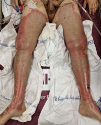
Methods: A 45-year-old truck driver sustained a 35% total body surface area (TBSA) burn from an accidental phenolic acid injury. After initial treatment and decontamination with a polyethylene glycol solution, he arrived at the hospital intubated and on vasopressor support. No other traumatic injuries were found. Local assessment revealed a 35% TBSA burn of partial and full thickness on the perineal region, legs, right arm, and a small area of the abdomen. His Revised Baux score was 97. During his recovery, the patient developed hepatic insufficiency followed by renal failure, which required dialysis. A few days later, a total body CT scan revealed multiple ischemic cerebral areas. After 12 days from admission and episodes of bronchial bleeding, the patient died due to a multiorgan failure. A skin biopsy and toxicological analysis for phenol levels were performed during his hospital stay.
Results: In addition to the clinical case analysis, a review of the medical literature regarding cases of death following phenol burns, a histological analysis of the biopsy tissue collected and an analysis of the blood levels of phenol and its derivative o-cresol were conducted.
Conclusions: This case report illustrates the lethality of phenol chemical burns and emphasizes the critical role of multidisciplinary management in addressing multi-organ complications
Alberto Schiarillo1, Anna Pensa1, Filippo Mariano2,3, Francesco Lupariello4, Catalina Ciocan5,6*, Daniela Risso1 and Maurizio Navissano1


