Case Report | Volume 9 - Issue 1 | Article DOI :
Download PDF
Mohammad Abidali¹*, Graeme Benzie¹, Hassan Abidali², Phillip Pearson³, Deniz Dolun¹, Corey Steinman¹, Jessica Jahoda¹, and Mohamed Aziz⁴
¹American University of the Caribbean Medical School, Dept of Pathology, USA
²Arizona State University, USA
³Philadelphia College of Osteopathic Medicine, Georgia, Dept of Biomedical Sciences, USA
?siParadigm Diagnostic Informatics, NJ, USA
Corresponding Author:
Mohammad Abidali *, Department of Pathology, American University of the Caribbean School of Medicine, AUC, 1 University Drive at Jordan Road Cupecoy, Dutch St. Maarten, USA
Keywords
Dedifferentiated chondrosarcoma; Biphasic; Aggressive; Genetic Alteration.
Abstract
A subtype of aggressive chondrosarcoma known as dedifferentiated chondrosarcoma (DDCS) accounts for about 10% of all chondrosarcomas. The characteristic feature is a low-grade cartilaginous tumor juxtaposed with a high-grade sarcoma with a low 5-year survival rate and a high local recurrence rate. With a 20% 2-year survival rate, DDCS typically strikes older men. There is currently limited literature on the pathogenesis of DDCS, and more investigations are required to improve prognostic factors and treatment options. Our case study’s subject is dedifferentiated chondrosarcoma (DDCS) arising from a single enchondroma. Although it is uncommon, the malignant transformation of a single enchondroma has been documented. Our case already has a pre-existing enchondroma, which supports that DDCS can arise from an enchondroma.
Citation
Abidali M, Benzie G, Abidali H, Pearson P, Dolun D, et al. (2023) Dedifferentiated Chondrosarcoma Developing in Solitary Enchondroma, Case Report of Rare Tumor with Uncommon Presentation and Brief Review of the Literature. SM J Case Rep 9: 5.
ABBREVIATION
Dedifferentiated chondrosarcoma (DDCS), Immunohisto chemistry studies (IHC), The European over 40 Bone Sarcoma Study (EURO-B.O.S.S)
INTRODUCTION
Conventional chondrosarcoma accounts for nearly 90% of all chondrosarcomas. Besides DDCS, other types of chondrosarcomas include clear-cell chondrosarcoma, mesenchymal chondrosarcoma, juxtacortical chondrosarcoma, and secondary chondrosarcoma [19]. Dedifferentiated chondrosarcoma (DDCS) is a rare, highly aggressive tumor with invasive behaviors and a poor prognosis. Dahlin and Beabout first described DDCS in 1971 as a high-grade sarcoma adjacent to low or intermediate chondrosarcoma with a sharp boundary between these two components [2]. The age of onset most frequently ranges from 50 to 60 years old, with a slight predominance of men, and the most involved sites are the femur, pelvis, and humerus [15]. The dedifferentiated component may exhibit numerous mesenchymal neoplasms with various features, such as osteosarcoma, fibrosarcoma, and leiomyosarcoma, among others [4]. DDCS exhibit aggressive behavior with a high local recurrence rate leading to poor survival. DDCS accounts for 10% of chondrosarcomas, most commonly present in the mid 50s, and carries a 2-year survival rate of 20% [2,3,4]. Due to its rarity, there is limited literature regarding the prognosis of DDCS. The preferred treatment remains surgical resection with disease-free margins. However, approximately 27% of cases do not have adequate surgical margins, with axial tumors exhibiting an increase in inadequate margins compared to appendicular lesions [4].
Although rare, the malignant transformation of an enchondroma has been reported [19]. DDCS has demonstrated a high local recurrence rate even after wide-margin surgical resection, indicating that chemotherapy may also be required. Further research and advancements are required to understand the pathophysiology and treatment options for DDCS. We present a case of dedifferentiated chondrosarcoma (DDCS) in a 52-year-old woman that developed from a pre-existing solitary lower femur enchondroma, and we provide a brief review of the literature.
CASE PRESENTATION
A 52-year-old woman presented to the ER with a lower femur fracture resulting from a fall from the steps. Her family provided a history of biopsy diagnosed small enchondroma at the lower end of the shaft of the femur four years before the current presentation. Due to the small size of the benign enchondroma, the patient was observed without specific treatment. The patient or the family gave no other significant medical history. X-ray of the lower femur at the current presentation showed a pathological fracture through the enchondroma. At that time, the fracture was corrected using an intramedullary retrograde rode locked proximally and distally (Figure-1 A). Unfortunately, the fracture did not heal, and the patient continued to have pain and swelling with a rapidly growing mass at the fracture site. Physical examination and lab tests proved an absence of inflammation. Serial X-rays showed a non-healed fracture and significant scalloped areas with cortical erosion. MRI confirmed a large tumor mass at the fracture site measuring 8x4 cm with extension into the surrounding soft tissue.
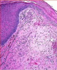
Microscopic examination of a biopsy taken from the mass showed two distinct histologic components. One cartilaginous component showed features of low-grade chondrosarcoma. The second component showed features of high-grade malignant neoplasm with pleomorphic and spindle cell features. There was an abrupt demarcation between the two components (Figure 1 B,C,D). Immunohistochemistry studies (IHC) studies were utilized for the definitive diagnosis of the non-cartilaginous tumor component. The tumor was only strongly positive for Vimentin, with focal positive staining with CD99 and S-100. The tumor cells were negative for all standard markers for hematologic, epithelial, neural, neuroendocrine, germ cells, or specific mesenchymal differentiation. Molecular studies showed genetic mutations in IDH1 and IDH2 in both the cartilaginous and the non-cartilaginous components of the tumor. The histomorphology, together with the IHC findings, were diagnostic of dedifferentiated chondrosarcoma with malignant fibrous histiocytoma (MFH) histomorphologic pattern.
A multidisciplinary tumor board meeting recommended neoadjuvant chemotherapy and complete surgical resection followed by adjuvant chemotherapy. The patient received a modified protocol using cisplatin, doxorubicin, and ifosfamide. Chemotherapeutics were administered as a single agent sequentially and not in combination, followed by complete surgical resection of the tumor and prosthetic knee joint reconstruction. Pathologic evaluation of the resection revealed a tumor mass of DDCS measuring 8 × 4 cm in size with a 70% necrosis of the dedifferentiated component. The tumor appeared to occur in the background of lobules of benign hyaline cartilage encased by reactive bone and covered by perichondrium consistent with surrounding enchondroma (Figure-1 E)

Figure 1: X-ray of the pathological fracture and pathological examination of the tumor
1A: X-ray of the pathological fracture of the lower femur. The fracture is corrected using an intramedullary retrograde rode locked proximally and distally
1B: Dedifferentiated chondrosarcoma showing cartilaginous and non-cartilaginous components with sharp demarcation (H &E stain X20)
1C: The non-cartilaginous component showing high-grade MFH-like histomorphology (H &E stain X60)
1D: The cartilaginous component showing malignant chondrocytes of low-grade chondrosarcoma (H&E stain X60)
1E: Lobules of benign hyaline cartilage encased by reactive bone and covered by perichondrium consistent with surrounding enchondroma (H &E stain X20)
Postoperative adjuvant chemotherapy included doxorubicin, cisplatin, and ifosfamide. The patient was monitored for recurrence and metastatic disease with physical examination, radiographic imaging, chest imaging, and whole-body bone scans. She was followed for thirty-six months with no evidence of recurrence or metastasis.
DISCUSSION
Cartilaginous tumors exist in benign or malignant forms. Chondrosarcoma, the malignant form, is a heterogenous tumor class that includes conventional, clear cell, mesenchymal, or dedifferentiated types [14]. Dedifferentiated chondrosarcoma (DDCS) is an uncommon and aggressive chondrosarcoma variant. In 1971, Dahlin and Beabout were the first to characterize the clinical and pathological aspects [5]. The characteristic histology and radiographic features of DDCS include a biphasic tumor comprised of conventional chondrosarcoma with an abrupt shift to a high-grade mesenchymal component. The tumor cells in both components seem to originate from a single precursor, but there are a substantial number of genetic alterations in the anaplastic component [12]. The potential for multiple differentiations of precursor cells is supported by the fact that a mutated human embryonic muscle cell line exhibits features of dedifferentiated chondrosarcomas [12].
Despite aggressive multimodal therapy, dedifferentiated chondrosarcoma has a significantly high mortality rate, with lung metastasis being the most common cause of death [6]. Strotman et al. analyzed 159 cases of DDCS (from 2001-2011) and concluded that the overall prognosis of dedifferentiated chondrosarcoma is poor, with a 5-year overall survival of 18%. Patients with a primary tumor located in the chest wall had a better prognosis. Tumors larger than 8 cm, with metastases at diagnosis, and treatment without surgical resection were significant predictors of mortality [13]. More recently, Gusho et al. analyzed 16 cases of DDCS from 2000 to 2018 and reported that their findings confirm the poor prognosis of DDCS patients, with a five-year estimate of 32%. They stated that with existing literature, their data might help enable future strategic recommendations for these patients [14]. In a study by Liu et al., they retrospectively analyzed twenty three patients with dedifferentiated chondrosarcoma confirmed by pathology from 2008 to 2015. They concluded that axial bone location, lung metastasis at diagnosis, inadequate surgical margin, incorrect diagnosis before surgery, and pathological fractures were related to poorer outcomes. They also concluded that pre- or postoperative chemotherapy had no definitive effect on improved survival [15].
Clinical presentations may include local edema, pain, pathological fracture, and sometimes with acute disease presentation [7,8]. Therefore, it is imperative to identify imaging features of dedifferentiation underlying chondroid lesions. Radiographs demonstrating aggressive osseous destruction or cortical infiltration, large unmineralized soft tissue mass, pathological fracture, areas with osteoid matrix, and rapid progression were suggestive of dedifferentiation [11]. Many studies based the radiographic diagnosis on the biphasic nature of the tumor; however, in some large series studies, the tumor showed a biphasic nature on only one-third of radiographs, one-third of MRI images, and one-half of CT scans [15]. Documentation of various imaging features is crucial to help guide the preoperative biopsy site or multifocal sampling at initial diagnosis because the dedifferentiated component determines the growth of lesions, metastasis, and prognosis of patients [8].
The pathogenesis of DDCS remains unknown. Evidence now suggests that DDCS has a monoclonal origin and splits into two components with either chondrogenic or non-chondrogenic origins. Differences in the growth rates of the two components show that the divergence occurs late in the precursor cell.The typical chondrosarcoma and the high-grade sarcomatous components of DDCS include many genetic alterations. In most DDCSs, genetic mutations in IDH1, IDH2, and COL2A1 are found in both components. According to DNA studies, additional genetic mutations, such as TP53 and RB, have been discovered solely in the dedifferentiated component [9]. Lucas et al., in a detailed study of 11 cases of DDCS, concluded that all the DDCSs cases (11 of 11; 100%) capture the genomic profile at a single time point of a biphasic tumor with the assumption that the two components of DDCS represent the temporal progression of conventional chondrosarcoma to a high-grade tumor. Such an assumption is acceptable in many malignancies where the temporal progression is well studied (e.g., colon cancer) but is less substantiated in bone sarcomas. Additional studies of metachronous DDCSs will be necessary to explore that constraint [11] further.
CD44 is a transmembrane glycoprotein that regulates growth, differentiation, survival, and cell motility. Upregulation of CD44 in the anaplastic component was noted compared with the chondrogenic component of DDCS. Unregulated CD44, resulting in the expression of different splice variants, is related to poor prognosis in many cancers and is also seen in high grade secondary peripheral chondrosarcomas [8]. Other studies reported that the underlying mechanism of dedifferentiation is unknown, but cell cycle regulators p16, p53, and retinoblastoma appear to have important roles in tumor development and dedifferentiation [12]. Single point mutations in the Ras genes play a role in tumor development by eliminating dependence on GTPase-activation of protein regulation. A few cases revealed H-Ras mutations in dedifferentiated chondrosarcoma but not in low-grade conventional chondrosarcomas, suggesting that the mutation may be associated with the aggressive nature of the disease rather than dedifferentiation [12]. Actin markers are observed in most of the dedifferentiated peripheral chondrosarcomas. Prognostic markers such as PAI-1 were identified in the chondrogenic component of the dedifferentiated peripheral chondrosarcoma and correlated with a good prognosis. In 2021, Lucas et al. reported, to their knowledge, to be the first to show that TERT promoter mutations are common in both components of DDCS. They explained that the TERT gene encodes the reverse-transcriptase subunit of the telomerase enzyme [11]. Recurrent C228T and C250T promoter mutations, which lead to TERT overexpression and telomerase activity, have been reported in a wide range of human cancers but, apart from myxoid liposarcoma, are rare in sarcomas [11].
The optimal treatment for DDCS remains unclear. Surgical resection of the tumor with wide or radical margins is the most crucial procedure [11,12,14,15]. McCarthy and Dorfman concluded that once dedifferentiation has occurred, a cure was unrealistic, regardless of the type of therapy employed. In contrast to classical osteosarcomas and Ewing’s sarcomas, the value of neoadjuvant or adjuvant therapeutical strategies remains uncertain and is still under investigation [6]. A recent study by Homeland et al. reported the Outcome of dedifferentiated chondrosarcoma for patients treated with multimodal therapy from several European countries (The European over 40 Bone Sarcoma Study (EURO-B.O.S.S). They concluded that adding intensive chemotherapy to surgery to treat DDCS is feasible and shows favorable survival data compared with previous reports. With the limitations of data from a non-controlled trial, they conclude that chemotherapy could be considered in managing patients, especially aged >40 years [16]. Their analysis of 451 sarcomas included in the EURO-B.O.S.S. study claims that this is the first study to prospectively explore the benefit of adding chemotherapy to treating patients with DDCS. They reported that the estimated 5-year overall survival was 39%, which is better than that previously reported from retrospective studies. The chemotherapy toxicity was considerable but manageable, and no toxic deaths were reported [16,17,18].
In our instance, the presence of an earlier enchondroma supports that DDCS can originate from an enchondroma. If only a small biopsy sample is taken, it may be difficult to microscopically distinguish the dedifferentiated component of DDCS from other fibroblastic and myofibroblastic sarcomas. Summers and co reported a bone case of low-grade myofibroblastic sarcoma with a dedifferentiated high-grade sarcoma recurrence. They detailed the importance of IHC and imaging studies to make such differentiation [20]. Possible malignant transformation of benign bone tumors has been reported. Our case showed a possible transformation of a benign cartilaginous lesion, enchondroma, to a chondrosarcoma. The malignant transformation of benign bone osteoma to osteosarcoma has also been reported [21].
Despite various histological features in the high-grade dedifferentiated chondrosarcomas component, they are resistant to chemotherapy. Due to the poor prognosis of DCCS and insufficient clinical outcome data, studies are encouraged to describe the natural course of the disease. We will better understand this uncommon tumor by reporting additional cases, and ongoing research will lead to the development of more accurate diagnoses and safe treatments that will improve patient outcomes.
ACKNOWLEDGMENT
Special thanks to Garrett Harris, Daniel Silva, and Priya Patel, MD candidates at the American University of the Caribbean, for their assistance in preparing manuscript images and reviewing the final manuscript.
REFERENCES
1. Anract P, Tomeno B, Forest M. Chondrosarcomes dédifférenciés. Etude de treize cas cliniques et revue de la littérature [Dedifferentiated chondrosarcoma. A study of 13 clinical cases and review of the literature]. Rev Chir Orthop Reparatrice Appar Mot. 1994;80(8):669-80. French. PMID: 7638395.
2. Liu C, Xi Y, Li M, Jiao Q, Zhang H, Yang Q, Yao W. Dedifferentiated chondrosarcoma: Radiological features, prognostic factors and survival statistics in 23 patients. PLoS One. 2017 Mar 16;12(3):e0173665. doi: 10.1371/journal.pone.0173665. PMID: 28301537; PMCID:PMC5354284. www.ncbi.nlm.nih.gov/pmc/articles/PMC5354284/.
3. Gong L-H, Su Y-B, Zhang W, Liu W-F, Dong R-F, Sun X-Q, Zhang M and Ding Y (2021) Dedifferentiated Central Chondrosarcoma: A Clinical, Histopathological, and Immunohistochemical Analysis of 57 Cases. Front. Med. 8:746909. doi: 10.3389/fmed.2021.746909”
4. Liu C, Xi Y, Li M, Jiao Q, Zhang H, Yang Q, Yao W. Dedifferentiated chondrosarcoma: Radiological features, prognostic factors and survival statistics in 23 patients. PLoS One. 2017 Mar 16;12(3):e0173665. doi: 10.1371/journal.pone.0173665. PMID: 28301537; PMCID: PMC5354284.
5. Bharath G, Burrah R, Shivakumar K, Manjunath S, Bhanumathi R. Dedifferentiated chondrosarcoma: An aggressive variant of chondrosarcoma. Asian Cardiovascular and Thoracic Annals. 2015;23(2):221-223. doi:10.1177/0218492314522253
6. Bruns, J., Fiedler, W., Werner, M. et al. Dedifferentiated chondrosarcoma—a fatal disease. J Cancer Res Clin Oncol 131, 333–339 (2005). https://doi.org/10.1007/s00432-004-0648-6
7. Cao, Lei, et al. “Dedifferentiated chondrosarcoma: a clinicopathologic analysis of 25 cases.” BMC Musculoskeletal Disorders, vol. 22, no. 1, 15 Feb. 2021, p. NA. Gale Academic OneFile, link.gale.com/apps/doc/ A653602311/AONE?u=anon~9e5d66f6&sid=googleScholar&xid=66 32d085. Accessed 23 Nov. 2022.
8. Rozeman, L., de Bruijn, I., Bacchini, P. et al. Dedifferentiated peripheral chondrosarcomas: regulation of EXT-downstream molecules and differentiation-related genes. Mod Pathol 22, 1489–1498 (2009). https://doi.org/10.1038/modpathol.2009.120
9. Dhinsa B, S, DeLisa M, Pollock R, Flanagan A, M, Whelan J, Gregory J: Dedifferentiated Chondrosarcoma Demonstrating Osteosarcomatous Differentiation. Oncol Res Treat 2018;41:456-460. doi: 10.1159/000487803
10. Calixto-Hope G. Lucas, MD; James P. Grenert, MD, PhD; Andrew Horvai, MD, PhD. “Targeted next-generation sequencing identifies molecular and genetic events in Dedifferentiated Chondrosarcoma Arch Pathol Lab Med—Vol 145, August 2021
11. Sakamoto A. The molecular pathogenesis of dedifferentiated chondrosarcoma. Indian J Orthop. 2014 May;48(3):262-5. doi: 10.4103/0019-5413.132506. PMID: 24932031; PMCID: PMC4052024.
12. Patrick K. Strotman MD,Taylor J. Reif MD,Stephanie A. Kliethermes PhD,Jasmin K. Sandhu PhD,Lukas M. Nystrom MD. Dedifferentiated chondrosarcoma: A survival analysis of 159 cases from the SEER database (2001-2011). J Surg Oncol.2017 Aug;116(2):252-257. doi: 10.1002/jso.24650. Epub 2017 Apr 18.
13. Gusho CA, Lee L, Zavras A, Seikel Z, Miller I, Colman MW, Gitelis S, Blank AT. Dedifferentiated Chondrosarcoma: A Case Series and Review of the Literature. Orthop Rev (Pavia). 2022 May 31;14(4):35448. doi: 10.52965/001c.35448. PMID: 35769663; PMCID: PMC9235431.
14. Liu C, Xi Y, Li M, Jiao Q, Zhang H, Yang Q, Yao W. Dedifferentiated chondrosarcoma: Radiological features, prognostic factors and survival statistics in 23 patients. PLoS One. 2017 Mar 16;12(3):e0173665. doi: 10.1371/journal.pone.0173665. PMID: 28301537; PMCID: PMC5354284.
15. Ivar Hompland a, Stefano Ferrari b, Stefan Bielack c, Emanuela Palmerini b, Kirsten S. Hall, et al. Outcome in dedifferentiated chondrosarcoma for patients treated with multimodal therapy: Results from the European Bone Over 40 Sarcoma Study. European Journal of Cancer 151 (2021) 150e158
16. Grimer et al. Dedifferentiated chondrosarcoma: Prognostic factors and Outcome from a European group. European Journal of Cancer. Volume 43, Issue 14, September 2007, Pages 2060-2065
17. Amer at al. Survival and Prognosis of Chondrosarcoma Subtypes: SEER Database Analysis. Wiley Online Library (wileyonlinelibrary. com). DOI 10.1002/jor.24463. JOURNAL OF ORTHOPAEDIC RESEARCH®FEBRUARY 2020312AMER ET AL.
18. Joseph B, Eskander H, Matar G, Grisel B, Webking S, Aziz M et al. (2021) Extraskeletal Myxoid Chondrosarcomas of the Ankle, Case Report of Uncommon Tumor and Brief Review of the Literature. SM J Clin Med 6: 4
19. Yonezawa H, Yamamoto N, Hayashi K, Takeuchi A, Miwa S, Igarashi K, Morinaga S, Asano Y, Saito S, Tome Y, Ikeda H, Nojima T, Tsuchiya H. Dedifferentiated chondrosarcoma of the middle finger arising from a solitary enchondroma: A case report. World J Clin Cases 2022; 10(10): 3297-3305 [PMID: 35647122 DOI: 10.12998/wjcc.v10.i10.3297]
20. Summers J Jr., Allison E, Millon J, Wallace A, Jahoda J, Aziz M.et al. (2021) Low Grade Myofibroblastic Sarcoma of the Bone with Recurrence as a High Grade Sarcoma: Case Report and Review of the Literature. SM Musculoskelet Disord 5: 4.
21. Latif A, Stastka P, Jackson G, White M, Khalefih S, Aziz M, et al. (2020) Parosteal osteosarcoma arising from the site of a prior excised osteoid osteoma: A rare progression, or an uncommon coincidence? Report of a case with brief review of the literature. JSM Clin Cytol Pathol 2: 4.