Deepa JP¹, Samriddhi S², Puri G³, Aravinda K⁴, Dixit A⁵, Gupta R⁶, and Aanchal G⁶*
¹ Reader, MDS, Department of Oral Medicine, Diagnosis, and Radiology, Swami Devi Dyal Dental College and Hospital, Golpura, India
² Postgraduate Student, BDS (MDS), Department of Oral Medicine, Diagnosis, and Radiology, Swami Devi Dyal Dental College and Hospital, Golpura, India
³ Professor and HOD, MDS, Department of Oral Medicine, Diagnosis, and Radiology, Swami Devi Dyal Dental College and Hospital, Golpura, India
? Professor, MDS, Department of Oral Medicine, Diagnosis, and Radiology, Swami Devi Dyal Dental College and Hospital, Golpura, India
? Reader, MDS, Department of Oral Medicine, Diagnosis, and Radiology, Swami Devi Dyal Dental College and Hospital, Golpura, India
? Senior Lecturer, MDS, Department of Oral Medicine, Diagnosis, and Radiology, Swami Devi Dyal Dental College and Hospital, Golpura, India
Corresponding Author:
Deepa Jatti, Department of Oral Medicine, Diagnosis and Radiology, Swami Devi Dyal Dental College and Hospital Golpura, India, Email: iafdeepa@gmail.com
Keywords
Monostotic fibrous dysplasia;
Postmenopausal female; Ground glass
appearance
Abstract
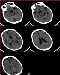
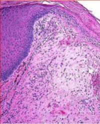
Fibrous dysplasia is a condition characterized by excessive proliferation of bone-forming mesenchymal cells. It can affect one bone (monostotic type) or multiple bones (polyostotic type). It is usually observed in adolescents and young adults and comprises 7% of benign bone tumors. The etiology is not clear but genetic predisposition is suspected. It has a predilection for long bones as well as the craniofacial skeleton. The maxilla is the most commonly affected facial bone, with facial asymmetry being the usual complaint. The diagnosis is based on radiological and histopathological examination. There are different treatment approaches including monitoring, medical treatment or surgery. A 45-year-old female reported with a complaint of painless swelling on the left side of maxilla since 1 year. A diffuse intraoral bony hard, non tender swelling was seen in the left maxilla involving the premolar-molar region. Plain film radiographs and Computed tomography revealed ground glass appearance of the left maxilla. The lesion was excised and on histopathology showed features of fibrous dysplasia. Very few cases of Fibrous dysplasia manifesting in the older and postmenopausal age group are reported in the literature. Once diagnosed, routine follow-up should be done on a yearly basis with x-ray examination
Citation
Deepa JP, Samriddhi S, Puri G, Aravinda K, Dixit A, Gupta R, et al. Monostotic Fibrous Dysplasia of Maxilla in a Postmenopausal Female- A Rare Case Report with Review of Literature. SM J Case Rep. 2016; 2(2): 1025