Case Report | Volume 10 - Issue 1 | Article DOI :
Download PDF
Riccardo Fornaro², Giorgio Incarbone¹,²*, Andrea Montalbetti¹,², Luca Emanuele¹,², Federica Anna Fasano¹,², Marta Sacchetti³, Emanuela Crobeddu², and Christian Cossandi²
¹Department of Clinical Surgical Diagnostic and Pediatric Sciences, University of Pavia, Italy
²Department of Neurosurgery, A.O.U. Maggiore della Carità University Hospital, Italy
³Department of Psychology, A.O.U. Maggiore della Carità University Hospital, Italy
Corresponding Author:
Giorgio Incarbone, Department of Clinical Surgical Diagnostic and Pediatric Sciences, University of Pavia, Italy
Keywords
VNS Therapy; Epilepsy; Post-operative complication; Re-intervention
Abstract
Background: In recent years, internet penetration in the island of Mauritius has increased substantially. However, increasing internet use is also associated with adverse effects especially amongst the young internet users and there is a scarcity of research in this area.
Objectives: To assess the prevalence and characteristics of internet addiction in the student population of the University of Mauritius.
Methodology: A cross-sectional study using a shortened and contextualized Internet Addiction Test (IAT) (Young, 1998) was carried out amongst 372 participants.
Results: It was observed that 5.1% of all students (4.9% males and 5.4% females) showed features of severe internet addiction. Although 42.5% perceived themselves to be addicted to the internet, they did not have significantly higher IAT scores than those who did not. There was no statistically significant difference between the prevalence of internet addiction in males and females. Main online activities comprised preferentially of information searching (94.9%) chatting/communicating (90.6%) and downloading media content (87.9%). The mean daily time online was 4.9 hours (standard deviation=3.1 hours). The mean years of internet use was 7.1 years (standard deviation=2.7 years). Results of simple linear regression showed a negative correlation and a small effect size of years of internet use on IAT scores and daily time online was positively correlated with and had a mild effect on IAT scores.
Conclusion: This study provides useful information on prevalence rates and characteristics of internet addiction in young people in Mauritius. Further studies are required to follow-up the evolution of this condition to assess its impact on young people’s health over time.
Citation
Fornaro R, Incarbone G, Montalbetti A, Emanuele L, Anna Fa sano F et al. (2024) VNS Therapy, Validation of Efficiency of Therapy: Case Report. SM J Case Rep 10: 4.
Introduction
Epilepsy affect 1% of the worldwide population. Antiepileptic drugs (AEDs) are the first option of treatment, but approximately one third of patients have seizure that do not respond to pharmacologic therapy [1,2].
This is defined as a “failure of adequate trials of two (or more) tolerated, appropriately chosen and appropriately used antiepileptic drugs regimens to achieve freedom free seizure” [3].
For drug resistant epilepsy people, the first choice of treatment is the resective surgery of the epileptic focus, when this is not possible or refused by the patients themselves other options are ketogenic diet, deep brain stimulation (DBS) and vagus nerve stimulation (VNS) Therapy [4].
VNS therapy™is composed of a pulse generator, surgically implanted under the left clavicle (or in the axilla for a more aesthetic result) connected to a lead wire with two helicoidal stimulating electrode wrapped around the left vagus nerve [5].
At this level action potentials are generated on the vagus nerve that propagate mostly afferently. Recently, has been speculated that those action potentials activate the vagal afferent network with an anti-ictal and anti-epileptogenic effect in patients with drug resistant epilepsy (DRE). Indeed, VNS Therapy ™is indicated as adjunctive therapy for the treatment of focal and generalized seizure in DRE in EU since 1994 (Physician’s Manual) [6].
Case Description
We report the case of a 46-years-old female patient with seizures onset at the age of 14. The first seizure was characterized by a strange taste followed by confusion lasted for a few hours. She was admitted to the pediatric ward, but no AEDs had been administrated and she had been dismissed after 48 hours. The second episode happened one year after. Since 1991 to 1995 5 seizure attacks were characterized by sialorrhea, activation of swallowing reflex, tachycardia and “break of contents”. Initially, Phenobarbital has been the first choice of treatment and after the first generalized seizures, she started a treatment with Vigabatrin 2 g/die and phenobarbital 300 mg/die. She kept suffering of partial and generalized seizures every 2 or 3 years. Thus, she received a polytherapy with a combination of Valproic acid, Levetiracetam, and Lamotrigine, with non-significant benefits in terms of seizure control at long term. In 2010 she suffered from ovarian cancer treated with surgery and chemotherapy. Two attacks of generalized seizure were reported during chemotherapy. In 2015 she also had a car accident with a following seizure attack while driving [7].
During the last neurological examination (March 2021), she reported that seizures occur twice every 15 days, described as breathing difficulties followed by confusion, speaking impairment and resolution after few minutes with heat sensation and residual aphasia and paraphasia. (Therapy Gardemale, Tolep, Zonisamide, Nubriveo) [8].
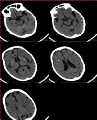
In this case surgery was not suitable because the electroencephalogram (EEG) did not show any epileptogenic foci. Furthermore, the magnetic resonance (MR) didn’t show any structural lesion possibly responsible for seizures [9].
After being evaluated by our neuro modulation team, composed by neurosurgeon, neurologist and neuropsychologist, the implantation of a VNS Therapy system™ was considered suitable for this patient and the operation was performed on 31 May 2021 [10].
Two months after the surgical implantation of the generator, lead and electrodes, she suffered from shorter and a smaller number of seizure and passed through a short seizure-free period. Stimulation parameters: 1,75 mA, 20Hz. Suddenly, three months after the implant, she experienced seizures with greater intensity and duration as compared to the pre implantation period. Moreover, she referred an accidental fall with possible direct trauma to the VNS generator [11]. Indeed, an increase of the impedance was found after checking the system. She underwent a neck CT scan that showed a displacement and a probable rupture of the helicoidal stimulating electrode around the vagus nerve. On 6th December 2021 the patient underwent revision surgery with repositioning of the stimulating electrode and on the follow up she went back to a seizure free state with the same stimulation parameters of the first implantation. The patient is still under poly pharmacological therapy, but drugs are progressively decreasing [12].
Discussion
The mechanism of action of VNS is still not well understood. Locus coeruleus and raphe nuclei, respectively the main center of norepinephrine and serotonin production, seem to play an important role due to the connection with the nucleus tractus solitaries [13]. Positron emission tomography shows thalamic involvement and functional MRI altered metabolism in the orbitofrontal cortex, temporal poles, insula and hypothalamus [14]. Finally VNS-responder patients have less synchronization on recordings with scalp and invasive electroencephalography [15].
Up until now, there have been few randomized control studies on VNS efficacy. Four trials [16-19], compared high versus low stimulation paradigm and the meta-analysis of these studies [20], found that patients treated with high stimulation paradigm more easily achieved at least 50% reduction of seizure frequencies. One trial includes three modalities of stimulation and found similar proportion of at least 50% reduction of seizure frequencies in the three groups [21].
Selection of candidates is of utmost importance. In the United State, VNS therapy is indicated for adult and children over 4 year of age with refractory partial-onset seizure. In Europe there is no age limitation and seizure can be partial or generalized [22]. Age, sex, type of seizure, frequency of seizure and etiology do not predict response to VNS therapy [23]. In general, VNS should be proposed in those patients with: 1- drug- resistant epilepsy; 2- adequate trials of at least two tolerated, appropriately chosen and appropriately used antiepileptic drugs; 3- exclusion of non-epileptic events; and 4- not indication for epilepsy surgery. Surgery, when feasible, should be preferred due to its superiority in achieving a seizure-free state [24]. Contraindications are very few: VNS cannot be performed in patients with prior bilateral or left vagotomy. Safety of VNS is not yet established in patients with cardiac arrhythmias, vasovagal syncope and respiratory problem. In the past, the need of regular brain MRI was considered a relative contraindication, but modern VNS systems have overcome this problem.
In our case indication was appropriate because seizures considerably improved after the implantation of VNS system, return as they were before after the displacement of the stimulating electrode and improved again after the revision surgery. Moreover, the patient was studied with EEG and MRI which do not show any epileptogenic foci or structural lesion possibly responsible for seizure [Figure 1].

Figure 1: SentTiva Generator by LivaNova: device, wireless wand and tablet
Indication of surgery also has to go through a neuropsychological evaluation. Our patient was absolutely aware of her condition showing no behavioral problem. She had a regular work and was completely independent in her daily life. Only mood was found slightly depressed.
The first test administered was the Montreal Cognitive Assessment (MoCA) test. This test showed normal results in all the item: attention, short, long and working memory, language, constructional praxia and executive function. Mood was analyzed using the Minnesota Multiphasic Personality Inventory-2 Restructured Form (MMPI-2-RF) and (BDI) that demonstrated slight depression, basically for feelings of inadequacy, and anger. A mild anxiety was found with the Hamilton Anxiety Scale (HAM-A). Severe insomnia and awakening problems with the Epworth Sleepiness Scale. As already told; these features did not represent a contraindication to VNS surgery [Table 1].
Table 1: Table shows different Epileptic Drugs and their common side effects during the treatment of Epilepsy.

One month after VNS implantation, all the tests were administered to the patient finding reduction of anxiety and better sleep especially because of the absence of premature awakening. Follow-up at 3 and 6 months confirmed this good outcome. In this particular case, we also wanted to highlight the fact that, after the displacement of the stimulating electrode, seizures came back as before VNS implantation and improved again after revision surgery, demonstrating the proper indication of VNS surgery.
Conclusion
VNS surgery is an important option in the treatment of patients with drug-resistant epilepsy. Advantages are not only associated with better seizure control, but also with the improvement of the quality of life. Patients’ selection with an extensive evaluation by a neuro modulation team of utmost importance in order to give the patient the best treatment option.
Compliance With Ethical Standard
All authors declare no conflict of interest.
All procedures performed in studies involving human participants were in accordance with the ethical standards of the institutional and/or national research committee and with the 1964 Helsinki declaration and its later amendments or comparable ethical standards.
Informed consent was obtained from all individual participants included in the study. No founding was received for this research.
References
1. Kwan P, Brodie MJ. Early identification of refractory epilepsy. N Engl J Med. 2000; 342: 314-319.
2. Mohanraj R, Brodie MJ. Diagnosing refractory epilepsy: response to sequential treatment schedules. Eur J Neurol. 2006; 13: 277-282.
3. Kwan P, Schachter SC, Brodie MJ. Drug-Resistant epilepsy. N Engl J Med. 2011; 365: 919-926.
4. Pellock JM, Pippenger CE. Adverse effects of antiepileptic drugs. In: Dodson WE, Pellock JM, editors. Pediatricepilepsy: diagnosis and therapy. New York: Demos; 1993; 253-264.
5. Schmidt D. Adverse effects of antiepileptic drugs. New York: Raven Press. 1982.
6. Camfield P, Camfield C. Acute and chronic toxicity of antiepileptic medications: a selective review. Can J Neurol Sci. 1994; 21: 7-11.
7. Faught E, Duh MS, Weiner JR, Guerin A, Cunnington MC. Nonadherence to antiepileptic drugs and increased mortality: findings from the RANSOM study. Neurology. 2008; 71: 1572-1578.
8. Vining EP, Freeman JM, Ballaban-Gil K, Camfield CS, Camfield PR, Holmes GL, et al. A multicenter study of the efficacy of the ketogenic diet. Arch Neurol. 1998; 55: 1433-1437.
9. Rugg-Gunn F, Miserocchi A, McEvoy A. Epilepsy surgery. Pract Neurol. 2020; 20: 4-14.
10. Giordano F, Zicca A, Barba C, Guerrini R, Genitori L. Vagus nerve stimulation: surgical technique of implantation and revision and related morbidity. Epilepsia. 2017; 58: 85-90.
11. Schachter SC, Saper CB. Vagus nerve stimulation. Epilepsia. 1998; 39: 677-686.
12. Ben-Menachem E, Hamberger A, Hedner T, Hammond EJ, Uthman BM, Slater J, et al. Effects of vagus nerve stimulation on amino acids and other metabolites in the CSF of patients with partial seizures. Epilepsy Res. 1995; 20: 221-227.
13. Majoie HJM, Rijkers K, Berfelo MW, Hulsman JA, Myint A, Schwarz M, et al. Vagus nerve stimulation in refractory epilepsy: effects on pro- and anti-inflammatory cytokines in peripheral blood. Neuroimmunomodulation. 2011; 18: 52-56.
14. Perez-Carbonell L, Faulkner H, Higgins S, Koutroumanidis M, Leschziner G. Vagus nerve stimulation for drug-resistant epilepsy. Pract Neurol. 2020; 20: 189-198.
15. Bodin C, Aubert S, Daquin G, Carron R, Scavarda D, McGonigal A, et al. Responders to vagus nerve stimulation (VNS) in refractory epilepsy have reduced interictal cortical synchronicity on scalp EEG. Epilepsy Res. 2015; 113: 98-103.
16. Michael JE, Wegener K, Barnes DW. Vagus nerve stimulation for intractable seizures: one year follow-up. J Neurosci Nurs. 1993; 25: 362-366.
17. A randomized controlled trial of chronic vagus nerve stimulation for treatment of medically intractable seizures: the vagus nerve stimulation study Group. Neurol. 1995; 45: 224-230.
18. Handforth A, DeGiorgio CM, Schachter SC, Uthman BM, Naritoku DK, Tecoma ES, et al. Vagus nerve stimulation therapy for partial- onset seizures: a randomized active-control trial. Neurol. 1998; 51: 48-55.
19. Klinkenberg S, Aalbers MW, Vles JSH, Cornips EM, Rijkers K, Leenen L, et al. Vagus nerve stimulation in children with intractable epilepsy: a randomized controlled trial. Dev Med Child Neurol. 2012; 54: 855-861.
20. Chambers A, Bowen JM. Electrical stimulation for drug-resistant epilepsy: an evidence-based analysis. Ont Health Technol Assess Ser. 2013; 13: 1-37.
21. DeGiorgio C, Heck C, Bunch S, Britton J, Green P, Lancman M, et al. Vagus nerve stimulation for epilepsy: randomized comparison of three stimulation paradigms. Neurol. 2005; 65: 317-319.
22. Liva Nova. VNS therapy system physician’s manual. London, UK: LivaNova; 2018.
23. Labar DR. Antiepileptic drug use during the first 12 months of vagus nerve stimulation therapy: a registry study. Neurol. 2002; 59: 38-43.
24. Van Ness PC. Surgical outcome for neocortical (extra hippocampal) focal epilepsy. In: Luders HO, editor. Epilepsy surgery. New York: Raven Press; 1992; 213-624.