Back to Journal

SM Vascular Medicine

Arrhythmogenic Right Ventricular Cardiomyopathy: From Diagnosis to Treatment

[ ISSN : 3067-9958 ]

Abstract
Details

Received: 19-Sep-2016

Accepted: 20-Sep-2016

Published: 21-Sep-2016

Gregory Dendramis1*, M.D.

1Cardiovascular Division, University Hospital “Paolo Giaccone”, Palermo, Italy

Corresponding Author:

Gregory Dendramis, C.da S.Caterina,

snc, CAP 90037, Piana degli

Albanesi, Palermo, Italy,

Keywords

Arrhythmogenic right ventricular cardiomyopathy; Diagnosis; Prognosis; Treatment

Abstract

Arrhythmogenic Right Ventricular Cardiomyopathy/dysplasia (ARVC) is a genetic form of cardiomyopathy affecting primarily the Right Ventricle (RV) but also may involves the Left Ventricle (LV) and may culminate in life-threatening ventricular arrhythmias, Sudden Cardiac Death (SCD) and/or heart failure. In most cases it is transmitted with an autosomal dominant pattern of inheritance, with incomplete penetrance and variable expression, but there are also some rare autosomal recessive forms as Naxos disease and Carvajal syndrome.

The term “cardiomyopathy” should be preferred to “dysplasia”, as already suggested by WHO/ International Society and Federation of Cardiology in 1996 and Maron, about classification of cardiomyopathy in 2006 [1,2].

Citation

Dendramis G. Arrhythmogenic Right Ventricular Cardiomyopathy: From Diagnosis to Treatment. SM Vasc Med. 2016; 1(1): 1005.

Other Articles

Article Image 1

Successful Retrieval of an Embolized Left Atrial Appendage Closure Device Using the

An 85-year-old gentleman presented with chest pain and sustained ventricular tachycardia and underwent emergency Percutaneous Coronary Intervention (PCI) of his right coronary artery using drug eluting stents.

A staged PCI was planned for the Left Anterior Descending Artery (LAD) three days later. The patient was known to have atrial fibrillation for many years and we planned to occlude the Left Atrial Appendage (LAA) in the same sitting in order to avoid long-term triple therapy.

Peiman Jamshidi1* and Florim Cuculi1


Article Image 1

Effect of Phosphatidylinositol-3-Kinase Gamma (PI3K?) Inhibitor CAY10505 in Nicotine and Monocrotaline induced Pulmonary Vascular Endothelium Dysfunction

This study has been designed to investigate the role of phosphatidylinositidyl-3-kinase gamma (PI3Kγ) in Mono Cro Taline (MCT)-and nicotine-induced endothelial dysfunction in pulmonary arteries. MCT (60 mg/ kg, s.c, once in 4 weeks) and nicotine (2 mg/kg, i.p, regularly) were administered in respective groups to produce pulmonary vascular endothelium dysfunction. Vascular endothelial dysfunction was assessed in terms of attenuation of acetylcholine-induced endothelium-dependent relaxation, sodium nitroprusside-induced endothelium-independent vasorelaxation (Isolated pulmonary artery ring preparation), decrease in serum nitrate/ nitrite level, mRNA expression of eNOS (rtPCR) and increased oxidative stress (superoxide anions and TBARS level), mean arterial blood pressure and right ventricular hypertrophy. PI3Kγ inhibitor CAY10505 (0.6 mg/kg, p.o) [(5-[5-4-fluorophenyl)-2-furanyl]-2, 4-thiazolidinedione] and atorvastatin (30 mg/kg, p.o, positive control) treatment for 7 days (21st-28th day) significantly improved, mean arterial blood pressure and right ventricular hypertrophy in both nicotine and Mono Cro Taline-induced pulmonary vascular endothelial dysfunction. Therefore, it may be concluded that CAY10505, a specific inhibitor of PI3Kγ up regulates eNOS expression and nitric oxide production to improve Mono Cro Taline and nicotine-induced pulmonary vascular endothelial dysfunction.

Priyanka Arora1 , Manpreet Kaur1 , Shweta Sharma1 and Saurabh Sharma1*


Article Image 1

A Review of Pneumatic Compression Therapy in the Treatment of Lymphedema

Lymphedema, a condition which typically affects the extremities, is characterized by accumulation of protein-rich fluid in the soft tissues due to malfunction of the lymphatic system. The normal role of the lymphatic system is to passively convey and actively pump interstitial fluid back into the blood stream. Primary (congenital) lymphedema arises from poorly understood factors, while secondary lymphedema is caused by another known disease. Most often, upper extremity secondary lymphedema is the result of breast cancer, with an incidence of 15-20% among female breast-cancer patients who have undergone a mastectomy or radiation as part of therapy [1]. It has been estimated that primary lymphedema affects 1.15 out of 100,000 children in North America [2].

Muluk SL1* and Taffe E2


Article Image 1

Endovascular Repair of Acquired Aortopulmonary Shunt after Shotgun Injury

Lymphedema, a condition which typically affects the extremities, is characterized by accumulation of protein-rich fluid in the soft tissues due to malfunction of the lymphatic system. The normal role of the lymphatic system is to passively convey and actively pump interstitial fluid back into the blood stream. Primary (congenital) lymphedema arises from poorly understood factors, while secondary lymphedema is caused by another known disease. Most often, upper extremity secondary lymphedema is the result of breast cancer, with an incidence of 15-20% among female breast-cancer patients who have undergone a mastectomy or radiation as part of therapy [1]. It has been estimated that primary lymphedema affects 1.15 out of 100,000 children in North America [2].

Enrico Mancuso1 , Vasco Gama Ribeiro2 , Cesare Quarto1 and Christoph A Nienaber1*


Article Image 1

Correlation of Near Infrared Spectroscopy Measurements of Tissue Oxygen Saturation with Transcutaneous pO2 in Patients with Chronic Wounds

Background: Assessment of tissue oxygenation and vascular function is an integral component in the management of chronic wounds. Current practice in wound centers is screening with Pulse Volume Recording (PVR) or Transcutaneous Oxygen Saturation (TCO2 ) to evaluate safety of compression wraps, utility for vascular intervention and expected response to Hyperbaric Oxygen Therapy (HBOT).

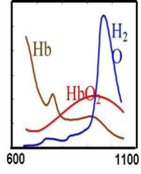
A near infrared (NIR) spectroscopy device (Kent, Calgary) using 4 wavelengths of light to determine Osaturation of hemoglobin (Hgb) in cutaneous tissue has been developed. The low absorption of light by H2O and melanin in the NIR range allows for measurement of absorption of deoxygenated and oxygenated hemoglobin and for the calculation of O2 saturation. The purpose of this study is to determine the correlation between the gold standard TCO2 and NIR spectroscopy in measuring cutaneous O2 .

Materials and methods: 20 patients with Fitzpatrick skin types I-III who had measurements of TCO2 (Perimed, Stockholm) also had simultaneous measurements obtained by NIR spectroscopy. The investigation was reviewed and approved by Institutional Review Board for Human Subjects at WVU. The Hemoglobin/ O2 dissociation curve at 370 degrees C, pH=7.4 and pCO2 =40 was used to calculate O2 saturations from measurement of TCO2 .

Results: Data pairs were analyzed using linear regression and the relationship y=0.93 x+5.35 with a correlation coefficient=0.92 and r2 =0.84 was calculated.

Conclusions: There was a significant correlation between measurements of tissue oxygenation using TCO2 and NIR spectroscopy. NIR spectroscopy has the advantage of not requiring skin contact and measurements can be taken even in the wound bed. Other advantages include: 1). Time (2 minutes vs. 90 minutes) and 2). Disposable cost ($150 –TCO2 vs. $0-NIR spectroscopy). 3). The ability to perform serial studies over time. Further studies are warranted to determine if this information is useful in determining response to HBOT or in predicting wound-healing trajectory

Robert E. Bowen, M.D.1*, Ginna Treadwell RN1 and Mark Goodwin RRT1


Article Image 1

Long-Term Survival of Korean Aortic Aneurysm Patients

The study aim is to determine factors affecting the long-term survival of subjects with Aortic Aneurysm (AA). The sample included 294 Korean patients aged ≥ 30 years who were hospitalized from 1994 through 2004. Diagnosis was confirmed in 294 AA subjects (18.4% in affected Coronary Artery Disease (CAD); 75.8% with abdominal only AA (AAA) and 24.2% with Thoracic AA (TAA)) by computed tomography angiography in Samsung Medical Center, Seoul, Korea. AA repair direct operation or percutaneous endovascular AA repair (Revascularized group) was performed for 60.3% of the patients. Death data were obtained from all participants between 1994 and 2009. The mean age of AA subjects was 68.7 (±8.1) years. The proportion of males was 82%. Five- and 10-year survival rates were 89.8% and 82.6%, respectively. The 5-and 10-year survival rates were 92.3% and 84.9% in revascularized group and 86.4% and 79.5% in non-revascularized group, respectively. Adjusted hazard ratios were 1.14 (95% Confidence Interval (CI) 1.06-1.22) in ages and 2.94 (95% CI 1.23-7.38) in smoking for AA. Age and smoking contributed to death in Korean AA patients. And the 10-year survival rate for AA patients in Korea was over 80%. Revascularized group shows higher survival rate than non-revascularized group.

Shin Yi Jang* and Duk-Kyung Kim


Article Image 1

Comparison of Flow Measurement by 4D Flow Magnetic Resonance Imaging and by Particles Image Velocimetry on Phantom of Abdominal Aortic Aneurysm

Predicting the rupture of Aortic Aneurysms is a complex problem that interests, from several decades, many researchers. The works on this issue are very complex, involving both the study of mechanical behavior of the artery as the flow of blood. The Magnetic Resonance Imaging (MRI) technique allows to obtain anatomic information of the arteries, than the flow inside thereof. The goal of this study is an inter comparison between flow data from MRI and those obtained by Particle Image Velocimetry (PIV). An experimental device simulating hemodynamic circulation is used. Initially in order to validate the device, the flow in a cylindrical glass tube is measured by these two techniques and then compared to a theoretical model. Secondly, the flow in a phantom in silicone, with an axisymmetric aneurysm, is evaluated with 4D flow MRI sequences and the measurements are compared with those obtained by PIV with good agreement. The ability of the MRI technique to measure the flow thus makes an essential device for the study of cardiovascular disease.

Yufei Wang1 , David Joannic1,2, Patrick Juillion1 , Alan Keromnes3 , Aurélien Monnet4 , Alain Lalande5 and Jean-François Fontaine1,2*


Article Image 1

Insights into Ovarian Follicle Angiogenesis: Morphological and Chronological Vascular Remodeling from Primordial to Ovulating Follicles

Ovarian function is dependent on the establishment and continual remodeling of a complex vascular system. The present review focuses on the ovarian morphological angiogenic processes that involve swine ovarian folliculogenesis and their modulatory molecular mechanisms: from the primordial follicles recruitment to the antral periovulatory stage. The process of angiogenesis, in particular, during the final stage of follicle development (from antral to periovulatory follicular phase) is deepened with a kinetic approach adopted by using an experimental prepubertal model where antral follicle’s selection, growth and ovulation are pharmacologically and timely controlled. Since the cyclicity of follicular angiogenesis is inducible under experimental conditions in several experimental and domestic animals using validated hormonal treatments, it may become a reproducible model to study in vivo molecular mechanisms and pathways involved in angiogenesis and blood vessel remodeling.

The present review has also focused the attention on the regulatory role of vascular endothelial growth factor A in controlling ovarian follicular development as orchestra conductor of the angiogenic process. The main animal model considered in the present review is the pig. The reproductive processes of this domestic animal is of a high translational value by considering the similarity with women of the long periovulatory window (40-44 hours from luteinizing hormone surge to ovulation). This animal model could become, for this reason, a valuable model in understanding the regulatory pathways involved in the final stage of follicular maturation.

Martelli A*, Russo V, Mauro A, Di Giacinto O, Nardinocchi D, Mattioli M, Barboni B and Berardinelli P


Article Image 1

Superficial Vein Thrombosis and Thromboembolic Events in a Patient Cohort undergoing Small Saphenous Vein Surgery

Background: The primary aim was to retrospectively assess the incidence of superficial vein thrombosis in a patient cohort that had undergone surgery of the Small Saphenous Vein (SSV) due to Chronic Venous Disease (CVD).

Material and Methods: 76 consecutive, unselected patients who had undergone primary SSV surgery for CVD over a four year time span were included in this retrospective study. Diagnosis of superficial vein thrombosis of the SSV and of Deep Venous Thrombosis (DVT) was made by means of patient history and preoperative Duplex Ultrasonography Findings (DUS).

Results: In 28 patients (36.8%) (36 legs) a condition after superficial vein thrombosis was diagnosed. Thromboembolic complications such as deep venous thrombosis and Pulmonary Embolism (PE) occurred in 11 of 76 patients (14.4%). All of these patients suffered from superficial vein thrombosis.

Conclusion: We conclude that studies on superficial vein thrombosis of the SSV and possible thromboembolic complications are rarely found in the literature. The morbidity associated with SSV insufficiency may be underestimated.

Heil Gerlinde S, Steinmueller Nancy, Mouton Kim and Mouton Wolfgang G*


Article Image 1

Management and Outcome of Abdominal Aortic Aneurysms in EndStage Renal Disease Patients: A Case Series

Aim: End-Stage Renal Disease (ESRD) patients on renal replacement therapy have increased cardiovascular morbidity and mortality rate and are at high risk for the development of Abdominal Aortic Aneurysms (ΑΑΑs). The aim of the present study was to investigate the management and outcome of ΑΑΑs in chronic hemodialysis patients.

Patients-Methods: During the period 2010-2015, an AAA was diagnosed in 19 ESRD patients. Thirteen out of them underwent Endovascular Aneurysm Repair (EVAR), 1 patient underwent Open Surgical Repair (OSR) and 5 patients were on regular follow-up.

Results: During the 6 year follow-up period, an endoleak was recorded in 7 out of the 13 aneurysms (36.8%). Endoleak was an early complication in one patient (14.3%) and late in six (85.7%) and was successfully managed with a repetitive EVAR in all cases. Five patients (26.3%) died including the patient who underwent OSR. The causes of death were sepsis in 3 patients (60%), device migration and aneurysm rupture in 1 patient (20%) and cerebrovascular accident in 1 patient (20%).

Conclusions: In end-stage renal disease patients with abdominal aortic aneurysms, EVAR is a safe therapeutic option with low both peri-operative and long-term morbidity and mortality.

E Liakou1,2, A Τsiantoulas1,2, S Fragidis2 , P Giamalis1 , M Tsiatsiou1, Κ Papazoglou3  and Α Papagianni1*