Yufei Wang1 , David Joannic1,2, Patrick Juillion1 , Alan Keromnes3 , Aurélien Monnet4 , Alain Lalande5 and Jean-François Fontaine1,2*
1Le2i FRE2005, CNRS, Arts et Métiers, Univ. Bourgogne-Franche Comté, France
2IUT Dijon-Auxerre, France
3DRIVE, Univ. Bourgogne-Franche Comté, France
4Siemens Healthcare, France
5Faculty of Medicine, University of Bourgogne-Franche Comte, France
Corresponding Author:
Jean-François Fontaine, Laboratory
of electronics, data processing and
image, University of Burgundy, IUT
Dijon-Auxerre, France,
Keywords
Abdominal Aorta Aneurysm;
Aorta phantom; Fluid velocity
measurement; Metrology in medical
Imaging; PIV; MRI
Abstract
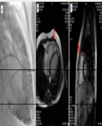
Predicting the rupture of Aortic Aneurysms is a complex problem that interests, from several decades, many researchers. The works on this issue are very complex, involving both the study of mechanical behavior of the artery as the flow of blood. The Magnetic Resonance Imaging (MRI) technique allows to obtain anatomic information of the arteries, than the flow inside thereof. The goal of this study is an inter comparison between flow data from MRI and those obtained by Particle Image Velocimetry (PIV). An experimental device simulating hemodynamic circulation is used. Initially in order to validate the device, the flow in a cylindrical glass tube is measured by these two techniques and then compared to a theoretical model. Secondly, the flow in a phantom in silicone, with an axisymmetric aneurysm, is evaluated with 4D flow MRI sequences and the measurements are compared with those obtained by PIV with good agreement. The ability of the MRI technique to measure the flow thus makes an essential device for the study of cardiovascular disease.
Citation
Wang Y, Joannic D, Juillion P, Keromnes A, Monnet A, Lalande A, et al.Comparison of Flow Measurement by 4D Flow Magnetic Resonance Imaging and by Particles Image Velocimetry on Phantom of Abdominal Aortic Aneurysm. SM Vasc Med. 2016; 1(2): 1008.