SM Case Reports

Archive Articles

Article Image 1

Unprecedented Brain Imaging Findings in a Case of Acute Disseminated Encephalomyelitis

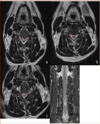
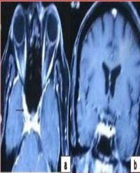
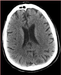
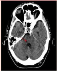
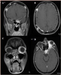
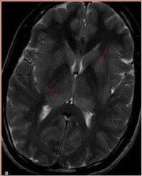
Acute Disseminated Encephalomyelitis (ADEM) is an acute widespread autoimmune demyelinating disease affecting central nervous system. It is characterized by multifocal white matter lesions on neuroimaging. Grey matter can also be affected, particularly basal ganglia, thalami, and brainstem. Variant Creutzfeldt - Jakob disease (vCJD) is a rare and fatal human neurodegenerative condition affecting younger patients. Pulvinar sign on Magnetic Resonance Imaging (MRI) is considered to be a strong indicator of variant CJD. We report a young patient who presented with febrile illness and altered mental state. Brain imaging revealed hyperintensity in bilateral medial and posterior thalamus. These imaging abnormalities are similar to those seen in variant CJD.

Mahale R¹, Mehta A¹, and Rangasetty S¹*


Article Image 1

Adult-onset progressive spastic paraparesis occurs commonly due to spinal cord pathology. Here, we report a middle-aged man who presented with adult-onset progressive spastic paraparesis of 10 year duration and MRI showed ‘snake-eye’ appearance in cervical cord posterior horn region. The probable cause for spastic paraparesis was sporadic Hereditary Spastic Paraparesis (HSP)

Rohan Mahale¹, Anish Mehta¹, Kiran Buddaraju¹, Abhinandan K Shankar¹, and Rangasetty Srinivasa¹*


Article Image 1

Cavernous Sinus Syndrome Causing Complete Ophthalmoplegia in Acute Herpes Zoster Ophthalmicus: A Rare Occurrence

Herpes Zoster Ophthalmicus (HZO) is associated with variety of ocular manifestations like episcleritis, keratitis, glaucoma, and cataracts. Extraocular muscle palsies are rare complications occurring in 7-31% of HZO. Complete unilateral ophthalmoplegia in HZO is rare. We report an elderly patient with left HZO who developed progressive left complete ophthalmoplegia with complete ptosis within 10 days of onset of a zosteriform rash. Magnetic resonance imaging brain showed contrast enhancement in cavernous sinus which has not been reported so far

Mahale R¹, Mehta A¹, and Rangasetty S¹*


Article Image 1

Delirium Tremens and Cardiovascular Collapse: A Case Report

Acute alcohol withdrawal syndrome leads to fluctuating changes in mental status. It also may cause cardiovascular collapse and death occasionally. We report a patient who dies due to cardiovascular complications of delirium tremens. Early clinical interventions such as hydration, sedation, preventing seizures by using benzodiazepines [1], infusion of dextrose and thiamine can be lifesaver. Hospitalization to Intensive Care Unit (ICU) must be provided immediately. Death is sometimes inevitable even if physicians perform an entire multidisciplinary clinical intervention. In this case, the situation of the patient, who suffers from alcohol withdrawal, worsens rapidly and he dies in two days during the follow-up in ICU.

Musa Kaya¹, Arif Duran¹, Mansur Kursad Erkuran¹, and Tarık Ocak²*


Article Image 1

Benign Vestibular Schwannoma: a Rare Cause of Nonaneurysmal Subarachnoid Hemorrhage

Background: Subarachnoid hemorrhage most commonly occurs secondary to trauma, aneurysms, or aggressive tumors. Benign tumors such as vestibular schwannomas are an uncommon cause of subarachnoid hemorrhage. Here we present a case of acute neurologic decline secondary to subarachnoid hemorrhage arising from a benign vestibular schwannoma.

Methods: The methods in this study include case presentation with operative findings and literature review.

Results: A 39 year old male presented with acute hearing loss, severe immediate headache, dizziness, facial numbness, and meningismus. Non-contrast head Computed Tomography (CT) demonstrated subarachnoid hemorrhage. CT angiogram and magnetic resonance imaging revealed a vascular tumor of the cerebellopontine angle with widening of the internal auditory canal, consistent with a vestibular schwannoma. The tumor was approached by retrosigmoid transmeatal craniotomy with complete surgical resection. The pathology revealed vestibular schwannoma.

Conclusion: Although extremely uncommon, benign tumors such as vestibular schwannomas may have an unusual presentation such as spontaneous subarachnoid hemorrhage.

Yusef Mosley¹, Angela Downes², Jamie Van Gompel³, and A Samy Youssef²*


Article Image 1

D

Background: The thalamic syndrome was described in 1906 by Déjerine and Roussy. It is characterized by transient hemiparesis, hemianaesthesia, allodynia, mild hemiataxia and severe pain on the affected side. The syndrome is related to lesions in the posterior nuclei of the thalamus, usually of vascular aetiology. The aim of this work is to report a patient with a central pain Déjerine-Roussy-like syndrome due to an ischemic lesion in the dorsal pons

Case report: A 69-year-old hypertense male was admitted in May 2015 with mild left hemiparesis and hemihypoesthesia. The brain Computed Tomography (CT) scan showed a small hypodense lesion in the tegmentum of the right hemipons. Seven months later, the patient was admitted again due to intense painful paresthesias on the left side of the body, including the face. A new brain CT scan showed the same pontine infarct. The patient was successfully treated with pregabalin (75 mg twice a day)

Discussion: The reported patient presented a Déjerine-Roussy like syndrome with a pontine topography of the vascular lesion. It is probable that the ischemic damage partially affected the medial lemniscus and the lateral spinothalamic tract. The diagnosis of the central neuropathic pain is fundamental to select the most adequate treatment.

Alvarez-Perez FJ*


Article Image 1

A Rare Case of Endometrial Adenocarcinoma with both Left and Right Cardiac Metastasis

Endometrial adenocarcinoma rarely metastasizes to the heart. Few reports in the literature have discussed such situations with this type of carcinoma. Most metastases to the heart originate from lung, breast, thyroid, or skin (melanoma). In most cases, only one heart cavity is involved. We report a rare case of a 66 year old woman with endometrial adenocarcinoma who has metastases to both the right and left ventricles

Ying T Sia*


Article Image 1

Monostotic Fibrous Dysplasia of Maxilla in a Postmenopausal Female- A Rare Case Report with Review of Literature

Fibrous dysplasia is a condition characterized by excessive proliferation of bone-forming mesenchymal cells. It can affect one bone (monostotic type) or multiple bones (polyostotic type). It is usually observed in adolescents and young adults and comprises 7% of benign bone tumors. The etiology is not clear but genetic predisposition is suspected. It has a predilection for long bones as well as the craniofacial skeleton. The maxilla is the most commonly affected facial bone, with facial asymmetry being the usual complaint. The diagnosis is based on radiological and histopathological examination. There are different treatment approaches including monitoring, medical treatment or surgery. A 45-year-old female reported with a complaint of painless swelling on the left side of maxilla since 1 year. A diffuse intraoral bony hard, non tender swelling was seen in the left maxilla involving the premolar-molar region. Plain film radiographs and Computed tomography revealed ground glass appearance of the left maxilla. The lesion was excised and on histopathology showed features of fibrous dysplasia. Very few cases of Fibrous dysplasia manifesting in the older and postmenopausal age group are reported in the literature. Once diagnosed, routine follow-up should be done on a yearly basis with x-ray examination

Deepa JP¹, Samriddhi S², Puri G³, Aravinda K⁴, Dixit A⁵, Gupta R⁶, and Aanchal G⁶*


Article Image 1

Sleep Paralysis and Decompression Sickness

We are presenting the case of a 38-year-oldpatientwith no significant medical history. He used to work as a professional diver but currently only partakes in recreational diving activities. Thirteen years ago, the patient reported self-limited para-paresis (24 hours) after diving, which improved after administering nor-mobaric oxygen and corticosteroids. This process was diagnosed as probable neurological Decompression Sickness (DCS).

Ortega-Albás JJ¹, Gomis Devesa AJ¹, Martínez A¹, and Salvador-Marín M²**


Article Image 1

Sarcoidosis Coexisting With Dermatomyositis

Sarcoidosis, a multisystem noncaseating granulomatous disease of unknown cause, has occasionally been associated with connective tissue diseases. African Americans and women have higher rates of sarcoidosis [1,2]. Its association with dermatomyositis is uncommon and most of cases are described in Japanese patients [3-13]. We report a patient with sarcoidosis who developed dermatomyositis

Rosa Gimenez- Garcia*


Article Image 1

Fabry

Fabry’s disease is a rare X-linked lysosomal storage disorder associated with potential multiorgan dysfunction. We describe Fabry’s disease in an adult patient presenting to the Rheumatology clinic, St John’s Hospital, West Lothian, Scotland with “Raynaud’s- like” phenomenon, rash and renal impairment.

Stephen Boyle¹ and Neil Douglas McKay¹*


Article Image 1

Vasculitis Presenting with Lower Extremity Myalgia

Giant Cell Arteritis (GCA) is the most common form of vasculitis in the adult population. It classically presents with new-onset headache in a patient older than 50 years, and reflects predominantly-intracranial vessel pathology. We describe a 67-year old man who presented with subacute myalgia of the lower extremities and constitutional symptoms, and was later found to have an extra-cephalic presentation of GCA. Magnetic Resonance Imaging of the lower extremities demonstrated diffusely enhanced signal intensity in the distribution of the quadriceps muscle, initially leading us to suspect an inflammatory myopathy. Muscle biopsy under radiographic guidance, however, was normal. Confronted with a middle-aged gentleman with non-specific symptoms and elevated laboratory markers of systemic inflammation, we performed a biopsy of the temporal artery and nailed the atypical, ‘silent’ form of GCA. The patient responded favorably to treatment with prednisone

Ohad Oren¹, Giris Jacob², Michal Oren³, Valerie Aloush⁴, and Jacob N. Ablin⁴*


Article Image 1

Erlotinib Induced Ischemic Colitis

Ischemic Colitis (IC) is a clinical entity that includes all secondary lesions due to circulatory anoxia of the wall of the colon and/or rectum. This complication may occur in connection with the use of many drugs. Rare forms were described in combination with chemotherapy are rather secondary to neutropenia than to chemotherapy itself. Ischemic colitis induced by Erlotinib use is an exceptional form. We describe in this report a case of ischemic colitis during treatment with Erlotinib.

Rachid Tanz¹, Tarik El Adddioui³, Mohammed Reda Khmamouch¹, Rachida Saouab⁴, Tarik Mahfoud¹, Bazine Aziz¹, Hassan Errihani², and Mohamed Ichou¹*


Article Image 1

Distinguishing Characteristics of Primary Retinal Vasculitis from Eales Disease

Purpose: To report on a case of a patient with primary retinal vasculitis.

Methods: Retrospective single case report.

Patient: A 27-year-old female presented with decreased vision in her left eye for the past year. On exam, her visual acuity for the left eye was 20/50. She has had trouble accessing medical care but denies any illness, infections or trauma.

Results: Upon ophthalmic examination, retinal abnormalities were seen such as the presence of peripheral vessel sheathing, peripheral non-perfusion and extensive neovascularization. Fluorescein angiography revealed significant late leakage of the retina vessels with greater severity in the left eye. Laboratory findings were unremarkable. After diagnosis of primary retinal vasculitis, the patient was treated with photocoagulation in both eyes. She also received two treatments of intravitreal bevacizumab for the neovascularization

Conclusion: Primary retinal vasculitis is often hard to diagnose due to disease course variation. The cause of retinal vasculitis must first be established as either an isolated condition or due to an inflammatory systemic condition. A thorough review of the patient’s medical history, review of systems, ophthalmic and physical examination, along with proper laboratory tests must be performed in order to ensure accurate diagnosis and treatment.

Courtney Crawford¹ and Yihharn Hwang²*


Article Image 1

Organophosphate (OP) Poisoning a Common Cause of Self-Harm: A Case Report

Now-a-days Organophosphate (OP) poisoning is a most common socio-medical issue in the evolving world. In 2012 suicide accounted for 1.4% of all deaths worldwide, making it the 15th leading cause of death. The Deliberate Self-Harm (DSH) tendency is the major reason for organophosphate poisoning. Organophosphate poisoning results from exposure to organophosphates (OPs) which extensively used as pesticides for agriculture in India. A case of OP poisoning was admitted in hospital and was treated with Atropine infusion for 10 days and Pralidoxime 9 days, after therapy for 12 days patient was survived. Based on patient illness, appropriate medication was given, patient signs and symptoms were reduced, and vital signs were controlled and discharged with good health.

Buchi Reddy¹, Maneesha Erraboina², Prathyusha Rani Thunla², Banothu Vishnu Priya², Beemarthi Navya², and Suresh Bandari³*


Article Image 1

Difficulty in Lumbar Puncture of a Patient With an Intrathecal Baclofen Pump and Possible Meningitis

Background: Patients with intrathecal baclofen pumps have a known increased risk of meningitis. Optimal antibiotic treatment targets the bacteria found in the Cerebro Spinal Fluid (CSF). However a lumbar puncture in patients with an intrathecal pump may result in damage to the spinal catheter by the needle and treatment failure.

Aims of the study: We report at case of a 32-year-old man with possible meningitis. He had spastic hemiplegia and epilepsy after a head trauma and was treated with baclofen via an intrathecal pump.

Methods: A CSF sample was needed for a specific diagnosis but the location of the spinal catheter hindered a safe lumbar puncture. A search of the current literature did not reveal that special equipment for drawing CSF directly from the pump was available. Therefore the patient was treated with antibiotics according to best practice.

Results: The lack of available literature on the subject and special equipment resulted in a suboptimal diagnosis. As a CSF sample was never obtained targeted antibiotic treatment was not possible.

Conclusion: Preferably the patient should be referred to a specialized hospital where a CSF sample can be drawn directly from the pump. Local hospitals should have the necessary equipment available or a standard procedure for referral to a specialist department for complicated cases. If this is not possible the authors suggest that patients with intrathecal pumps have an x-ray which may visualize the location of the spinal catheter during a lumbar puncture and use a small gauge needle. A post discharge pump checkup is also advised.

Elisabeth Arndal*


Article Image 1

Cystic and Calcified Retroperitoneal Sarcoma: A Very Rare Occurrence

Retroperitoneal liposarcoma is a primary malignant retroperitoneal tumor of mesenchymal cell origin mostly occurring after 40 yr of age. It is one of the most common primary retroperitoneal neoplasms. However retroperitoneal sarcomas are almost always solid, with rare case reports of cystic nature and extensive calcification in a cystic liposarcoma is still very rare. Here by presenting a rare case of Cystic and Calcified retroperitoneal liposarcoma patient who underwent exploratory laparotomy with excision of the lesion and on regular follow-up and disease-free since last 3 years.

Dillip Kumar Muduly¹, Kalinga Ketan Naik², Deepak Gupta³, and Atul Agarwal³*


Article Image 1

Unusual Presentation of Metastatic Bladder Cancer: Carcinomatous Meningitis

Classical metastasis sites of bladder transitional cell carcinoma are nodes, liver, lung and bone. The meningeal infiltration is exceptional and it’s reported only few case reports in the literature and the majority shows that many patients had the complication occur after successful treatment of their systemic disease. We report her new case report of meningitis carcinomatous revealing bladder cancer in young woman.

I. ElAlami¹, R. Tanz¹, M.A. Azami², M.A. Mokhlis¹, C. Elm’hadi¹, H. Errihani³, and M. Ichou¹*