Osteosynthesis of the Humerus: Indications, Techniques and Results in a Precarious Environment
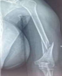
Introduction: Lesions of the humerus are relatively frequent and pose no diagnostic problem with the help of radiography. The real controversies concern the therapeutic indications, pitting the advocates of orthopaedic treatment against those of surgical treatment. Despite the risk of complications, surgery is an effective treatment for humeral damage. The aim of this work is to study the indications, techniques and results of osteosynthesis of humerus fractures and to share the results of our experience.
Methodology: This was a descriptive cross-sectional study collected retrospectively over a period of 4 years and 6 months in the Orthopaedic Traumatology Department of the CHU Yalgado Ouedraogo (Ouagadougou - B.F). It concerned all patients aged at least 15 years who had undergone osteosynthesis of the humerus for a proven injury, whether recent or long-standing. Anatomical results were assessed on the basis of radiographs. Functional results were assessed using the modified Constant, Stewart and Hundley scores, the MEPS and the patient’s subjective assessment. Ninety-three (93) patients were selected. Results: The average age was 37.86, with a sex ratio of 2.1. Road traffic accidents were the most frequent cause of death. The indications for osteosynthesis were dominated by recent fractures in 78.49% of cases. The majority of lesions were diaphyseal, accounting for 58.06% of cases (54 patients). The average operating time was 4.5 days. General anaesthesia was used in 83 patients. The screw plate was used in 60 cases. Forty-seven patients were able to undergo rehabilitation sessions. We recorded 31 cases of complications. Consolidation was achieved in 82 patients with an average consolidation time of 3.5 months. The anatomical result in our analysis was good in 78 cases, acceptable in 11 cases and poor in 4 cases.
Conclusion: The indications for osteosynthesis are becoming increasingly frequent. In our study, osteosynthesis of the humerus demonstrated its effectiveness, with appreciable anatomical and functional results.
Dabire MN2, Tinto S3, Ouedraogo I1*, Korsaga AS3, Ouedraogo Aji4, Sawadogo M1, And Tall M5