Research Article | Volume 2 - Issue 1 | Article DOI :
Download PDF
Troy H Maetani1 *, Stacy E Smith2 and Barbara N Weissman2
1 Department of Radiology, University of North Carolina-Chapel Hill School of Medicine, USA
2 Department of Radiology, Harvard Medical School, USA
Corresponding Author:
Troy H Maetani, Division of Musculoskeletal Imaging and Intervention, Department of Radiology, University of North Carolina-Chapel Hill School of Medicine, 101 Manning Drive, CB#7510 Old Clinic Bldg, Chapel Hill, NC 27515, USA, Tel: (919) 966-6646; E-Mail: troy_maetani@med.unc.edu
Keywords
Bisphosphonate;
Denosumab; Femur; Fracture;
Radiography
Abstract
Introduction: The study objective was to assess lateral femoral cortex variants that may mimic prefracture findings of drug-associated atypical femoral fractures (AFF) among hip radiographs.
Materials and Methods: Bilateral hip radiographs of 1493 consecutive patients (mean age 67.7, 804 women) were reviewed. Hips were positive if localized lateral subtrochanteric femoral cortical thickening (LSFCT) was present. Positive studies were divided into a medication group if history of bisphosphonate or denosumab use was present or a variant group. The medication group was subcategorized into a prefracture group if classing beaking LSFCT or a contralateral AFF was presentor a non-prefracture group. The LSFCT width, femoral head and lesser subtrochanteric distances were measured. Analysis of Variance (ANOVA) was performed (p <0.01) to compare the three groups, with post hoc Tukey HSD evaluation. Cross-sectional imaging for each group was reviewed.
Results: Of the1493 exams, 1079 were included. In the 24 patients with LSFCT, 8 patients were assigned to the medication group and 16 to the variant group. Of the 8, 3 met criteria for the prefracture group and 5 were subcategorized to the non-prefracture group. Differences among the prefracture versus the non-prefracture and variant groups were statistically significant (p<0.01). Cross-sectional imaging of the correlated LSFCT with the third Trochanter posterolaterally in the variant group and laterally for the prefracture group
Conclusions: The third trochanter is an anatomic variant that may mimic prefracture findings of drugassociated AFF. The third trochanter can be differentiated radiographically from prefracture AFF findings with a < 3 mm width and < 3mm lesser subtrochanteric distance.
Citation
Maetani TH, Smith SE and Weissman BN. Assessment of an Anatomic Variant That May Mimic Prefracture Findings of Drug-Associated Atypical Femoral Fractures on Conventional Radiographs: The Third Trochanter. SM J Clin Anat. 2018; 2(1): 1005.
Introduction
Bisphosphonates and Denosumab are very effective therapies for osteoporosis that reduce the risk of fragility fractures [1,2]. Drug-associated atypical femoral fractures (AFF) in the lateral subtrochanteric femur and diaphysis of the femur are potential rare adverse events of bisphosphonate and denosumab therapy, but they have also been documented in adults who had no exposure to these medications [3-7]. Due to the increased morbidity and mortality associated with drug associated AFF [8], the Task Force of the American Society of Bone and Mineral Research (ASBMR) established a clinical and radiographic definition for drug-associated AFF to delineate them from osteoporotic fractures [9]. To meet criteria, the fracture must be located along the femoral diaphysis distal to the lesser trochanter and proximal to the supracondylar flare and must meet four out of the f ive major features (Table 1)
Table 1: American Society of Bone and Mineral Research Task Force Case Definition of Atypical Femoral Fractures [9].
| S.No |
2013 Revised Major Features of Atypical Femoral Fractures |
| 1 |
The fracture is associated with minimal or no trauma, as in a fall from a standing height or less |
| 2 |
The fracture line originates at the lateral cortex and is substantially transverse in its orientation, although it may become oblique as it progresses |
| medially across the femur |
| 3 |
Complete fractures extend through both cortices and may be associated with a medial spike; incomplete fractures involve only the lateral cortex |
| 4 |
The fracture is noncomminuted or minimally comminuted |
| 5 |
Localized periosteal or endosteal thickening of the lateral cortex is present at the fracture site (“beaking” or “flaring”) |
Central to the major criteria features of drug-associated AFF is a fracture line originating in the lateral cortex and cortical (endosteal or periosteal) thickening [10-13] in patients with a history of bisphosphonate use.
Independent radiographic findings that do not meet the prescribed ASBMR criteria, such as cortical thickening without a fracture line, have been shown to likely represent an evolution of f indings that may develop into a drug-associated AFF over time [12,14-20]. Several studies have described these as prefracture findings of drug-associated AFF [9,21,22] that may lead to preemptive discontinuation of drug therapy. Moreover, if there is a cortical lucency and an incomplete fracture with pain, prophylactic nail fixation has been used [23,24]. Therefore, conditions that may mimic these findings are of clinical importance to prevent the need for more advanced imaging and therapeutic decisions related to the care of an AFF [25].
We have found lateral subtrochanteric femoral cortical thickening (LSFCT) in patient’s naïve to bisphosphonate and denosumab therapy. We hypothesized that the anatomic third trochanter (Figure 1),

Figure 1: 3D volume rendered CT image of the Third Trochanter (white arrow).
a bony prominence at the gluteus maximus tendon attachment on to the superior gluteal tuberosity, may mimic an AFF. The purpose of our study, therefore, was to assess the incidence and anatomic causes for LSFCT in patients with or without these treatments that may mimic prefracture findings of drug-associated AFF.
Materials and Methods
After Institutional Review Board approval, we retrospectively reviewed the bilateral hip radiographs of 1493 consecutive patients (age range 18 to 91, 804 women) referred to our hospital for radiographic hip evaluation from 2002 to 2014. The major indication for requested studies was “pain” (92%), with the remaining 8% of studies requested to evaluate for limited range of motion, infection, metabolic disorders, and metastatic work-up. Patients with a history of prior hip surgery, primary or metastatic bone lesions, or bone diseases that distorted femoral architecture (e.g. Paget’s disease, fibrous dysplasia) were excluded. For all patients, an anteroposterior (AP) and a frog lateral radiograph of the bilateral hips were obtained. For the AP view, standard technique included attempted internal rotation of the leg by about 15 degrees
A positive study was defined as any degree of localized LSFCT (Figure 2).

Figure 2: Categorization of patients found to have lateral subtrochanteric femoral cortical thickening.
For the purpose of this study, the LSFCT was considered to be a third trochanter, which was defined as an osseous protuberance or tubercle at the superior portion of the gluteal tuberosity [26]. All positive studies were confirmed by consensus of two experienced musculoskeletal radiologists and one musculoskeletal radiology fellow. All positive studies were then assigned to a medication group if the patient had a history of bisphosphonate or denosumab use or to a variant group if no medication history was present. Within the medication group, patients were subcategorized to a Prefracture group if the patient had radiographic findings of either classic beaking LSFCT or a contralateral drug-associated AFF as per ASBMR criteria (Figure 3)

Figure 3: Right hip radiographs of a 72-year-old woman with 6-year history of bisphosphonate use, with classic beaking cortical thickening (white arrow), assigned to the prefracture group.
or to a non-prefracture group if these findings were not present (Figure 4).

Figure 4: Right hip radiographs of a64-year-old woman with 5-year history of bisphosphonate use, non-beaking lateral subtrochanteric femoral cortical thickening (white arrow), and no contralateral drug-associated atypical femoral fracture, assigned to the non-prefracture group.
To characterize the shape and location of areas of lateral cortical thickening, the width of LSFCT was measured from the peripheral margin of the cortical prominence perpendicularly to a line drawn along the lateral margin of the distal normal lateral cortex (Figure 5). The distances from the superior margin of the femoral head to the midpoint of lateral cortical thickening and from the level of the bottom of the lesser trochanter to the midpoint of the cortical prominence were measured (Figure 5).

Figure 5: Coronal right hip CT. Measurements of the lateral subtrochanteric femoral cortical thickening width (*) and superior margin of the femoral head to mid cortical thickening (a) and level of the lower lesser trochanter to mid cortical thickening (b) distances
A one-way analysis of variance (ANOVA) was conducted to compare the three groups with post hoc comparisons performed with the Tukey HSD test.
Patient demographics were obtained from medical records with the type, dose,and duration of therapy in cases of bisphosphonate or denosumab use. All available cross-sectional imaging for positive studies were reviewed and anatomical causes of the radiographic
findings were determined in consensus by the same three radiologists. The location of the cortical thickening was noted and the presence of an attached tendon was determined. A third trochanter was identified on cross-sectional imaging as cortical thickening at the posterior or posterolateral aspect of the subtrochanteric proximal femur with clear attachment of the gluteus maximus tendon.
Results
Of the 1493 patient exams (2986 hip radiographs) reviewed, 1079 patients (2158 hip radiographs) met inclusion criteria. One thousand f ifty five patients showed no lateral cortical abnormality and were designated negative studies (97.8%). Twenty-four patients (2.2%) were found to have LSFCT (34 total hips; 14 unilateral, 10 bilateral) and were labeled as positive studies.
Sixteen of the 24 patients with LSFCT did not have a history of bisphosphonate or denosumab use and were assigned to the variant group (23 total hips; 9 unilateral, 7 bilateral). The remaining 8 patients had a history of drug therapy; 6 patients had a history of bisphosphonate use (mean 5.67 years) and 2 had used denosumab (total of 2 and 7 doses, respectively), with the 2 patients with a denosumab history having had no prior history of bisphosphonate therapy. Only 3 patients out of the 8 with a history of drug therapy were found to have a contralateral drug-associated AFF or classic beaking cortical thickening, (2 on bisphosphonate and 1 on denosumab therapy) and were assigned to the prefracture group, with a group total of 3 unilateral hips. The remaining 5 patients within the medication arm did not have a contralateral drug-associated AFF or classic beaking LSFCT and were assigned to the non-prefracture group (8 total hips; 2 unilateral, 3 bilateral)
Within the medication arm, the Prefracture group had a mean LSFCT width of 4.3 ± 1.2 mm, mean femoral head distance of 161.7 ± 3.5 mm and lesser subtrochanteric distance of 58.3 ± 11.6 mm. The non-prefracture group had a mean LSFCT width of 2.4 ± 1.2 mm, mean femoral head distance of 96.9 ± 7.1 mm and lesser subtrochanteric distance of 1.0 ± 2.8 mm.
In the variant group, the mean LSFCT width was 2.7 ± 0.8 mm,the femoral head and lesser subtrochanteric distances were 105.3 ± 17.3 mm and 3.9 ± 14.4 mm, respectively (Table 2)
Table 2: Comparison of Patients with Lateral Subtrochanteric Femoral Cortical Thickening
| |
Lateral Subtrochanteric Femoral Cortical Thickening |
| |
Reviewed |
Included |
Present |
Absent |
| # Of Patients |
1493 |
1079 |
24 |
1055 |
| # Of Hips |
2986 |
2158 |
34 |
2124 |
| (14 unilateral, 10 bilateral) |
| |
| |
Patient Drug Therapy History |
Total |
Measurements |
| All Positive Studies N=24) |
Bisphosphonate |
Denosumab |
None |
Patients |
Hips |
LSFCT |
Femoral Head |
Lesser Subtrochanteric |
| Width (mm) |
Distance (mm) |
Distance (mm) |
| |
Prefracture Group |
|
|
|
|
|
|
|
|
| Medication |
- Classic Beaking LSFCT, or |
2 |
1 |
0 |
3 |
3 (3 unilateral) |
4.3 ± 1.2† |
161.7 ± 3.5† |
58.3 ± 11.6† |
| |
Contralateral AFF |
|
(4 years duration) |
|
|
|
|
|
|
| |
Non-Prefracture |
4 |
1 |
0 |
5 |
8 (2 unilateral, |
2.4 ± 1.2† |
96.9 ± 7.1† |
1.0 ± 2.8† |
| |
Group |
(1 year duration) |
3 bilateral) |
| Non- Medication |
|
|
|
|
|
|
|
|
|
| Variant Group |
0 |
0 |
16 |
16 |
23 (9 unilateral, |
2.7 ± 0.8† |
105.3 ± 17.3† |
3.9 ± 14.4† |
| |
|
|
|
|
7 bilateral) |
|
|
|
Cross sectional imaging was available in 3 of the 3 patients in the prefracture group (3 total hips), 3 of the 5 patients in the non prefracture group (6 total hips) and 6 of the 16 patients (12 total hips) in the variant group. All cross-sectional imaging of the variant and non-prefracture groups showed that the localized cortical thickening corresponded to the superior aspect of the gluteal tuberosity (Figure 1), previously described as the third trochanter.
Based upon the ANOVA results, there was a statistically significant difference between the LSFCT [F(2,31) = 4.9, p = 0.01], femoral head distance [F(2,31) = 21.8, p = 0.01], and lesser subtrochanteric distance [F(2,31) = 26.7, p = 0.01], between the prefracture, non-prefracture and the variant groups. Post hoc comparisons using the Tukey HSD test indicated that the mean scores for the LSFCT and femoral head and lesser subtrochanteric distances were statistically significantly different between the variant and pre-fracture groups and the non prefracture and the pre-fracture groups, but the difference between the variant and non-prefracture groups was statistically insignificant. Taken together, these results suggest that the LSFCT within the medication non-prefracture group likely represent anatomic variation like the LSFCT seen in the non-medication variant group, independent of bisphosphonate or denosumab history and not indicative of the risk of AFF.
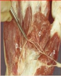
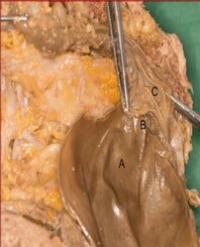
In all variant and non-prefracture group LSFCT cases, the third trochanter was located at the posterior or posterolateral aspect of the proximal femur with a distinct tendon insertion (Figure 6),

Figure 6: A. Axial CT and B. Axial MRI (Proton Density) showing localized cortical thickening at the superior aspect of the gluteal tuberosity at the ascending gluteus maximus tendon insertion (black arrow).
whereas LSFCT in the 3 hips from patients in the Prefracture group were all seen at the lateral aspect of the proximal femur, with no discernible tendon insertion (Figure 7).

Figure 7: Zone A. Posterior to Posterolateral region of third trochanter. Zone B. Lateral region of prefracture cortical thickening
The incidence of this third trochanter was 1.9% on radiographic examination
Discussion
The current recommended guidelines for drug-associated AFF include discontinuation of anti-resorptive medication, limited weight bearing and avoidance of vigorous activity [9], if bone edema is present in these patients. These recommendations are based on several publications that have described the evolution of drug associated AFF in patients who present with periosteal thickening without a fracture line, who then develop a fracture line or the “dreaded black line” [16] within the cortical thickening, that can lead to an incomplete or complete AFF [12,14,16-18,27]. Thus, in patients with LSFCT without a fracture line, as was true in all of our positive studies designated to the Prefracture group, cross-sectional magnetic resonance imaging (MRI) is recommended as a gold standard to evaluate for an associated fracture line or for bone marrow edema [28].
Diagnosis of an AFF can be challenging and is usually made by a combination of imaging findings and clinical history. Clinical symptoms of groin or femur pain and concurrent use of bisphosphonates or denosumab may prompt a physician to obtain skeletal imaging to assess the uncommon presence of an AFF. As shown in this study, a normal variant (the third trochanter) may importantly mimic prefracture findings of AFF. As one of the major criteria for drug associated AFF designated by the ASBMR is cortical thickening of the lateral subtrochanteric femur, it is essential to distinguish this abnormal cortical thickening from normal variation. In our cases, an area of cortical thickening provided a mimic for the changes of impending fracture and was seen in 1.9% of consecutive femur radiographs. This oblong, rounded or conical bony excrescence, described as the “third trochanter”, is located at the superior aspect of the gluteal tuberosity [26,29-31], where the ascending slip of the gluteus maximus tendon attaches (Figure 1). Awareness of the third trochanter can help distinguish between LSFCT that is an anatomic variant and cortical thickening that is a pathologic entity in order to guide patient care. The incidence of the third trochanter is 1.9% in our series and 6.2%-6.6% in other series [26,29] and is felt to increase the surface area of tendon attachment which results in greater efficiency of gluteus maximus muscle contraction [29]. This mechanical loading can alter the surface morphology, leading to variations of the third trochanter surface such as bony crests, ridges and tuberosities.
Limitations of this study are inherent in the retrospective analysis design and small numbers of positive cases. Given the retrospective review, patient position and degree of internal rotation on radiographic evaluation was variable which potentially altered visualization of the third trochanter even if present. It is anticipated that greater degrees of internal rotation would provide improved visualization of the third trochanter. Therefore, the true incidence of this finding is difficult to determine based on this study. Also, Bolanowski et al. defined the third trochanter as a tubercle located lateral to the line connecting the top of the greater trochanter with the superior bifurcation of the linea aspera and measurable features including a length/width ratio ≤ 5,and a height/width ratio > 0.05. Given the two dimensional nature of the conventional radiographs, the third trochanter could not be delineated in relation to the linea aspera as cross-sectional imaging was not available in all cases, and the height could not be measured. Future studies with three-dimensional capabilities such as computed tomography (CT) could be utilized to meet these criteria and to obtain these ratios.
The gluteus maximus tendon insertion at the gluteal tuberosity is also a possible location for other conditions that may be painful or produce abnormal morphology. Calcific tendinitis has been shown to affect the subtrochanteric femur posteriorly along the linea aspera at the level of or up to 6 cm below the lesser Trochanter [32]. Although solid periosteal reaction can be seen in calcific tendinitis, no other commonly associated radiographic findings such as soft tissue calcifications, tendinopathy or osseous erosions [32,33] were seen in the positive cases in the variant and non-prefracture groups in the current study to suggest that this was a result of calcific tendinitis rather than an anatomic variant
Conclusion
One entity that may mimic prefracture findings of AFF as described in bisphosphonate and denosumabtreated patients as well as adults without exposure to these medications is the anatomic variant of the third trochanter. Based upon our results, the third trochanter is characterized by its posterolateral location, a width less than 3mm and a distance of less than 3mm from the lesser trochanter, whereas prefracture findings of atypical femoral fractures are typically laterally positioned, with a width greater than 3 mm and more distal to the lesser trochanter. In questionable cases, MR imaging will clearly differentiate between these entities.
References
1. Black DM, Cummings SR, Karpf DB, Cauley JA, Thompson DE, Nevitt MC, et al. Randomised trial of effect of alendronate on risk of fracture in women with existing vertebral fractures. The Lancet. 1996; 348: 1535-1541.
2. Nakamura T, Matsumoto T, Sugimoto T, Hosoi T, Miki T, Gorai I, et al. Clinical Trials Express: fracture risk reduction with denosumab in Japanese postmenopausal women and men with osteoporosis: denosumab fracture intervention randomized placebo controlled trial (DIRECT). The Journal of Clinical Endocrinology and Metabolism. 2014; 99: 2599-2607.
3. Capeci CM, Tejwani NC. Bilateral Low-Energy Simultaneous or Sequential Femoral Fractures in Patients on Long-Term Alendronate Therapy. Journal of Bone and Joint Surgery. 2009; 91: 2556-2561.
4. Giusti A, Hamdy NAT, Papapoulos SE. Atypical fractures of the femur and bisphosphonate therapy: A systematic review of case/case series studies. Bone. 2010; 47: 169-180.
5. Goh SK, Yang KY, Koh JSB, Wong MK, Chua SY, Chua DTC, et al. Subtrochanteric insufficiency fractures in patients on alendronate therapy: a caution. The Journal of Bone and Joint Surgery. 2007; 89: 349-353.
6. Kwek EBK, Goh SK, Koh JSB, Png MA, Howe T Sen. An emerging pattern of subtrochanteric stress fractures: a long-term complication of alendronate therapy? Injury. 2008; 39: 224-231.
7. Neviaser AS, Lane JM, Lenart BA, Edobor-Osula F, Lorich DG. Low energy femoral shaft fractures associated with alendronate use. Journal of Orthopaedic Trauma. 2008; 22: 346-350.
8. Lo JC, Huang SY, Lee GA, Khandelwal S, Khandewal S, Provus J, et al. Clinical correlates of atypical femoral fracture. Bone. 2012; 51: 181-184.
9. Shane E, Burr D, Abrahamsen B, Adler RA, Brown TD, Cheung AM, et al. Atypical subtrochanteric and diaphyseal femoral fractures: second report of a task force of the American Society for Bone and Mineral Research. Journal of Bone and Mineral Research. 2014; 29: 1-23.
10. Feldstein AC, Black D, Perrin N, Rosales AG, Friess D, Boardman D, et al. Incidence and demography of femur fractures with and without atypical features. Journal of Bone and Mineral Research. 2012; 27: 977-986.
11. Koeppen VA, Schilcher J, Aspenberg P. Atypical fractures do not have a thicker cortex. Osteoporosis International. 2012; 23: 2893-2896.
12. Schilcher J, Aspenberg P. Incidence of stress fractures of the femoral shaft in women treated with bisphosphonate. Acta Orthopaedica. 2009; 80: 413-415.
13. Schilcher J, Michaëlsson K, Aspenberg P. Bisphosphonate use and atypical fractures of the femoral shaft. The New England Journal of Medicine. 2011; 364: 1728-1737.
14. Ahlman MA, Rissing MS, Gordon L. Evolution of bisphosphonate-related atypical fracture retrospectively observed with DXA scanning. Journal of Bone and Mineral Research. 2012; 27: 496-498.
15. Koh J, Goh S, Png M. Distribution of atypical fractures and cortical stress lesions in the femur: implications on pathophysiology. Singapore Med J. 2011; 52: 77-80.
16. Koh JSB, Goh SK, Png MA, Kwek EBK, Howe T Sen. Femoral cortical stress lesions in long-term bisphosphonate therapy: a herald of impending fracture? Journal of Orthopaedic Trauma. 2010; 24: 75-81.
17. McKiernan FE. Atypical femoral diaphyseal fractures documented by serial DXA. Journal of Clinical Densitometry. 2010; 13: 102-103.
18. Powell D, Bowler C, Roberts T, Garton M, Matthews C, McCall I, et al. Incidence of serious side effects with intravenous bisphosphonate: a clinical audit. QJM : Monthly Journal of the Association of Physicians. 2012; 105: 965-971.
19. Sayed-Noor AS, Sjödén GO. Case reports: two femoral insufficiency fractures after long-term alendronate therapy. Clinical Orthopaedics and Related Research. 2009; 467: 1921-1926.
20. Ward WG, Carter CJ, Wilson SC, Emory CL. Femoral stress fractures associated with long-term bisphosphonate treatment. Clinical Orthopaedics and Related Research. 2012; 470: 759-765.
21. Chan SS, Rosenberg ZS, Chan K, Capeci C. Subtrochanteric femoral fractures in patients receiving long-term alendronate therapy: imaging features. AJR: American Journal of Roentgenology. 2010; 194: 1581-1586.
22. Porrino JA, Kohl CA, Taljanovic M, Rogers LF. Diagnosis of proximal femoral insufficiency fractures in patients receiving bisphosphonate therapy. AJR: American Journal of Roentgenology. 2010: 194: 1061-1064.
23. Banffy MB, Vrahas MS, Ready JE, Abraham JA. Nonoperative versus prophylactic treatment of bisphosphonate-associated femoral stress fractures. Clinical Orthopaedics and Related Research. 2011; 469: 2028-2034.
24. Oh CW, Oh JK, Park KC, Kim JW, Yoon YC. Prophylactic nailing of incomplete atypical femoral fractures. The Scientific World Journal. 2013.
25. Shane E, Burr D, Ebeling PR, Abrahamsen B, Adler RA, Brown TD, et al. Atypical subtrochanteric and diaphyseal femoral fractures: Report of a Task Force of the American Society for Bone and Mineral Research. Journal of Bone and Mineral Research. 2010; 25: 2267-2294.
26. Bolanowski W, Śmiszkiewicz-Skwarska A, Polguj M, Jédrzejewski KS. The occurrence of the third trochanter and its correlation to certain anthropometric parameters of the human femur. Folia Morphologica. 2005.
27. Mohan PC, Howe TS, Koh JSB, Png MA. Radiographic features of multifocal endosteal thickening of the femur in patients on long-term bisphosphonate therapy. European Radiology. 2013; 23: 222-227.
28. Blood T, Feller RJ, Cohen E, Born CT, Hayda R. Atypical Fractures of the Femur: Evaluation and Treatment. JBJS Reviews. 2015; 3.
29. Ghosh S, Sethi M, Vasudeva N. Incidence of third trochanter and hypotrochanteric fossa in human femora in Indian population. OA Case Reports. 2014; 3: 14.
30. Hrdlička A. The Gluteal Ridge and Gluteal Tuberosities (3rd Trochanters). American Journal of Physical Anthropology. 1937; 23: 127-198.
31. Lozanoff S, Sciulli PW, Schneider KN. Third trochanter incidence and metric trait covariation in the human femur. Journal of Anatomy. 1985; 143: 149-159.
32. Flemming DJ, Murphey MD, Shekitka KM, Temple HT, Jelinek JJ, Kransdorf MJ. Osseous involvement in calcific tendinitis: A retrospective review of 50 cases. American Journal of Roentgenology. 2003; 181: 965-972.
33. Hottat N, Fumière E, Delcour C. Calcific tendinitis of the gluteus maximus tendon: CT findings. European Radiology. 1999; 9: 1104-1106