Back to Journal

Annals of Burns and Trauma

Eikenella corrodens Wound Infection Secondary to Self-Bite: Case Report

[ ISSN : 3068-0905 ]

Abstract Citation Introduction Case Report Discussion Acknowledgement References
Details

Received: 16-Dec-2017

Accepted: 30-Dec-2017

Published: 02-Jan-2018

Kuzucu EA, Çalışkan E, Bayrak ZS, Öcal D, Çağatay M, Saltaş H and Erdem G*

Department of Medical Microbiology, University of Health Sciences, Turkey

Corresponding Author:

Erdem G, Department of Medical Microbiology, University of Health Sciences, D??kap? Y?ld?r?m Beyaz?t Training and Research Hospital, Sehit Omer Halisdemir Caddesi, No:12, 06110, Altindag, Ankara, Turkey, Tel: +90 312 5962680; Email: gul-bahar93@hotmail.com

Keywords

Eikenella corrodens; Human Bite; Wound Infection

Abstract

Since Eikenella corrodens is part of the normal oral flora, it can be isolated from fist fight and human-bite injuries. Suspicion of the clinician, collection of adequate specimen, and proper culture technique will improve its isolation in the laboratories. Here we report a case of wound infection due to Eikenella corrodens in a 27-year-old female patient after biting herself during examination at the emergency ward.

Citation

Kuzucu EA, Çalışkan E, Bayrak ZS, Öcal D, Çağatay M, Saltaş H, et al. Eikenella corrodens Wound Infection Secondary to Self-Bite: Case Report. Ann Burns and Trauma. 2018; 2(1): 1005.

Introduction

Eikenella corrodens is a slow-growing, fastidious, nonmotile, facultative anaerobic gram negative bacillus. It is normally found in the flora of oral cavity, upper respiratory, gastrointestinal and genitourinary tracts [1,2]. Although previously believed to be non pathogenic,Eikenella species have been shown to cause various infections in human such as; pulmonary infection, empyema, arthritis, endocarditis, pancreatic abscesses, human and animal bite wound infection [1,3,4]. In many studies E.corrodens has been detected as the causative agent resulting in infections due to human bites. Although human bites are less common than animal bites, injuries caused by human bites must not be underestimated due to high risk of serious complications [1,5]. The bacteriology of human bite wounds reflects aerobic and anaerobic normal oral flora [6].

Because of the overgrowth of fast growing organisms those may be found on the surface of the wounds,Eikenella species can be missed. So during examination of wounds or abscesses after human bites, it must be kept in mind that E.corrodens may be the suspected agent and it should be treated immediately.

Here we report a case of wound infection due to E.corrodens in a patient, with psychiatric disorder, after biting herself during examination at the emergency department.

Case Report

A 27-year-old female patient with no previous history or medication of psychiatric disorder applied to the emergency service of University of Health Sciences, Dışkapı Yıldırım Beyazıt Training and Research Hospital with complaints of nonsense speech, nervousness, self-harm and harm to surrounding people. During examination at the emergency service, she bit the tip of her left thumb seriously. After several blood analyses and radiologic examinations she was hospitalized at psychiatry clinic.

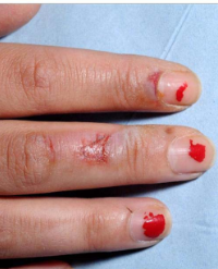
On the second day of hospitalization a malodorous discharge developed on the bite wound. Plastic surgery was consulted (Figure 1A,1B).

Figure 1A: Patients thumb on the second day.

Figure 1B: Patients X Ray on the second day.

Plastic surgery planned reconstructive surgery, dressing was done, elevation and tetanus prophylaxis were suggested, amoxicillin-clavulanic acid 1000 mg orally every 12 hours was started as antimicrobial therapy.

Because of the lack of healing in the lesion, infectious diseases and plastic surgery sections were again consulted. Wound culture swab specimens, which were not collected in the first visit, were collected by recommendation of infectious diseases clinician and ampicillin-sulbactam (2 g ampicillin+1g sulbactam) IVq 6hr for 5 days regimen was started.

Gram-stained smears of the swab specimens revealed; few epithelial cells, few Polymorphonuclear Leukocytes (PMNL), few gram positive cocci and gram negative bacilli.

The specimen was plated on Columbia agar with 5% sheep blood (BDTM,Becton Dickinson), Eosin-Methylene-Blue (EMB, BDTM,) and chocolate agars (BDTM), at the microbiology laboratory, and incubated for 48 hrs at 5-10% CO2 ambient air at 37°C.

At the end of 48 hrs incubation, no growth was observed on EMB agar plate, on the other hand small pitting colonies with bleach like odor and greenish discoloration around, were grown on blood agar plates. Gram negative thin bacilli were seen on Gram stain (Figure 1C,1D).

Figure 1C: Small pitting and oxidase positive colonies.

Figure 1D: Gram negative bacilli on Gram stain.

Results of the biochemical tests were as follows; catalase, indole, urease, and motility tests negative, oxidase test positive. Identification of the strain to the species level was done by both BD Phoenix™(Becton Dickinson,USA) automated system and MALDI-TOF mass spectrometry (Bruker,Germany), and detected as E.corrodens which was also suspected before, with its pitting and bleach like odor colonies. After ampicillin-sulbactam (2 g ampicillin+1g sulbactam) IV q 6hr for 5 days regimen, treatment was continued with 10 days of amoxicillin-clavulanic acid (1000mg orally every 12 hours) therapy.

The patient whose psychiatric symptoms receded was discharged with a diagnosis of dissociative disorder and proposal of infectious diseases, psychiatry, neurology outpatient clinic control.

Oral consent was approved for use of patient’s photos.

Discussion

Although being an inhabitant of the oral cavity and upper respiratory tract, E.corrodens may lead to serious infections after injuries, particularly human bites and fist fight injuries. Stoloff et al., published a series of 33 cases,18(54.5%) of which were due to human bites and fist injuries. In our case, E.corrodens was isolated also from a wound as a result of human bite [2].

Although declined, still 5% of the patients undergo amputation because of serious complications in human bite traumas [6].

To avoid a delay in diagnosis, the clinician must suspect of an infection of E. corrodens if the history of the patient harbours such truma. Isolation depends on collection of adequate specimen, and carefull evaluation of the plates of wound cultures at least after 48hrs of incubation [2].

E.corrodens belong to “AACEK” (formerly “HACEK”) group organisms. This abbreviation stands for Aggregatibacter aphrophilus (formerly Haemophilus aphrophilus and H.paraphrophilus), Aggregatibacter actinomycetemcomitans (formerly Actinobacillus), Cardiobacterium hominis, E.corrodens, and Kingella kingae and other Kingella spp [7]. Since HACEK microorganisms are fastidious, antimicrobial susceptibility testing is impractical for many laboratories, and published reports and guidelines usually guide antimicrobial selection of the clinican [8]. The organism is typically susceptible to many antibiotics, including penicillin G,ceftriaxone, ampicillin, amoxicillin–clavulanic acid, trimethoprim sulfamethoxazole, and fluoroquinolones, but resistant to oxacillin,cefazolin,clindamycin, and erythromycin [3,6,7,9].

E.corrodens is less fastidious, compared with the other organisms in the AACEK group, and may generally be recovered within 24 to 48 hrs on routine blood and chocolate agar media, as in our case [7]. Despite initiation of a broad spectrum, amoxicillin-clavulanic acid, therapy, covering both aerobic and anaerobic flora members of oral cavity, as the severity of symptoms increased, to prevent a possible complication of osteomyelitis, treatment was changed to parenteral ampicillin-sulbactam in this case.

Management of such injuries include immediate multidisciplinary approach; surgery, infectious diseases and laboratory disciplines must act together as in this case [1,10]. Surgical debridement, isolation of the organism and administration of accurate antimicrobial therapy improved the wound healing of our patient.

Acknowledgement

This study is presented at 10th Balkan Congress of Microbiology,16-18 November 2017, Bulgaria.

References

1. Sula B, Tekin R. Soft tissue abscess due to Eikenella corrodens after human bite. Journal of Microbiology and Infectious Diseases. 2016; 6: 36-37.

2. Stoloff AL, Gillies ML. Infections with Eikenella corrodens in a general hospital: A report of 33 cases. Reviews of Infectious Diseases. 1986; 8: 50-53. 

3. Paul K, Patel SS. Eikenella corrodens infections in children and adolescents: case reports and review of the literature. Clinical Infectious Diseases. 2001; 33: 54-61.

4. Yakut N, Kepenekli Kadayıfçı E, Karaaslan A, Atici S, Akkoc G, Ocal Demir S, et al. Deep neck infection caused by Eikenella corrodens coinfection with Streptococcus mitis and Streptococcus oralis in an infant. Turkish Journal of Pediatric Disease. 2017; 3: 203-205.

5. Raval P, Khan W, Haddad B, Mahapatra AN. Bite injuries to the hand - review of the literature. The Open Orthopaedics Journal. 2014; 8: 204-208.

6. Goldstein EJC. Bite wounds and infection. Clinical Infectious Diseases. 1992; 14: 633-640.

7. Mandell, Douglas, and Bennett’s Principles and Practice of Infectious Diseases (Eighth Edition). Eikenella corrodens. 2015.

8. Coburn B, Toye B, Rawte B, Jamieson FB, Farrell DJ, Patel SN. Antimicrobial susceptibilities of clinical isolates of HACEK organisms. Antimicrobial Agents and Chemotherapy. 2013; 57: 1989-1991.

9. Billingsley EM. Paronychia Organism-Specific Therapy. Medscape Drug Reference. 2016.

10. Tricarda T, Bunda L, Alhefzia A, Lemoine JP, Schneider L, Karger C, et al. Eikenella corrodens bone and hipjoint infection. A case report and literature review. Arch Pediatr. 2016; 23: 1146-1149.

Other Articles

Article Image 1

Skin Reaction on Face Following the Use of Cloves (Syzygium aromaticum)

Clove (Syzygium aromaticum) (Figure 1) is a plant-derived spice that has been traditionally used for centuries as food preservative and as medicinal plants [1].

The anti-inflammatory, antioxidant and bactericide activities of clove are mainly due to its major components, which is the eugenol with a concentration rate ranging from to (77-95%) [2,3].

Clove is generally safe when taken in foods in lower concentrations [4], however, it is not recommended as a topical application on skin due to insufficiency of safety and toxicity data [5]; it was found to be highly cytotoxic for human fibroblasts and endothelial cells [6] leading to allergic skin reactions (burning, hives, itching, irritation, rash…), ulcer formation and/or tissue necrosis [7].

Samir El Mazouz1, Abdelmoughit Echchaoui1*, Narjis Badrane2 and Majda
Askour3


Article Image 1

Risk Factors for Infection and Mortality in Burned Children: Where are we going?

Burns are very frequent and affect approximately 1% of the general population every year. The
immunocompromising effects of burns, hospital stay; diagnostic and therapeutic procedures put
those patients at increased risk of morbidity and mortality. Pediatric burn patients are susceptible
to a broad spectrum of infections representing the most common and severe complication in this
population.

Over the last few years patient survival after burn injury has increased; however, despite advances
in the management of burn patients, infections remain the most common cause of morbidity and
mortality.

Rosanova MT*, Stamboulian D and Lede R


Article Image 1

Chemical Burn Injury Secondary to Airbag Deployment: A Case Report and Literature Review

Background: Burn wounds caused by airbags deployment are infrequent, and with the increasing use of these safety feature in cars the documentation and sharing of the information related to these injuries is important to help improve the design of airbags and give the treating clinicians some background knowledge when treating injuries of similar nature.

Case presentation: This is a case report of lady who sustained a chemical burn injury to her finger following the deployment of airbags when she had a frontal car collision. The pH of the wound has helped to establish the pathophysiology of the wound.

Conclusion: Thorough history and examination help to detect minor injuries which can be overlooked otherwise. Airbags are associated with both friction and chemical burns, and pH of the burn wound helped in differentiating between them, and decides on first aid and appropriate management and follow ups.

Diaa Othman*, Farhan Akram and Mohammad Anwar


Article Image 1

MRI-Induced Burns from ECG Leads: Thermal or Electrical Burns?

Burns are a common global cause of injury; however iatrogenic burns are rare and preventable mode of injury. We report two cases who sustained superficial partial thickness burns from ECG monitoring leads during Magnetic Resonance Imaging (MRI). The two patients have sustained 0.2% and <1% superficial partial thickness burn of their Total Body Surface Area (TBSA) respectively. Common features included a relatively long duration of spine MR scanning (90 and 30 minutes) and high Body Mass Index (BMI). Both patients made uneventful recovery and were discharged within 24 hours of admission. It is still uncertain whether injuries were thermal or electrically-induced, however several mechanisms have been implied. These include electromagnetic inductionheating, the antenna effect and current induction through a closed loop. In this report, we discuss the proposed mechanisms of injury and highlight the fundamental need for staff education, and preparing national guidelines and safety checklists to prevent similar injuries in the future.

Othman D*, Abdel-Rehim S and OBoyle C


Article Image 1

Epidemiology of Burns Patients in a Tertiary Care Hospital in South India -A Retrospective Analysis

Burns are major health hazards which are associated with high mortality and morbidity rates. Burns management in developing countries is a major challenge due to inadequate access to burn care facility along with poorly equipped health care system. Although we are faced with a high burden of burns in our country, appropriate research on burns is still inadequate. We present our data of burns patients from a single center tertiary care hospital in south India.

Aim: To analyze various demographic characteristics, clinical and microbiological profile along with outcome of all burns patients admitted to our hospital.

Methods: This study was a retrospective analysis of burns patients admitted to the Critical Care Unit of Apollo Speciality Hospital, Vanagaram and a tertiary care facility in Chennai over a period of 3 years. Data such as age, gender, co-morbidities, type and degree of burns, percentage of burns and length of stay, mortality rate and infection rate were analyzed.

Results: There were a total of 94 burns patients included in the study. Amongst these, 61 patients (65%) were males and 33 patients (35%) were females. A majority of our burns population, 72% (n=68), belonged to age group from 21-50 years with a mean age of 40.50 years (SD±17.18).Mean total body surface area involved among burns was 48.56 (SD±21.08). Thermal burns were the commonest type of burns seen in 70%patients (n=66). These included flame burns in 49% patients (n=46) and scald burns in21% patients (n=20).Patients who presented to us within 6 hours post burns were 46% (n=43). Around 60% (n=56) patients had a hospital stay duration of ≤ 2 weeks. Infection rate among our patients was 62.8% and a mortality rate of 37% (n=35) was observed.

Conclusion: We highlight key demographic, clinical and microbiological data of all burns patients from a single center tertiary hospital in south India. This knowledge should help develop better strategies for management and prevention of mortality due to burns.

Ebenezer R1*, Rohit V1 , Isabella P2 , Nagarajan Ramakrishnan1 and Ganapathy

Krishnan3


Article Image 1

Pattern of the Burn Wounds Infections in Bahrain Defence Force Military Hospital

Background: Loss of skin as the first line of defence suppress immune system, extend hospital stay and makes burn patients more vulnerable to acquire healthcare associated infections. Such infections become the reason for mortality and morbidity in burn patients. Therefore, continuous monitoring of the pattern of nosocomial infection and drug resistance is necessary for assuring patient safety and healthcare quality improvement.

Aim: To describe the microbial profile of infections among burn patients admitted to Bahrain Defence Force Military Hospital.

Method: This is a retrospective cohort study involving 295burn patients. Wound swab method was used to isolate the microorganisms first on admission and then on suspicion of infection. The overall percentage of wound culture positive cases was 11.8% and the wound infection percentage was 5% of the total number of patients admitted to the unitA total of 126 swabs was performed from the burn wounds.

Results: Gram+ bacteria S.aureus was predominant in initial cultures. It was however, succeeded by Gram- bacteria, Pseudomonas, from third culture onwards. Fungal infection with Candida was most prevalent (n=21,16.7%). With TBSA >30% polymicrobial growth was observed which tend to increase with increase length of hospitalization. A greater proportion of patients (42.9%, n=15) had acquired healthcare associated infections and was negatively associated with increased length of hospitalization(r=-0.418, p=0.012).

Conclusion: 5% of the total patient population was categorized as health care associated infection cases. Gram negative bacteria, Pseudomonas were the prevalent microorganism isolated from wound infection of the burn patients.

Mohammad Abdulaziz Dahag, Nayef A Louri*, Nigamananda Dey and Siji Susan Philip


Article Image 1

Mesenchymal Stem Cells and Regenerative Quantum Medicine: The Novel A, B and C of Burns Treatment

Large burns are extensive soft tissue injuries with severe deep skin damage associated with high mortality [1-2]. This type of lesions can also lead to mental distress and chronic ilness but specially to scars, contractures, limitation of motion and bad quality of life [3-5]. Treatment of large burns as well as chronic nonhealing wounds have always been difficult medical problems and many different methods have been used to treat such injuries [6-7]. Current wound treatments are ineffective in many cases, so alternative novel types of therapy are needed and should urgently be understood and correctly explored. Abnormal damaged body environments can promote wound healing and tissue regeneration by local biochemical “alarm signals”, but usually these innate mechanisms are not enough to reach complete restoration of normal skin, specially in an acceptable time [8-12].When these impairments in wound healing arise, it is also often followed by an increased susceptibility to infection, further complications, failure of other organs and death [13-14]. In these circumstances, time management should be a crucial fundamental issue to be considered. In the near future we will not only ask for better novel therapeutic approaches but also for those that could lead to complete skin healing, restoration of function and acceptable aesthetics in the shortest period of time [ 15-16]. The development of tissue engineering and living skin substitutes started more than 40 years ago with the in vitro culture of keratinocytes as established by Howard Green in 1975 [17-18]. His outstanding discoveries led to the possibility of replacing the epidermis of extensively burned patients by using cultured keratinocytes autografts [19].

Mansilla E1*, Drago H2 , Marín GH1 , Jorrat R2 and Sturla F2


Article Image 1

Fatal Multi-Organ Failure Following Occupational Chemical Burn after Exposure to Phenolic Acid: A Case Report and Literature Review

Aim: Describe the complications and management of a rare case of burn injury by phenolic acid.

Methods: A 45-year-old truck driver sustained a 35% total body surface area (TBSA) burn from an accidental phenolic acid injury. After initial treatment and decontamination with a polyethylene glycol solution, he arrived at the hospital intubated and on vasopressor support. No other traumatic injuries were found. Local assessment revealed a 35% TBSA burn of partial and full thickness on the perineal region, legs, right arm, and a small area of the abdomen. His Revised Baux score was 97. During his recovery, the patient developed hepatic insufficiency followed by renal failure, which required dialysis. A few days later, a total body CT scan revealed multiple ischemic cerebral areas. After 12 days from admission and episodes of bronchial bleeding, the patient died due to a multiorgan failure. A skin biopsy and toxicological analysis for phenol levels were performed during his hospital stay.

Results: In addition to the clinical case analysis, a review of the medical literature regarding cases of death following phenol burns, a histological analysis of the biopsy tissue collected and an analysis of the blood levels of phenol and its derivative o-cresol were conducted.

Conclusions: This case report illustrates the lethality of phenol chemical burns and emphasizes the critical role of multidisciplinary management in addressing multi-organ complications

Alberto Schiarillo1, Anna Pensa1, Filippo Mariano2,3, Francesco Lupariello4, Catalina Ciocan5,6*, Daniela Risso1 and Maurizio Navissano1