Back to Journal

Annals of Burns and Trauma

Chemical Burn Injury Secondary to Airbag Deployment: A Case Report and Literature Review

[ ISSN : 3068-0905 ]

Abstract Citation Introduction Case Presentation Outcome and Follow-Up Discussion Conclusion References
Details

Received: 18-Jan-2038

Accepted: 27-Sep-2017

Published: 04-Oct-2017

Diaa Othman*, Farhan Akram and Mohammad Anwar

Burns and Plastic Surgery Unit, Pinderfields General Hospital, UK

Corresponding Author:

Diaa Othman, Burns and Plastic Surgery

Unit, Pinderfields General Hospital, UK,

Tel: +44 844 811 8110;

Keywords

Airbag Injuries; Airbag Burns; Chemical Burns

Abstract

Background: Burn wounds caused by airbags deployment are infrequent, and with the increasing use of these safety feature in cars the documentation and sharing of the information related to these injuries is important to help improve the design of airbags and give the treating clinicians some background knowledge when treating injuries of similar nature.

Case presentation: This is a case report of lady who sustained a chemical burn injury to her finger following the deployment of airbags when she had a frontal car collision. The pH of the wound has helped to establish the pathophysiology of the wound.

Conclusion: Thorough history and examination help to detect minor injuries which can be overlooked otherwise. Airbags are associated with both friction and chemical burns, and pH of the burn wound helped in differentiating between them, and decides on first aid and appropriate management and follow ups.

Citation

Othman D, Akram F and Anwar M. Chemical Burn Injury Secondary to Airbag Deployment: A Case Report and Literature Review. Ann Burns and Trauma. 2017; 1(1): 1003.

Introduction

With the increasing use of airbags, the improved safety of passengers is undisputed, with the combination of seatbelt with airbags preventing mortality and reducing gravity of injuries [1]. However, the airbags can cause injuries directly attributable to their deployment, with the spectrum of these injuries ranging from simple abrasions to rarely fatal head injury [2]. Many of the injuries are so infrequent and minor that we usually do not find clinical data related to them.

Case Presentation

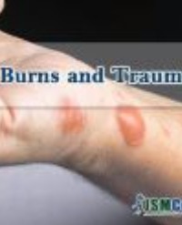
A 26-year-old lady self-presented to the Accidents and Emergency Department at Pinderfields General Hospital,Wakefield, following a frontal collision driving at 25 miles/hour, complaining of burning pain of her hand dorsum. After being cleared from ATLS point of view, hand examination showed partial thickness burn over the eponychial fold of her index finger and full thickness burn over her middle finger middle phalanx,with surrounding erythema.The pH of these wounds was found to be 10.These features are consistent with chemical burns; where they appear well demarcated with sharp edges and with a splash shape (Figure 1).

Figure 1: Airbags deployment chemical burns of the index and middle fingers.

After assessment, the wounds were irrigated with saline until the pH normalized to 7, and antiseptic dressing was applied. She was discharged on simple oral analgesia and follow up appointment was arranged in the regional burns unit.

Outcome and Follow-Up

The patient was reviewed later at the Burns Unit, and her burn completely healed within 2 weeks without any complications, and was discharged with appropriate scar care few weeks later.

Discussion

Air bags in cars are inserted for protection against injuries to the passengers. With the use of seatbelts combined with airbags, the number of mortalities and serious injuries has been markedly reduced. Zador and Ciccone re- ported that the combination of the air bag with seat belt reduced the incidence of fatal injury in front impact crashes in cars by 28% compared to that with seat belt only [3].

Despite their importance with regards to improved safety is undisputed, infrequently air bags deployments result in injuries.These injuries range from irritant dermatitis to an unlikely event of death.The spectrums of these injuries are determined by position of passenger, use of seatbelt restraint and for ocular injuries wearing of glasses [4]. According to Ulrich, the most common type of injury was abrasion (63.6%), followed by contusion, laceration, and burn comprising 7.8% of all injuries [5]. Chemical ophthalmic burns are important to diagnose early and characterized reduced visual acuity, with the eye giving alkaline pH measurement and complete corneal uptake of fluorescein.The eye should be irrigated with normal saline [6].

Hallock proposed classification of burns caused by air bag deployment, and classified thermal burns into two types: direct burn from high temperature gases and indirect burn due to melting of clothing [3]. The former is a burn related to contact with the expelled hot gases. These cases usually involve the upper extremities; as the exhaust vents are located on the rear of the bag [4].

Chemical burns incurred by airbags are mostly of minor severity and most affected body parts are upper extremities, as was the case with patient, followed by face and chest [6].

Airbags are made of nylon and are retained in the steering wheel central area. Rapid deceleration activates electrical mechanism that triggers a series of chemical reactions that ignite a sodium azide pellet, with the pellet reacting with potassium nitrate leading to the very rapid production of a large amount of high-temperature nitrogen gas, as well as sodium hydroxide and other gases, causing the bag to inflate within 20 hundredths of a second (300 km/h) [5]. Moments after the airbag deployment, the gases produced dissipate through the small pores of the fabric. However, deflation occurs within 2 seconds; allowing the occupant to move [7]. These high-temperature gases produce a corrosive alkaline aerosol able to cause thermal and alkali chemical burns [7]. However, Heimlich has indicated that these strong alkalis are no longer being produced, with recent model air bags are not expected to cause chemical burn [3].

Conclusion

A detailed history and examination of a patient should be done in case of road traffic accident to avoid missing injuries of minor or subtle nature. Airbags deployment can cause either thermal or chemical burns and the pH of the wound may help in establishing the diagnosis.The occurrence of such injuries resulting from safety equipment like airbags and seatbelts should never undermine their role in passenger’s safety, however, should encourage development of innovations lowering the risk of injuries associated with them. Measuring the burn pH can help establish the diagnoses and decide on appropriate treatment.

References

 1. Crandall CS, Olson LM, Sklar DP. Mortality Reduction with Air Bag and Seat Belt Use in Head-on Passenger Car Collisions. American Journal of Epidemiology. 2001; 153: 219-224.

2. Cunningham K, Brown TD, Gradwell E, Nee PA. Airbag associated fatal head injury: case report and review of the literature on airbag injuries. J Accid Emerg Med. 2000; 17: 139-142.

3. Hallock GG. Mechanisms of burn injury secondary to airbag deployment. Annals of Plastic Surgery. 1997; 39: 111-113.

4. Wallis LA, Greaves I. Injuries associated with airbag deployment. Emergency Medicine Journal. 2002; 19: 490-493.

5. Ulrich D, Noah EM, Fuchs P, Pallua N. Burn injuries caused by air bag deployment. Burns. 2001; 27: 196-199.

6. Virginia JM; Amber RL; Stefan DM. Analysis of Burn Injuries in Frontal Automobile Crashes. Journal of Burn Care & Rehabilitation. 2004; 25: 357 362.

7. Baruchin AM, Jakim I, Rosenberg L, Nahlieli O. On burn injuries related to airbag deployment. Burns 1999; 25: 49-52.

Other Articles

Article Image 1

Skin Reaction on Face Following the Use of Cloves (Syzygium aromaticum)

Clove (Syzygium aromaticum) (Figure 1) is a plant-derived spice that has been traditionally used for centuries as food preservative and as medicinal plants [1].

The anti-inflammatory, antioxidant and bactericide activities of clove are mainly due to its major components, which is the eugenol with a concentration rate ranging from to (77-95%) [2,3].

Clove is generally safe when taken in foods in lower concentrations [4], however, it is not recommended as a topical application on skin due to insufficiency of safety and toxicity data [5]; it was found to be highly cytotoxic for human fibroblasts and endothelial cells [6] leading to allergic skin reactions (burning, hives, itching, irritation, rash…), ulcer formation and/or tissue necrosis [7].

Samir El Mazouz1, Abdelmoughit Echchaoui1*, Narjis Badrane2 and Majda
Askour3


Article Image 1

Risk Factors for Infection and Mortality in Burned Children: Where are we going?

Burns are very frequent and affect approximately 1% of the general population every year. The
immunocompromising effects of burns, hospital stay; diagnostic and therapeutic procedures put
those patients at increased risk of morbidity and mortality. Pediatric burn patients are susceptible
to a broad spectrum of infections representing the most common and severe complication in this
population.

Over the last few years patient survival after burn injury has increased; however, despite advances
in the management of burn patients, infections remain the most common cause of morbidity and
mortality.

Rosanova MT*, Stamboulian D and Lede R


Article Image 1

MRI-Induced Burns from ECG Leads: Thermal or Electrical Burns?

Burns are a common global cause of injury; however iatrogenic burns are rare and preventable mode of injury. We report two cases who sustained superficial partial thickness burns from ECG monitoring leads during Magnetic Resonance Imaging (MRI). The two patients have sustained 0.2% and <1% superficial partial thickness burn of their Total Body Surface Area (TBSA) respectively. Common features included a relatively long duration of spine MR scanning (90 and 30 minutes) and high Body Mass Index (BMI). Both patients made uneventful recovery and were discharged within 24 hours of admission. It is still uncertain whether injuries were thermal or electrically-induced, however several mechanisms have been implied. These include electromagnetic inductionheating, the antenna effect and current induction through a closed loop. In this report, we discuss the proposed mechanisms of injury and highlight the fundamental need for staff education, and preparing national guidelines and safety checklists to prevent similar injuries in the future.

Othman D*, Abdel-Rehim S and OBoyle C


Article Image 1

Epidemiology of Burns Patients in a Tertiary Care Hospital in South India -A Retrospective Analysis

Burns are major health hazards which are associated with high mortality and morbidity rates. Burns management in developing countries is a major challenge due to inadequate access to burn care facility along with poorly equipped health care system. Although we are faced with a high burden of burns in our country, appropriate research on burns is still inadequate. We present our data of burns patients from a single center tertiary care hospital in south India.

Aim: To analyze various demographic characteristics, clinical and microbiological profile along with outcome of all burns patients admitted to our hospital.

Methods: This study was a retrospective analysis of burns patients admitted to the Critical Care Unit of Apollo Speciality Hospital, Vanagaram and a tertiary care facility in Chennai over a period of 3 years. Data such as age, gender, co-morbidities, type and degree of burns, percentage of burns and length of stay, mortality rate and infection rate were analyzed.

Results: There were a total of 94 burns patients included in the study. Amongst these, 61 patients (65%) were males and 33 patients (35%) were females. A majority of our burns population, 72% (n=68), belonged to age group from 21-50 years with a mean age of 40.50 years (SD±17.18).Mean total body surface area involved among burns was 48.56 (SD±21.08). Thermal burns were the commonest type of burns seen in 70%patients (n=66). These included flame burns in 49% patients (n=46) and scald burns in21% patients (n=20).Patients who presented to us within 6 hours post burns were 46% (n=43). Around 60% (n=56) patients had a hospital stay duration of ≤ 2 weeks. Infection rate among our patients was 62.8% and a mortality rate of 37% (n=35) was observed.

Conclusion: We highlight key demographic, clinical and microbiological data of all burns patients from a single center tertiary hospital in south India. This knowledge should help develop better strategies for management and prevention of mortality due to burns.

Ebenezer R1*, Rohit V1 , Isabella P2 , Nagarajan Ramakrishnan1 and Ganapathy

Krishnan3


Article Image 1

Pattern of the Burn Wounds Infections in Bahrain Defence Force Military Hospital

Background: Loss of skin as the first line of defence suppress immune system, extend hospital stay and makes burn patients more vulnerable to acquire healthcare associated infections. Such infections become the reason for mortality and morbidity in burn patients. Therefore, continuous monitoring of the pattern of nosocomial infection and drug resistance is necessary for assuring patient safety and healthcare quality improvement.

Aim: To describe the microbial profile of infections among burn patients admitted to Bahrain Defence Force Military Hospital.

Method: This is a retrospective cohort study involving 295burn patients. Wound swab method was used to isolate the microorganisms first on admission and then on suspicion of infection. The overall percentage of wound culture positive cases was 11.8% and the wound infection percentage was 5% of the total number of patients admitted to the unitA total of 126 swabs was performed from the burn wounds.

Results: Gram+ bacteria S.aureus was predominant in initial cultures. It was however, succeeded by Gram- bacteria, Pseudomonas, from third culture onwards. Fungal infection with Candida was most prevalent (n=21,16.7%). With TBSA >30% polymicrobial growth was observed which tend to increase with increase length of hospitalization. A greater proportion of patients (42.9%, n=15) had acquired healthcare associated infections and was negatively associated with increased length of hospitalization(r=-0.418, p=0.012).

Conclusion: 5% of the total patient population was categorized as health care associated infection cases. Gram negative bacteria, Pseudomonas were the prevalent microorganism isolated from wound infection of the burn patients.

Mohammad Abdulaziz Dahag, Nayef A Louri*, Nigamananda Dey and Siji Susan Philip


Article Image 1

Mesenchymal Stem Cells and Regenerative Quantum Medicine: The Novel A, B and C of Burns Treatment

Large burns are extensive soft tissue injuries with severe deep skin damage associated with high mortality [1-2]. This type of lesions can also lead to mental distress and chronic ilness but specially to scars, contractures, limitation of motion and bad quality of life [3-5]. Treatment of large burns as well as chronic nonhealing wounds have always been difficult medical problems and many different methods have been used to treat such injuries [6-7]. Current wound treatments are ineffective in many cases, so alternative novel types of therapy are needed and should urgently be understood and correctly explored. Abnormal damaged body environments can promote wound healing and tissue regeneration by local biochemical “alarm signals”, but usually these innate mechanisms are not enough to reach complete restoration of normal skin, specially in an acceptable time [8-12].When these impairments in wound healing arise, it is also often followed by an increased susceptibility to infection, further complications, failure of other organs and death [13-14]. In these circumstances, time management should be a crucial fundamental issue to be considered. In the near future we will not only ask for better novel therapeutic approaches but also for those that could lead to complete skin healing, restoration of function and acceptable aesthetics in the shortest period of time [ 15-16]. The development of tissue engineering and living skin substitutes started more than 40 years ago with the in vitro culture of keratinocytes as established by Howard Green in 1975 [17-18]. His outstanding discoveries led to the possibility of replacing the epidermis of extensively burned patients by using cultured keratinocytes autografts [19].

Mansilla E1*, Drago H2 , Marín GH1 , Jorrat R2 and Sturla F2


Article Image 1

Eikenella corrodens Wound Infection Secondary to Self-Bite: Case Report

Since Eikenella corrodens is part of the normal oral flora, it can be isolated from fist fight and human-bite injuries. Suspicion of the clinician, collection of adequate specimen, and proper culture technique will improve its isolation in the laboratories. Here we report a case of wound infection due to Eikenella corrodens in a 27-year-old female patient after biting herself during examination at the emergency ward.

Kuzucu EA, Çalışkan E, Bayrak ZS, Öcal D, Çağatay M, Saltaş H and Erdem G*


Article Image 1

Fatal Multi-Organ Failure Following Occupational Chemical Burn after Exposure to Phenolic Acid: A Case Report and Literature Review

Aim: Describe the complications and management of a rare case of burn injury by phenolic acid.

Methods: A 45-year-old truck driver sustained a 35% total body surface area (TBSA) burn from an accidental phenolic acid injury. After initial treatment and decontamination with a polyethylene glycol solution, he arrived at the hospital intubated and on vasopressor support. No other traumatic injuries were found. Local assessment revealed a 35% TBSA burn of partial and full thickness on the perineal region, legs, right arm, and a small area of the abdomen. His Revised Baux score was 97. During his recovery, the patient developed hepatic insufficiency followed by renal failure, which required dialysis. A few days later, a total body CT scan revealed multiple ischemic cerebral areas. After 12 days from admission and episodes of bronchial bleeding, the patient died due to a multiorgan failure. A skin biopsy and toxicological analysis for phenol levels were performed during his hospital stay.

Results: In addition to the clinical case analysis, a review of the medical literature regarding cases of death following phenol burns, a histological analysis of the biopsy tissue collected and an analysis of the blood levels of phenol and its derivative o-cresol were conducted.

Conclusions: This case report illustrates the lethality of phenol chemical burns and emphasizes the critical role of multidisciplinary management in addressing multi-organ complications

Alberto Schiarillo1, Anna Pensa1, Filippo Mariano2,3, Francesco Lupariello4, Catalina Ciocan5,6*, Daniela Risso1 and Maurizio Navissano1