Original Article | Volume 7 - Issue 1 | Article DOI : https://dx.doi.org/10.36876/jgm.1023
Download PDF
Lan Li1,2#, Ning Zhang3#*, Qianye Zhang3, Athanasios K. Petridis4, Konstantinos Gousias5, Zuzana Gazova6, Zuzana Bednarikova6, Thomas Eckert7,8, Gabriele Loers9, Ruiyan Zhang3, Imke Greving10, Elena Longo11, Helen Louton12, Anirban Bhunia13, Svenja Dannewitz14 and Hans-Christian Siebert1*
1RI-B-NT Research Institute of Bioinformatics and Nanotechnology, Germany
2Peking University Binhai Hospital, China
3Institute of BioPharmaceutical Research, Liaocheng University, China
4Medical School, Heinrich-Heine-Universität Düsseldorf, Department of Neurosurgery, St. Lukes Hospital, Thessaloniki, Greece
5Department of Neurosurgery, Athens Medical Center, Greece
6Department of Biophysics, Institute of Experimental Physics, Slovak Academy of Sciences, Slovakia
7Department of Chemistry and Biology, University of Applied Sciences Fresenius, Germany
8RISCC Research Institute for Scientific Computing and Consulting, Heuchelheim, Germany
9Research Group Neuronal and Cellular Signal Transduction, Institute of Human Genetics, University Medical Center Hamburg-Eppendorf, Germany
10Institute of Materials Physics, Helmholtz-Zentrum Hereon c/o DESY, Germany
11Elettra-Sincrotrone Trieste S.C.p.A., Italy
12Department of Veterinary Science, Chair of Animal Welfare, Behavioral Science, Animal Hygiene and Animal Husbandry, Germany
13Department of Chemical Sciences, Bose Institute, Unified Academic Campus, India
14Senara GmbH, Germany #L.L. and N.Z. have contributed equally to this work
Corresponding Author:
Hans-Christian Siebert, RI-B-NT Research Institute of Bioinformatics and Nanotechnology, Schauenburgerstr, 116, 24118 Kiel, Germany. Email: hcsiebert@aol.com
Keywords
Glioblastoma; Lectins; Quetiapine; Donkey Milk
Abstract
Glioblastoma multiforme (GBM) is the most aggressive and most common type of cancer originating in the brain. In this publication we discuss our approach to support the GBM standard therapy (surgery, chemotherapy and radiation) by the administration of donkey milk (200 ml per os and day) as well as by the application of the neuroleptic drug quetiapine. Our therapeutic strategy can easily be tested in a clinical context because quetiapine is often routinely applied during tumor therapy in order to suppress depressions. Donkey milk is known to stabilize the immune system which is strongly compromised in the case of GBM patients under a standard therapy. Since it is discussed in the literature that donkey milk and quetiapine have anti-tumor properties, especially, in the case of GBM, we focus on certain compounds in donkey milk and their potential interference with quetiapine. These compounds are proteins with a carbohydrate specificity, e. g. lectins which are able to bind galactose residues in a specific way, such as human galectin-3. Such lectins and their ligands are related to crucial tumor migration pathways. This can be achieved by a multimodal strategy combining molecular modeling with histological and radiological examinations. In addition, Atomic Force Microscopy (AFM) and X-ray nanotomography with synchrotron beams are extremely helpful because they enable a high-resolution three-dimensional imaging of cell- and tissue-probes. In combination with molecular modelling
Keywords
Glioblastoma; Lectins; Quetiapine; Donkey Milk
Abbreviations
GBM: Glioblastoma multiforme, AFM: Atomic Force Microscopy, hGal3: human galectin-3, ECM: extracellular matrix NMR: Nuclear Magnetic resonance, CRD: Carbohydrate Binding Domain, MRI: Magnetic Resonance Imaging, Nano-CT: Nanotomographie, TEM: Transmission Electron Microscopy, GFAP: Glial Fibrillary Acidic Protein, Idh1: isocitrate dehydrogenase 1, ROI: region of interest, H&E: hematoxylin and eosin staining, MD: molecular dynamics, DPC: dodecylphosphocholine, SPR: surface plasmon resonance, MS: mass spectrometry, NPT-Ensemble: isotherme-isobare Ensemble, RMSD: Root Mean Square Deviation, GPs: General Practitioners.
INTRODUCTION
Galectins are galactose-binding lectins from human and animal origin. They play a key-role in cell differentiation and de-differentiation processes. In relation to a lectin-based cancer therapy, it is a promising new approach to test receptor-specific neuroleptic molecules with a potential glycomimetic function as potential drugs which could suppress tumor-growth and spread as well as tumor resurrection. The carbohydrate binding pocket of human galectin-3 (hGal3) interacts in the nucleus with galactose-containing saccharide chains. In this context one could speculate that glycomimetics and psycho-active drugs like tegaserod, vortioxetine and quetiapine could potentially influence human galectin-3 (hGal3) interactions in the nucleus, in the cytoplasm, on the cell surface and together with integrins actions in the extracellular matrix (ECM) [1-5].
It has to be emphasized that glycomimetic molecules and lysozymes have an impact on lectin - carbohydrate interaction processes, especially, in the absence and in the presence of sialic acids. These processes can be influenced by tegaserod, cyclic and linear peptides or quetiapine showing structural similarities with certain carbohydrate residues in respect to their general shape, stacking forces and characteristic functional groups [6,7]. For example, tryptophan (Trp, W) and tyrosine (Tyr, Y) residues which are responsible for the carbohydrate recognition process exhibit these similarities in relation to psycho-active drugs and might be suited candidates for a new approach in glioblastoma multiforme (GBM) therapies. The structure-based interaction processes, especially, when sialic acids are involved, provide key-information related to infection diseases and cancer [8,9]. An innovative diagnostic strategy to monitor a therapeutical success might consists of a combination of synchrotron radiation X-ray nanotomography and Atomic Force Microscopy. It has to be emphasized that our therapeutic approach is completed by the use of lysozymes mainly from donkey milk origin as carrier material [10]. It has been highlighted that glioblastoma patients are highly immune compromised during a therapy. Donkey milk with its high level of DML and sialyllactose [11,12] can be considered as a promising option to support a therapy in which DML is complexed with quetiapine. These constructs shall be incorporated in the tumor hole after neurosurgical intervention or directly in satellite tumors which are not suited for any resection.
Various histological techniques (light microscopy, staining methods, electron microscopy, X-ray nanotomography with synchrotron beams [13] and atomic force microscopy (AFM) were used to assess GBM reference tissues, namely in absence of any treatment. The microscopic techniques were supported by molecular modeling tools which focus on the interaction of galectin-3 with glycomimetics. A clinical study in which quetiapine (brand name seroquel) and donkey milk are applied as a supporting approach to standard therapies has just started. Further results of our clinical study will be published in a follow-up article.
RESULTS
In order to analyze the interactions between human galectin-3 (hGal3) as well as quetiapine and serotonin we have designed a suited hGal3 model structure based on the 8ITZ.pdb data set (Figure 1). HGal3 is active inside and outside of the nucleus as well as on the outer cell membrane and also with integrins in the extracellular matrix (ECM). These findings document the suitability of this galectin for a glycobiological approach to combat glioblastomas in the light of recent glycobiological insights [14]. HGal3 shuttles between the nucleus and the cytoplasm where it interacts with different proteins and saccharides thereby generating an information transfer between various cell compartments.

Figure 1: a-d): Human galectin-3 (hGal3) structure in complex with a galactose-containing ligand and two other ligands in backbone presentations. (a): hGal3 in complex with a galactose-containing ligand. Trp181, the key-residue of this carbohydrate interaction is highlighted in green. Two key-residues responsible for the quetiapine interaction are colored as follows: Tyr79 – red, Ser96 – blue. (b): hGal3 in complex with a galactose containing ligand and quetiapine which is located at the low-structured part outside the Carbohydrate Binding Domain (CRD) of this galectin. (c): hGal3 in complex with a galactose-containing ligand and serotonin. Two key-residues which are stabilizing the hGal3 – serotonin complex, are colored as follows: Asp148 – orange, Ser237 – blue. (d): The binding site of serotonin is located inside the CRD of hGal3.
Our molecular modeling calculations with quetiapine and serotonin focus directly on the hGal3 based biochemical pathways. Serotonin, which is released after a quetiapine application shows structural similarities in comparison to tegaserod. With the strategic combination of biophysical and biochemical methods (e. g. NMR, AFM and immunostaining) supported by molecular modeling techniques it is possible to shed light on the role of hGal3 and its interactions with other biomacromolecules in the presence of quetiapine or serotonin. The corresponding complexes could be a potential target to suppress glioblastoma growth, tumor cell migration and resurrection by influencing apoptotic processes during a therapy with quetiapine. It is important to note that quetiapine acts on various neurotransmitter receptors, such as the serotonin and dopamine receptor. Therefore, the levels of serotonin and dopamine (molecules with a potential glycomimetic function) are altered in a certain way. Since nuclear functions of hGal3 are not fully understood we have carried out biochemical and biophysical experiments as well as molecular modeling calculations of glycans and lectins, especially, in the absence and in the presence of neuroleptic and glycomimetic molecules [15-17].
Quetiapine, a drug which is used as an atypical antipsychotic medication for the treatment of schizophrenia, bipolar disorder as well as major depressive disorder by balancing the levels of dopamine and serotonin in the brain can under special circumstances be used in glioblastoma therapies. In this context it is of interest that quetiapine is a glycomimetic molecule in relation to human lysozyme and a suited ligand for donkey milk lysozyme [7]. Since quetiapine neither fits into the predicted carbohydrate binding pocket of donkey milk lysozyme nor into the galactose binding pocket of human galectin-3 but forms stable complexes with both proteins in molecular dynamics simulations it makes sense to study this behavior in detail. Quetiapine interacts with hGal3 at its flexible region consisting of 120 amino acid residues linked to the carbohydrate recognition domain (CRD) of this chimeric galectin. From a glycobiological background it is a challenge to evaluate if the impact of hGal3 on apoptotic processes [3,5] can be influenced by the administration of quetiapine or other glycomimetics (e. g. tegaserod). Galectin-3 shuttles between the cytoplasm and the nucleus. In this context we assume that it is involved in apoptotic processes regulated within the nucleus [2,4]. HGal3 can therefore be considered also as a crucial biomedical regulator in the extracellular matrix (ECM) but furthermore, in the cytoplasm and in the nucleus. Quetiapine or serotonin which is significantly released by quetiapine are suited candidates for a promising approach in glioblastoma therapies. Quetiapine acts as a glycomimetic molecule in respect to the CRD of human lysozyme which means that it fits exactly in the predicted carbohydrate binding pocket [7]. This has encouraged us to compare lysozymes from different species (human, donkey, chicken) as potential nanocarriers in glioblastoma therapies. In addition, tegaserod is a glycomimtic molecule which mimics polysialic acid [6] while the glycomimetic properties of quetiapine have been described in relation to human lysozyme [7] and a bacterial oligosaccharide [10]. Molecular dynamics simulations provide interesting results when human hGal3 is analyzed in the presence of quetiapine as well as in the presences of serotonin which has structural similarities in comparison to tegaserod. In both complexes (hGal3 bound to a galactose-containing ligand and to quetiapine as well as to serotonin) the interactions between the ligands (quetiapine or serotonin) the are established by van der Waals interactions. In the case of quetiapine, the following amino acid residues are essential: Tyr79, Pro80, Gln94, Ser96, Pro 113, Leu114 und Asp242. In the case of serotonin, these amino acid residues are important: Pro117, Asn119, Asp148, Gln150, Val155, Lys233 und Ser237. Our findings are also of relevance when lysozyme – based nanocarriers have to be constructed in order to harbor the neuroleptic/ glycomimetic molecules under study.

Figure 1: a) Glial Fibrillary Acidic Protein (GFAP) antibodies were applied in order to obtain information about the cell’s cytoskeleton of a glioblastoma tissue obtained from a glioblastoma multiforme (GBM) i.e. a grade 4 glioma brain tumor. A Ki67 and a Idh1 monoclonal antibody staining (violet color) has been carried out in addition. b) Butterfly glioblastoma [20] with satellite tumors (with arrow) before the neurosurgical intervention as documented by MRI (Magnetic Resonance Imaging). c) Four months after the neurosurgical intervention. The white arrow points to a satellite tumor. Standard chemotherapy and radiation have been applied. In addition, 200 ml donkey milk per os and day was administered during this time period.
In order to document a potential therapeutic progress, it is essential to identify cutting-edge diagnostic tools beyond immunostaining (Figure 2a), Magnetic Resonance Imaging (MRI) (Figure 2b,2c) and Transmission Electron Microscopy (TEM) (Figure 3). On a nanomedical level a strategic combination of X-ray nanotomography (nanoCT) with synchrotron beams (Figure 4) and AFM analysis (Figure 5) is such a new diagnostic approach which is essential to evaluate brain tissue and tumor morphology at a cellular scale. AFM technologies are not only helpful to identify structural properties of glioblastoma tissues it can also be used to analyze the consistency of various cell tissue probes. The combination of X-ray nanotomography with synchrotron radiation and AFM could be considered as a new key approach to assess brain tissue architecture at high resolution scales [18,19].
Regarding the histological examination, the antibody staining shown in Figure 2a is used for a routine control to identify glioblastoma tissues. Mature astrocytes are detectable by GFAP antibodies. The maturation process is induced by galectin-1. A monoclonal antibody recognizes the Ki67 (Ki – Kiel) antigen, a protein that is suitable to mark dividing human cells. Another monoclonal antibody that specifically and sensitively recognizes the Idh1 (isocitrate dehydrogenase 1) protein carrying the R132H mutation is routinely processed in (formalin-fixed, paraffin embedded) tissues from the corresponding patients. Idh1 on the right side was found to be overexpressed in glioblastoma and is essential to tumor growth.

Figure 3: Transmission electron microscopy (TEM) images of cells from a glioblastoma multiforme (GBM) at two different sections. The small bars on the right side at the bottom correspond to 5 mm.

Figure 4: (a) X-ray nanotomography slice of GBM tissue. (b) Orthogonal view of the GBM tissue displayed in (a). (C) X-ray nanotomography slice of a GBM/infiltrated tissue.
A clinical case documenting a butterfly glioblastoma is displayed in Figure 2b and 2c. A Butterfly glioblastoma below to a rare subgroup of glioblastoma with a bihemispheric tumor crossing the corpus callosum, and is associated with a dismal prognosis [19-34]. The main tumor had been operated out by a neurosurgical intervention. Existing satellite tumors had to remain and further treated with a standard therapy (Temuzolomid and radiation at 64 Gy) supported by the administration of donkey milk, 200 ml per os and day. The administration of donkey milk has to be considered as a first attempt to test a potential effect of donkey milk lysozyme which is considered as an encapsulation and carrier substance for quetiapine. These constructs have to be placed in the tumor hole after a neurosurgical intervention and within the satellite tumors in order to combat a tumor progression. AFM technology seems to be a proper tool to monitor tissue probes obtained from such kind of tumors [18,19].

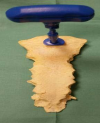
Figure 5: AFM images of glioblastoma cells. Left: Height in tapping mode. Right: Amplitude image in tapping mode.
Even if it is too early to jump into conclusions four months after the initial tumor surgery the size of the satellites did not change and the region around the gross total resection area of the butterfly tumor showed little tumor progress in the posterior resection area (Figure 2c). The cerebral blood flow as well as MRI spectroscopy indicates that the satellite tumors were still active but their size remained the same over four months. The mitotic index of the glioblastoma was 20-25% (Ki67). The patient died on an unrelated reason to the cause of the tumor. Indeed, the application of quetiapine and 200 ml donkey milk per os and day is not sufficient alone to achieve a significant therapeutic progress. Therefore, we use the data reported here for the construction of a nanomatrix consisting out of a DML which is able to harbor an adequate number of quetiapine molecules. In the following we report a series of imaging techniques that can be used for the of GBM tissues and hence also for future analysis in response to the described treatment. Therefore, we have to explain in which way AFM and X-ray nanotomography provides additional valuable information in comparison to standard microsvcopical techniques. With TEM techniques we can detect already the poor structural preservation of brain tumor cells. The images in Figure 4 were obtained following the region of interest (ROI) Zernike nanotomography (nanoCT) [13]. In this preliminary trial, four human GBM samples were imaged. The specimens were obtained after tumor surgical removal and specifically prepared for this nanoCT feasibility study. The images displayed in Figure 4 are selected views from the reconstructed tomographic volumes. In detail, a) and b) display virtual slices of a piece of GBM tissue and c) shows an area of interest of a GBM/infiltrating specimen. The images show a chaotic cell organization in more detail than images obtained by TEM and AFM. In the framework of a research project on the pH alterations of SARS CoV2 patients during their stay in intense care units also glioblastoma tissues from an infected patient was examined in order to test whether a direct impact of this virus is detectable in the brain. In order to analyze the impact of neuroleptic drugs on healthy tissues we evaluated probes from a long-term member of a chondroprotective food supplement study. The cartilage tissue of a 14,5-year-old dog which had been treated with ephedrine during the last months of its life was scrutinized with histological methods and AFM techniques [7- 21]. In the case of an AFM - based glioblastoma diagnosis (Figure 5) it is of highest importance to analyze the morphological and topological parameters of glioblastoma cells such as the shape of the nuclei and other cellular components including membrane structures in the dependence of the stage of the therapy. The canine tissue probe of the regio auris was taken from a 14,5-year old dog which has been treated with neuroleptics for therapeutic reasons. The probe consists of skin with subcutaneous connective tissue, muscles and glands, as well as some cartilage documenting the state of the extracellular matrix. The animal was a long-time member of a chondroprotective food supplements study published in 2021 [21]. Beside routinely applied diagnostic approaches (Figure 2 a-c), the multimodal combination of TEM (Figure 3), X-ray nanotomograpy using synchrotron beams and AFM (Figure 5,6) are additional supporting methods providing a complete three-dimensional view of the glioblastoma tissue under study. In summary, the AFM images recorded in contact mode are essential to obtain structural and morphological information in a complementary way to synchrotron-beam-based nanotomography. It has to be emphasized that the glioblastoma tissue probe which is shown in Figure 5 was taken from a patient which suffered also under a SARS CoV2 infection. It was of interest to compare such data with results obtained by X-ray nanotomography from glioblastoma tissue of a patient which had not been infected by SARS CoV2 (Figure 4). We did not detect any hints which argue in favor of a direct impact of the COVID-19 virus on these brain tissue probes when comparing the corresponding data sets (Figure 4 and Figure 5). Since the application of neuroleptic molecules in high doses is an essential building block of our therapeutic approach, we were interested to learn whether neuroleptic molecules could have an impact on distinct tissue regions of an organism. Therefore, we have carried out an AFM analysis of tissue probes from a domestic animal which had been treated with a neuroleptic drug for clinical reasons (Figure 6). It turned out that a canine tissue probe of the regio auris from a 14,5-year old dog which was a long-term member of a chondroprotective food supplement evaluation [21] is the ideal object of study. The dog had to be treated with ephedrinehydrochloride (brand name caninphedrin) during the last months of its life in order to manage urinary incontinence. This drug works by tightening the muscles in the bladder and urinary tract, helping to reduce involuntary urine leakage. We have not found any hints that the application of this neuroleptic drug had caused a harmful damage to the cells or the extracellular matrix under study.
With a NMR as well as a molecular modeling, especially, molecular dynamics (MD) and X-ray crystallography - supported analysis of galectins and neuroleptic drugs in combination with microscopic techniques (under special consideration of AFM and X-ray nanotomography with synchrotron beam), we have established a link between cell/ tissue analysis and molecular modeling. Especially, the application of glycomimetics as well as neuroleptic molecules and lectins (galectins and lysozymes with a lectin-function) turned out to be a promising approach in GBM therapies. In particular, the AFM analysis in the tapping mode in combination with synchrotron-beam-based X-ray nanotomography can be considered as a suited tool for monitoring the texture and organization of glioblastoma cells in relation to potential impacts of neuroleptic molecules and glycomimetics (here quetiapine). Lectin-like molecules (e. g. human or donkey milk lysozyme) will now be used as encapsulation substances which carry the neuroleptic molecules or glycomimetics under study to their targets, e. g. hGal3 in a tumor region.

Figure 6: AHematoxylin and eosin (H&E) staining was used for histological and cytological examination of tissue and cell preparations in dogs. The images show proteoglycan- and collagen-rich cartilage tissue at 250x magnification. The inset of an AFM image (compare also Figure. 5) shows the three-dimensional structure of a tissue section.
DISCUSSIONS AND CONCLUSIONS
General Practitioners (GPs) play a central, proactive role in patient monitoring. They provide longitudinal, continuous care to control side effect surveillance, especially, in cooperations with oncologists and psychiatrists when palliative care is needed during and after a glioblastoma standard therapy. Since donkey milk strengthens the immune system and has potentially supported by psychoactive drugs such as quetiapine or vortioxetine also a positive impact regarding tumor progression, it is of importance to use state-of-the-art nanomedical techniques to monitor the course of the disease on a sub-cellular and sub-molecular level.
Biochemical pathways in respect to carbohydrate - galectin - interactions on the cell-surface, with integrins in the ECM and inside the cell even inside the nucleus are not understood in detail up to now. The combination of microscopical methods (under special consideration of atomic force microscopy (AFM) and X-ray nanotomography using synchrotron beams) with molecular modeling, especially, molecular dynamics (MD) simulations, enables us to identify the potential benefits of certain neuroleptic molecules. We speculate that such molecules could act as receptor-dependent glycomimetic molecules suited for new approaches in a glioblastoma therapy. As already outlined for incretin-mimetics and donkey milk lysozyme, specific interactions with glycosylated and non-glycosylated dodecylphosphocholine (DPC) membrane-models are detectable by 1H- and 31P-NMR methods [7,8]. Therefore, it is of importance to identify suited techniques which enable us to study alterations on a sub-cellular and sub-molecular level [7,18]. In the case of an aggressive resurrection the use of incretin-mimetics could be supportive in respect of the healing process. In order to refine our therapeutic approach, the strategic combination of biophysical methods, as discussed here, is essential. With AFM experiments carried out in the phase/ contact/ tapping mode it is possible to gain valuable information about the consistency of the cell tissues under study. In the case of GBM it is of relevance that the consistency of the corresponding tumor tissue is coarse, rubbery and unorganized in comparison to healthy brain tissue probes. When AFM analysis and synchrotron - beam - based - nanotomography techniques are combined, it is feasible to detect malignant cells also in the peripheral regions of the tumor tissue. It has to be emphasized in this context that surface plasmon resonance (SPR) and mass spectrometry (MS) can also be used as supportive techniques for the evaluation of our DML lysozyme based glioblastoma therapy. In particular, other components of donkey milk e.g. sialidase and the galectin-3 binding protein could also open a new avenue to a cancer therapy based on glycobiological insights [22,23]. In this context, it is important to compare structural and functional properties of DML, hen egg white and human lysozyme with each other [24-26]. Further, we have to rely on sophisticated biophysical techniques [27-29] in relation to neuroleptics/ anti-psychotic drugs [30-33] and the results of a ketogenic diet [34] in mouse models.
Lysozymes, especially, DML used as encapsulation materials [24,25] will be discussed in a follow-up study. Especially, donkey milk lysozyme indicates suited properties for the storage of neuroleptic and glycomimetic drugs in the glioblastoma resection hole or within potential satellite tumors. Therefore, we can conclude that a neuroleptic drug like quetiapine is a promising tool not only as a potent drug in schizophrenia therapies. It can also be applied as crucial component in an improved GBM therapy because this drug seems to block glioblastoma cell migration in a hGal3 related way via the white matter fiber tracks in the brain [5-30]. In order to be more effective with our clinical approach related to an improved GBM therapy, we have to deliver quetiapine and/ or other glycomimetic molecules (e. g. tegaserod or specially designed peptides) using DML based nanocarriers (Figure 7).

Figure 7: Structural data concerning the molecular-modeling-based complex of human galectin-3 (hGal3) and the neuroleptic molecule quetiapine leading to an innovative diagnostic approach to analyze glioblastoma tissues by a combination of atomic force microscopy (AFM) and X-ray nanotomography with synchrotron beams. Our preventive care approach has started as a supporting glioblastoma treatment with donkey milk under special consideration of donkey milk lysozyme (DML).
In summary, the application of donkey milk [35] in combination with approved neuroactive drugs such a quetiapine or vortioxetine [36] have an important impact on primary care implications of glioblastoma patients. With a medical guidance these approaches can be carried out by the patients themselves. Food with strengthen the immune system of such patients and psycho active drugs belong in many cases to a palliative concept in glioblastoma treatment. However, this is not connected to any curative approach. Nevertheless, as lined out in this article, there is a good chance to identify compounds in donkey milk which have a potential to block the growth of tumor cells possibly supported by neuroactive drugs. In order to be more efficient, it is now of importance to focus on donkey milk exosomes and use them as nanocarriers [37,38] which could be implemented in the tumor hole, or in satellite tumors where a resection is impossible.
MATERIALS AND METHODS
Molecular Modeling
Galectin-3, nucleic acids and psychoactive drugs were further tested with in silico techniques as described [39]. The setup included an optimization of the hydrogen bonding network to increase the solute stability, and a pKa prediction to fine-tune the protonation states of protein residues at the chosen pH of 7.4. Na+ and Cl− ions were added with a physiological concentration of 0.9 %, with an excess of either Na+ and Cl− to neutralize the cell. After steepest descent and simulated annealing minimizations to remove clashes, the simulation was run for 100 ns using the AMBER14 force field [40] for the solute, GAFF2 and AM1BCC [41] for ligands, special amino acids (hydroxyprolin) and ions and TIP3P for water. The cutoff was set to 8 Å for Van der Waals forces (the default used by AMBER [42], no cutoff was applied to electrostatic forces (using the Particle Mesh Ewald algorithm)) [43]. The equations of motion were integrated with a multiple timestep of 1.25 fs for bonded interactions and 2.5 fs for non-bonded interactions at a temperature of 310 K, 0,993,249 g/ml density and a pressure of 1 atm (NPT ensemble) using algorithms described in detail previously [44]. After inspection of the solute RMSD as a function of simulation time, the first 10 ns were considered equilibration time and excluded from further analysis.
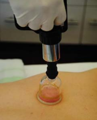
Atomic Force Microscopy (AFM)
Samples of glioblastoma tissue were sectioned using a microtome to produce 100 µm thin cuts. Canine tissue samples were sectioned using cryotome to 60 µm thin cuts. The slides (cuts) were directly glued to the cover slip glass, gently washed with 1× phosphate buffered saline solution and ultra-pure water, and left to dry completely on air. The sample’s surface was visualized using the atomic force microscope NTegra (Mt-mdt company) in tapping mode with SNL-10 cantilevers (Bruker AFM Probes, Camarillo) with nominal resonance frequency 65 kHz and spring constant 0.35 N/m. The height and phase images were scanned at 90 x90 µm scan size with 0.1-0.2 Hz speed and 512 pix resolution. The background correction was applied to all images using Gwyddion software 2.55.
X-Ray Zernike Nanotomography Setup
Small specimens of GBM tissue were derived from formalin fixed paraffin blocks and imaged by X-ray Zernike nanotomography at the imaging beamline P05 [45] at DESY (Hamburg, Germany) operated by the Helmholtz-Zentrum Hereon. The experimental setup is described in detail by Longo et al 2020 [13]. The images presented in this paper were collected with an effective pixel size of 85.8 nm x 85.8 nm [13].
Histological Analysis
Glial Fibrillary Acidic Protein (GFAP) antibodies as well as a Ki67 and a Idh1 monoclonal antibody staining were used for a histological staining. Paraffin embedded and cut in 3μm on a microtome, put on glass plates. Preparation: 5’ xylole, 5’min, alcohol 96%, 5’ alcohol 70%, 6’ heamtoxyline. Rinse in tap water 30’’, differentiation, rinsed in tap water, 4’ rosine riansed in tap water, 3’ alcohol 70%, 3’ alcohol 80%, 3’alcohol 96%, 2’ aceton, 5’xylole, 5’ xylole. Xylol: deparafiniosation: alcohol 96-70%: hydration of the slices and removal of Xylem to allow staining with hematoxyline. Hematoxyline differentiation: Solution of HCl and alcohol. Removal of unbound hematoxyline. Eosine: Alcohol 70-80-96%: dehydration of the slices after cosine staining. Aceton: differentiation of the slices, Xylol: Alcohol is removed in this way and substituted with Xylol which makes the slices transparent. Slices are mounted in Endella. Paraffin sections of 4 to 6 µm were obtained from the samples, de paraffined using toluene, re-hydrated and staining with hematoxylin and eosin solutions. After staining, the samples were dehydrated, mounted, and imaged using a 10x microscope.
ETHICAL STATEMENT
All blood and tissue probes from humans and animals used in this study were obtained after routine controls or necessary neurosurgical interventions. In the case of humans, the patients have agreed that their probes and MRI data can be taken for research purposes.
CREDIT AUTHORSHIP CONTRIBUTION STATEMENT:
Lan Li: Writing – original draft, Validation, Project administration, Methodology, Funding acquisition, Conceptualization. Ning Zhang: Writing – original draft, Validation, Project administration,Methodology, Funding acquisition, Conceptualization. Qianye Zhang: Validation, Software, Project administration, Formal analysis, Data curation. Athanasios K. Petridis: Supervision, Resources, Investigation, Data curation, Conceptualization. Konstantinos Gousias: Supervision, Resources, Investigation, Data curation, Conceptualization. Zuzana Gazova: Resources, Project administration, Methodology, Investigation, Data curation, Conceptualization – concerning Atomic Force Microscopy. Zuzana Bednarikova: Resources, Project administration, Methodology, Investigation, Data curation, Concptualization – concerning Atomic Force Microscopy Thomas Eckert: Visualization, Resources, Project administration, Methodology, Investigation, Data curation, Conceptualization - concerning Molecular Modelling. Gabriele Loers: Supervision, Project administration, Methodology, Investigation, Data curation, Conceptualization. Ruiyan Zhang: Writing – original draft, Visualization, Supervision, Methodology, Data curation, Conceptualization. Elena Longo: Resources, Project administration, Methodology, Investigation, Data curation, Conceptualization – concerning X-ray Nanotomography Imke Greving: Resources, Project administration, Methodology, Investigation, Data curation, Conceptualization – concerning X-ray Nanotomography Helen Louton: Supervision, Project administration, Methodology, Investigation, Data curation, Conceptualization.Anirban Bhunia: Supervision, Project administration, Methodology, Investigation, Data curation, Conceptualization. Svenja Dannewitz: Supervision, Project administration, Methodology, Investigation, Data curation, Conceptualization. Hans-Christian Siebert: Writing – review & editing, Writing – original draft, Visualization, Supervision, Software, Data curation, Conceptualization.
FUNDING
This work was supported by the Slovak Research and Development Agency under the Contract no. APVV-18-0284 and APVV-22-0598; Slovak Grant Agency VEGA grant 02/0176/21; Young Scholar Innovation Team of Liaocheng University (No. LCUGYTD2023-03).
ACKNOWLEDGMENTS
We thank our experts at the RI-B-NT: Marzieh Mohri (Bioinformatics) and Peter Engelhard (Paleontology). We are grateful to Eckhard Wolf (Faculty of Veterinary Medicine, Gene Center and Department of Biochemistry, Ludwig-Maximilians University, Munich, Germany), Melitta Schachner (Center for Molecular Neurobiology Hamburg, University Medical Center Hamburg-Eppendorf, University of Hamburg, Germany), Hubertus Maximilian Mehdorn (University Hospital of Schleswig-Holstein, Campus Kiel, Germany) for scientific support as well as Fabian and Philipp Siebert (RI-B-NT) for technical assistance. Roland Schauer, Hans-Joachim Gabius, Hans-Dieter Klenk and Tibor Kožár who unfortunately passed away in the years 2019, 2021 and 2024 were at many scientific meetings inspiring discussion partners over the years in respect to their pioneering work in the fields of sialic acid research, tumor lectinology, virology and computer-aided molecular modeling. Many thanks also to the Carl Friedrich-von-Weizsäcker Gesellschaft e. V. - Wissen und Verantwortung, to the team of the Arc Warder as well as to Andree Mehrens (KITZ - Kieler Innovations- und Technologiezentrum) for multiple support.
REFERENCES
- Gabius HJ, Manning JC, Kopitz J, André S, Kaltner H. Sweet complementarity: the functional pairing of glycans with lectins. Cell Mol Life Sci. 2016; 73: 1989-2016.
- Fritsch K, Mernberger M, Nist A, Stiewe T, Brehm A, Jacob R. Galectin-3 interacts with components of the nuclear ribonucleoprotein complex. BMC Cancer. 2016; 16: 502.
- Ajarrag S, St-Pierre Y. Galectins in Glioma: Current Roles in Cancer Progression and Future Directions for Improving Treatment. Cancers. 2021; 13: 5533.
- Gousias K, Theocharous T, Simon M. Mechanisms of Cell Cycle Arrest and Apoptosis in Glioblastoma. Biomedicines. 2022; 10: 564.
- Sedlář A, Trávníčková M, Bojarová P, Vlachová M, Slámová K, Křen V, et al. Interaction between Galectin-3 and Integrins Mediates Cell- Matrix Adhesion in Endothelial Cells and Mesenchymal Stem Cells. Int J Mol Sci. 2021; 22: 5144.
- Zhang R, Loers G, Schachner M, Boelens R, Wienk H, Siebert S, et al. Molecular Basis of the Receptor Interactions of Polysialic Acid (polySia), polySia Mimetics, and Sulfated Polysaccharides. ChemMedChem. 2016; 11: 990-1002.
- Zhang N, Li L, Mohri M, Siebert S, Lütteke T, Louton H, et al. Protein- carbohydrate interaction studies using domestic animals as role models support the search of new glycomimetic molecules. Int J Biol Macromol. 2024; 279: 134951.
- Siebert HC, Eckert T, Bhunia A, Klatte N, Mohri M, Siebert S, et al. Blood pH Analysis in Combination with Molecular Medical Tools in Relation to COVID-19 Symptoms. Biomedicines. 2023; 11: 1421.
- Schauer R, Kamerling JP. Exploration of the Sialic Acid World. Adv Carbohydr Chem Biochem. 2018; 75: 1-213.
- Zhang R, Wu L, Eckert T, Burg-Roderfeld M, Rojas-Macias MA, Lütteke T, et al. Lysozyme’s lectin-like characteristics facilitates its immune defense function. Q Rev Biophys. 2017; 50: e9.
- Živkov Baloš M, Ljubojević Pelić D, Jakšić S, Lazić S. Donkey Milk: An Overview of its Chemical Composition and Main Nutritional Properties or Human Health Benefit Properties. J Equine Vet Sci. 2023; 121: 104225.
- Licitra R, Li J, Liang X, Altomonte I, Salari F, Yan J, et al. Profile and content of sialylated oligosaccharides in donkey milk at early lactation. LWT. 2019; 115: 108437.
- Longo E, Sancey L, Flenner S, Kubec A, Bonnin A, David C, et al. X-ray Zernike phase contrast tomography: 3D ROI visualization of mm- sized mice organ tissues down to sub-cellular components. Biomed Opt Express. 2020; 11: 5506-5517.
- Nagaraj V, Mikhail M, Baronio M, Gatto A, Nayak A, Theis T, et al. Antagonistic L1 Adhesion Molecule Mimetic Compounds Inhibit Glioblastoma Cell Migration In Vitro. Biomolecules. 2022; 12: 439.
- Bertuzzi S, Quintana JI, Ardá A, Gimeno A, Jiménez-Barbero J.Targeting Galectins With Glycomimetics. Front Chem. 2020; 8: 593.
- Wang Y, Huang N, Li H, Liu S, Chen X, Yu S, et al. Promoting oligodendroglial-oriented differentiation of glioma stem cell: a repurposing of quetiapine for the treatment of malignant glioma. Oncotarget. 2017; 8: 37511-37524.
- Bhat K, Saki M, Cheng F, He L, Zhang L, Ioannidis A, et al. Dopamine Receptor Antagonists, Radiation, and Cholesterol Biosynthesis in Mouse Models of Glioblastoma. J Natl Cancer Inst. 2021; 113: 1094- 1104.
- D’Amico L, Svetlove A, Longo E, Meyer R, Senigagliesi B, Saccomano G, et al. Characterization of transient and progressive pulmonary fibrosis by spatially correlated phase contrast microCT, classical histopathology and atomic force microscopy. Comput Biol Med. 2024; 169: 107947.
- Van der Meeren L, Verduijn J, Krysko DV, Skirtach AG. AFM analysis enables differentiation between apoptosis, necroptosis, and ferroptosis in murine cancer cells. iScience. 2020; 23: 101816.
- Bjorland LS, Dæhli Kurz K, Fluge Ø, Gilje B, Mahesparan R, Sætran H, et al. Butterfly glioblastoma: Clinical characteristics, treatment strategies and outcomes in a population-based cohort. Neurooncol Adv. 2022; 4: vdac102.
- Eckert T, Jährling-Butkus M, Louton H, Burg-Roderfeld M, Zhang R, Zhang N, et al. Efficacy of Chondroprotective Food Supplements Based on Collagen Hydrolysate and Compounds Isolated from Marine Organisms. Mar Drugs. 2021; 19: 542.
- Zhang N, Liu Q, Wang D, Wang X, Pan Z, Han B, et al. Multifaceted roles of Galectins: from carbohydrate binding to targeted cancer therapy. Biomark Res. 2025; 13: 49.
- Loimaranta V, Hepojoki J, Laaksoaho O, Pulliainen AT. Galectin-3- binding protein: A multitask glycoprotein with innate immunity functions in viral and bacterial infections. J Leukoc Biol. 2018; 104: 777-786.
- Cao C, Tian L, Li J, Raveendran R, Stenzel MH. Mix and Shake: A Mild Way to Drug-Loaded Lysozyme Nanoparticles. ACS Appl Mater Interfaces. 2024; 16: 27177-27186.
- Mukhametova LI, Zherdev DO, Eremin SA, Levashov PA, Siebert HC, Tsvetkov YE, et al. Application of the Chitooligosaccharides and Fluorescence Polarization Technique for the Assay of Active Lysozyme in Hen Egg White. Biomolecules. 2024; 14: 1589.
- Zhang Q, Sun W, Zheng M, Wang Q, Liu G, Li L, et al. Donkey milk inhibits tumor growth by inducing apoptosis, pyroptosis and modulation of Th1/Th2 responses in a 4T1 murine breast cancer model. Journal of Functional Foods. 2024; 118: 106256.
- Bertuzzi S, Lete MG, Franconetti A, Diercks T, Delgado S, Oyenarte I, et al. Exploring Glycan-Lectin Interactions in Natural-Like Environments: A View Using NMR Experiments Inside Cell and on Cell Surface. Chemistry. 2025; 31: e202403102.
- Ferstl S, Schwaha T, Ruthensteiner B, Hehn L, Allner S, Müller M, et al. Nanoscopic X-ray tomography for correlative microscopy of a small meiofaunal sea-cucumber. Sci Rep. 2020; 10: 3960.
Citation
Li L, Zhang N, Zhang Q, Petridis AK, Gousias K et al, (2026) Im proved Diagnostic and Therapeutic Strategy to Treat Glioblastoma Symp toms in Preventive Care . J Gen Med 6(1): 1023.