Case Report | Volume 2 - Issue 1 | Article DOI :
Download PDF
Vijayram R Malladi1*, Rizwan Ishtiaq1 , Lily Wu2 , Lucy Chie2 , Daryl TY Lau1 and Aysha Aslam1
1Department of Gastroenterology, Harvard Medical School, USA
2Department of Obstetrics and Gynecology, Harvard Medical School, USA
Corresponding Author:
Vijayram Reddy Malladi, Department of
Gastroenterology, Beth Israel Deaconess
Medical Center, Harvard Medical School,
Liver Center, 110 Francis Street, Suite
4A, Boston, USA; Tel: 617632-1051; Fax:
617632-1125;
Keywords
Intrahepatic Cholestasis of Pregnancy; Chronic Hepatitis B; Pruritus;
Ursodeoxycholic acid
Abstract
Intrahepatic cholestasis of pregnancy is a specific liver condition. It usually occurs in the second or third trimester of pregnancy and is characterized by pruritus and elevated serum bile acid level. Timely diagnosis and management of this disease entity is necessary as increasing bile salts concentration can have adverse outcomes on the mother and the baby. A high index of suspicion is essential to diagnose intrahepatic cholestasis of pregnancy among patients with chronic liver diseases for their overlapping clinical manifestations. In this case report, we present the clinical course and management of a 35-year-old Asian female with intrahepatic cholestasis of pregnancy in the setting of chronic hepatitis B.
Citation
Malladi VR, Ishtiaq R, Wu L, Chie L, Lau DTY and Aslam A. Intrahepatic Cholestasis of Pregnancy in an Asian Woman with Chronic Hepatitis B. SM Virol. 2017; 2(1): 1013.
Introduction
Hepatitis B Virus (HBV) infection is a global health problem affecting approximately 350 million people worldwide [1]. Prevalence of hepatitis B among pregnant women in the endemic regions such as China and Thailand is about 7.6% and 6.2% respectively [2,3]. Perinatal transmission is the major route of acquiring hepatitis B infection in HBV endemic countries. The risk of HBV transmission increases with the maternal HBV DNA level [4].
During pregnancy, the woman’s immune response is suppressed with rising levels of hormones. T his immunological alteration can lead to increased viral replication and hepatitis flares [5]. Hepatitis flares with elevated serum aminotransferases are especially common during third trimester of pregnancy and postpartum period [6]. In a recent study conducted by Chang et al., HBV DNA f lares were observed in 9% of the women during pregnancy and 4% during post-partum [6]. Women with chronic hepatitis B should be observed closely during pregnancy and post-partum.
T here are a number of clinical entities unique to pregnancy that can affect the liver. Hyperemesis gravidarum, for example, usually occurs in the first 20 weeks of pregnancy. Intrahepatic cholestasis of pregnancy (ICP) usually develops during the second to third trimester of pregnancy. ICP is a common pregnancy related disorder with increased perinatal mortality and it is manifested by pruritus and elevated bile acids and transaminase levels [7]. ICP usually resolves after pregnancy. History of chronic hepatobiliary disease, family history of ICP, use of oral contraceptives and multiple gestations are reported risk factors for ICP [8]. Adverse effects on the fetus due to ICP include preterm birth, asphyxia, respiratory distress syndrome and in extreme cases, fetal death [9]. Acute fatty liver of pregnancy and HELLP syndrome typically manifest in the third trimester of pregnancy.
We herein present a case of intrahepatic cholestasis of pregnancy and active hepatitis during third trimester in a woman with chronic hepatitis B.
Case History
A 35-year-old Chinese female with documented HBeAg positive Chronic Hepatitis B (CHB) presented to our Liver Clinic at 27-week gestation with her second pregnancy. During her first pregnancy 6 years prior, she was in the immunotolerant phase of CHB, with high level of viremia and persistently normal serum aminotransferases, and was initiated on lamivudine during the third trimester to help prevent vertical transmission of HBV. She gave vaginal birth to a healthy child, and discontinued her lamivudine after delivery. Her first child received both HBV vaccine and HBIg at birth, and tested negative for HBV. Three years ago, the patient experienced active hepatitis B and was prescribed tenofovir 300mg daily which resulted in optimal HBV DNA suppression though HBeAg remained reactive. During the first trimester of this current pregnancy, patient experienced nausea and vomiting and had uncertainty regarding safety of the antiviral therapy during pregnancy, thus had self-discontinued the antiviral therapy.
During liver clinic visit at 27-week gestation, she complained of excessive itchiness on the extensor surface of her elbows and knees that kept her from sleeping. Excoriations on bilateral elbows and knees were noted. She did not have any signs of advanced liver disease such as leg swelling, ascites, or jaundice. Her HBV DNA was significantly elevated off antiviral therapy at 6 log10 IU/ml (or 1 million IU/ml). Her serum aminotransferases, total bilirubin, alkaline phosphatase, albumin and INR were all within the normal limit. Total Bile acids were elevated at 12.8 umol/L (cholic acid 4.5 umol/L, deoxycholic acid 4.9 umol/L, chenodeoxycholic acid 3.4 umol/L). Based on the history of pruritus and elevated bile acid level, diagnosis of Intrahepatic Cholestasis of Pregnancy (ICP) was made, and Ursodeoxycholic Acid (UDCA) was prescribed.
She was restarted on tenofovir 300mg due to high level of viremia. T he risk of vertical transmission has decreased significantly with the administration of HBV vaccine and HBIg to babies born to mother with chronic hepatitis B [10]. The risk of perinatal transmission, however, remains at 10-15% if the maternal HBV DNA is >200,000 IU/ml. Tenofovir is indicated in women with HBV DNA level >200,000 IU/ml during the third trimester to further reduce HBV vertical transmission [11]. Our patient was reassured about the safety profile of tenofovir with pregnancy.
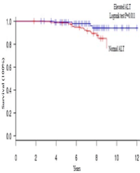
Her pruritus improved with UDCA. Her total bile acids, however, continued to rise to 20.8 umol/L five weeks later, with cholic acid increased to 8.3 umol/L (or 40%). In addition, her ALT increased to 110 U/L despite reduction of HBV DNA level to 3 log10 IU/ml (1,000 IU/ml) (Figure 1).

Figure 1: The clinical course of the patient with ICP in the setting of CHB. Patient developed hepatitis flare despite HBV DNA suppression by Tenofovir. The rise of ALT level occurred with the onset of pruritus and peaked with total bile acid level. ALT decreased abruptly within 48 hours after elective induction of labor and normalized subsequently. (The blue solid line indicates ALT levels and the red dotted line shows the HBV DNA trend).
Her platelet count, bilirubin and hepatic synthetic function remained normal. There was no evidence of preeclampsia. Her abdominal ultrasound was entirely normal without steatosis, gallstone or focal hepatic lesion.
The patient’s ALT level peaked at 123 U/L at 35 weeks 6 days’ gestation while HBV DNA continues to decrease to 100 IU/ ml. Fetus was closely monitored with twice weekly ultrasound surveillance. Decision was made to proceed with induction of labor at 36 weeks’ gestation due to diagnosis of ICP as well as rising serum aminotransferase. The baby experienced respiratory distress and needed intubation and surfactant treatment initially at NICU but recovered well. The baby received both HBV vaccine and HBIg shortly after birth. The mother’s ALT level decreased from 123 U/L to 83 U/L within 48 hours after delivery and normalized 4-weeks post-partum. She stopped UDCA after delivery without recurrence of pruritus. Her total bile acid levels decreased to 15.5 umol/L with significant reduction of cholic acid to 2.8 umol/L (18%) approximately 6-weeks post-partum. Her hepatitis B condition was stable on tenofovir with optimal HBV DNA suppression.
Discussion
ICP is one of the most common liver diseases during second and third trimester of pregnancy. It is significantly more frequent in South Asia (0.8%-1.46%) and South America (9.2%-15.6%) than in Europe (0.1% to 0.2%) [12]. The association of ICP with hepatitis B has not been established but coexistence of ICP with hepatitis B can have serious consequences [13]. The possible explanations for the more severe maternal outcomes of intrahepatic cholestasis in patients with hepatitis B include promotion of hepatocellular systemic inflammatory responses, acceleration of viral replication, and the additive effects of the two leading to deterioration of maternal hepatic function. Advanced maternal age, personal or family history of ICP or use of contraceptives are risk factors for ICP. ICP is also more common among patients with cholelithiasis, hepatitis C or nonalcoholic fatty liver disease [14]. Our patient did not report any of these risk factors except CHB.
A high index of suspicion is necessary to diagnose ICP, particularly if there is elevation of liver tests in pregnant women with CHB. Our patient was noted to have significant rise in HBV DNA level when she presented with pruritus in early third trimester of pregnancy. The high level of viremia could be secondary to a general reduction of immune response during pregnancy especially after she discontinued antiviral therapy. The rise in serum aminotransferases despite HBV DNA suppression with restarting antiviral therapy was concerning. In this case, the abrupt reduction of ALT levels within 48 hours after induction of labor suggests the association of hepatitis flare with ICP. Elevation in liver enzymes may be detected in up to 60% of the subjects with ICP. Hyperbilirubinemia, which rarely reaches 6 mg/ dL, is another common laboratory finding noted in about 25% of ICP cases [15].
Patients with ICP have a decrease in bile flow without any involvement of biliary obstruction or liver dysfunction during the second/third trimester, and it usually resolves during post-partum [16]. The pathogenesis of ICP is unclear, but various bile acid and phospholipid transporters are likely to be involved [17]. Some patients with ICP have heterozygosity for mutations in the genes encoding familial intrahepatic cholestasis protein-1 FIC1, Bile Salt Excretory Protein (BSEP), and Multidrug Resistance Protein 3 (MDR3) [18]. T he development of cholestasis in the setting of elevated female sex hormones during pregnancy suggests a hormonal influence on the pathogenesis [19, 20].
ICP should be considered in pregnant women with symptoms of pruritus and absence of diseases that have similar presentation.
Ultrasound is recommended to rule out cholelithiasis. Autoimmune cholangitis such as Primary Biliary Cholangitis (PBC), Primary Sclerosing Cholangitis (PSC) and use of herbal medications should be excluded. For patients with elevated serum aminotransferases, viral hepatitis, autoimmune hepatitis, preeclampsia, HELLP syndrome, and acute fatty liver of pregnancy also need to be considered. Elevated serum bile acids concentration (>11mmol/L) with cholestasis and pruritus in the third trimester of pregnancy constitute the diagnosis of ICP [21]. Typically, pruritus involves the palms and soles, and worsens at night. Our patient experienced pruritus in extensor surfaces of elbows and knees that did get worse at night. The symptoms of pruritus in third trimester, total bile acids level of 12.8 umol/L and absence of other causes of cholestasis confirmed the diagnosis of ICP in this case.
Bile acid concentration greater than 40 umol/L is associated with increased risk of fetal distress and mortality [22]. There is no general consensus on fetal surveillance. Fagan et al. suggested a weekly non-stress test, estimation of amniotic fluid volume, and umbilical artery doppler ultrasonographic examination, as well as regular growth scans from 30 week of gestation in the setting of ICP [23]. Early delivery at 36 to 37 weeks is recommended to decrease the risk for intrauterine deaths which are more common in the last month of pregnancy [24]. The rates of fetal malformations and abortions do not appear to increase, and fetal birth weight for gestational age appears to be adequate in ICP. A study conducted in China reported an increased rate of premature rupture of membranes, neonatal asphyxia, fetal distress and congenital disabilities in the newborns among women with CHB and ICP [25]. Those findings need to be validated by further observations. In our case, the maternal bile acid levels were lower than 40umol/L; the regular fetal monitoring and timely induction of labor ensure the health of the newborn.
Ursodeoxycholic Acid (UDCA) is the first line treatment for ICP as it is safe for both the mother and the fetus [26]. In one study, therapy with UDCA helped normalize levels of bile acids in babies with minimal accumulation in fetal blood and amniotic fluid [27]. A meta-analysis conducted by Bacq et al. found that women who received UDCA therapy had good fetal outcomes, less pruritus and improvement of liver enzymes [28].
Conclusion
ICP is a common liver condition unique in late second and third trimester of pregnancy that can lead to severe maternal and fetal adverse outcomes. A high index of suspicion is required to diagnose ICP especially in women with CHB for spontaneous HBV reactivation with elevation of serum aminotransferases is particularly common during third trimester of pregnancy. The flare of hepatitis B can overshadow the symptoms of ICP. Increased vigilance with frequent monitoring of pregnant women is essential for timely diagnosis and management of ICP to prevent adverse maternal and fetal outcomes. It is essential to consider delivery by 36 to 37 weeks gestation. Further studies are required to validate the increased severity of ICP among women with CHB.
References
1. Dienstag JL. Hepatitis B virus infection. N Engl J Med. 2008; 359: 1486-1500.
2. Shi G, Zhang SX. Meta-analysis on the positive rate of hepatitis B surface antigen among pregnant women in China. Chin Prev Med. 2013; 1: 26-30.
3. Sirilert S, Traisrisilp K, Sirivatanapa P, Tongsong T. Pregnancy outcomes among chronic carriers of hepatitis B virus. International Journal of Gynecology & Obstetrics. 2014; 126: 106-110.
4. Chen T, Wang J, Feng Y, Yan Z, Zhang T, Liu M, et.al. Dynamic changes of HBV markers and HBV DNA load in infants born to HBsAg (+) mothers: can positivity of HBsAg or HBV DNA at birth be an indicator for HBV infection of infants?. BMC Infect Dis. 2013; 13: 524.
5. Trowsdale J, Betz AG. Mother’s little helpers: mechanisms of maternal-fetal tolerance. Nat Immunol. 2006; 7: 241-246.
6. Chang CY, Aziz N, Poongkunran M, Javaid A, Trinh HN, Lau D, et.al. Serum alanine aminotransferase and hepatitis B DNA flares in pregnant and postpartum women with chronic hepatitis B. The American journal of gastroenterology. 2016; 111: 1410-1415.
7. Reyes H. Intrahepatic cholestasis. A puzzling disorder of pregnancy. Journal of gastroenterology and hepatology. 1997; 12: 211-216.
8. Hardikar W, Kansal S, Elferink RP, Angus P. Intrahepatic cholestasis of pregnancy: When should you look further?. World Journal of Gastroenterology. 2009;15: 1126-1129.
9. Egerman RS, Riely CA. Predicting fetal outcome in intrahepatic cholestasis of pregnancy: is the bile acid level sufficient?. Hepatology. 2004; 40: 287-288.
10. Poland GA, Jacobson RM. Prevention of hepatitis B with the hepatitis B vaccine. New England Journal of Medicine. 2004; 351: 2832-2838.
11. Terrault NA, Bzowej NH, Chang KM, Hwang JP, Jonas MM, Murad MH. AASLD guidelines for treatment of chronic hepatitis B. Hepatology. 2016; 63: 261-283.
12. Tan LK. Obstetric cholestasis: current opinions and management. Annals of the Academy of Medicine, Singapore. 2003; 32: 294-298.
13. Tan J, Liu X, Mao X, Yu J, Chen M, Li Y, et.al. HBsAg positivity during pregnancy and adverse maternal outcomes: a retrospective cohort analysis. Journal of viral hepatitis. 2016; 23: 812-819.
14. Ropponen A, Sund R, Riikonen S, Ylikorkala O, Aittomäki K. Intrahepatic cholestasis of pregnancy as an indicator of liver and biliary diseases: A population-based study. Hepatology. 2006; 43: 723-728.
15. Mullally BA, Hansen WF. Intrahepatic cholestasis of pregnancy: review of the literature. Obstetrical & gynecological survey. 2002; 57: 47-52.
16. Germain AM, Carvajal JA, Glasinovic JC, Williamson C. Intrahepatic cholestasis of pregnancy: an intriguing pregnancy-specific disorder. Journal of the Society for Gynecologic Investigation. 2002; 9: 10-14.
17. Pauli-Magnus C, Meier PJ, Stieger B. Genetic determinants of drug-induced cholestasis and intrahepatic cholestasis of pregnancy. In Seminars in liver disease 2010; 30: 147-159.
18. Chacko KR, Wolkoff AW. Intrahepatic Cholestasis of Pregnancy: New Diagnostic Insights. Annals of Hepatology. 2017; 16: 176-178.
19. Reyes H. Sex hormones and bile acids in intrahepatic cholestasis of pregnancy. Hepatology. 2008; 47: 376-379.
20. Glantz A, Reilly SJ, Benthin L, Lammert F, Mattsson LÅ, Marschall HU. Intrahepatic cholestasis of pregnancy: Amelioration of pruritus by UDCA is associated with decreased progesterone disulphates in urine. Hepatology. 2008; 47: 544-551.
21. Brites D. Intrahepatic cholestasis of pregnancy: changes in maternal-fetal bile acid balance and improvement by ursodeoxycholic acid. Annals of hepatology. 2002; 1: 20-28.
22. Garcia-Flores J, Cañamares M, Cruceyra M, Garicano A, Espada M, Lopez A, et.al. Clinical value of maternal bile acid quantification in intrahepatic cholestasis of pregnancy as an adverse perinatal outcome predictor. Gynecologic and obstetric investigation. 2015; 79: 222-228.
23. Fagan EA. Disorders of the liver, biliary system and pancreas. Medical Disorders in Obstetric Practice, Fourth Edition. 2008: 282-345.
24. Williamson C, Hems LM, Goulis DG, Walker I, Chambers J, Donaldson O, et.al. Clinical outcome in a series of cases of obstetric cholestasis identified via a patient support group. BJOG. 2004; 111: 676-681.
25. Hu Y, Ding YL, Yu L. The impact of intrahepatic cholestasis of pregnancy with hepatitis B virus infection on perinatal outcomes. Therapeutics and clinical risk management. 2014; 10: 381.
26. Tran TT, Ahn J, Reau NS. ACG clinical guideline: liver disease and pregnancy. The American journal of gastroenterology. 2016; 111: 176-194.
27. Mazzella G, Nicola R, Francesco A, Patrizia S, Luciano B, Anna M, et.al. Ursodeoxycholic acid administration in patients with cholestasis of pregnancy: effects on primary bile acids in babies and mothers. Hepatology. 2001; 33: 504-508.
28. Bacq Y, Sentilhes L, Reyes HB, Glantz A, Kondrackiene J, Binder T, et.al. Efficacy of ursodeoxycholic acid in treating intrahepatic cholestasis of pregnancy: a meta-analysis. Gastroenterology. 2012; 143: 1492-1501.