SM Case Reports

Archive Articles

Article Image 1

A Pediatric Case Series of Distal Ureteral Stones Clearance after Tamsulosin Treatment

Medical tamsulosin therapy in children has shown encouraging results in facilitating spontaneous clearance of lower ureteral calculi.

In this paper we report 5 paediatric cases of tamsulosin use in clearance of distal ureteral stones. All patients showed typical symptoms of sudden onset of abdominal/ flank acute pain and were diagnosed by imaging findings.

We report this case series to emphasize the importance of tamsulosin as initial option to treat urolithiasis in children at the emergency room


Paula Nunes1 *


Article Image 1

A Case Report on Prednisolone Induced Acute Pancreatitis

Acute pancreatitis is defined as an acute inflammation of the pancreas that may also involve peripancreatic tissues and/or remote organ systems. Although the common causes of acute pancreatitis are gall stones and alcohol consumption, drug induced cases are also reported frequently. Whereas steroids induced pancreatitis cases were less and rarely reported so the true incidence is unknown, quality of the evidence is limited since the availability of studies is very less. Prednisolone is a commonly used intermediate acting corticosteroid. It is a challenge to physicians to diagnose the drug induced pancreatitis than other causes. Here in this case report we describe a 22-year-old male patient who had received prednisolone tablet for Nephrotic syndrome. According to Naranjo’s ADR probability scale, the reaction was probable cause for acute pancreatitis.

Maneesha Erraboina¹ and Sandeep Reddy Jillella²*


Article Image 1

Pure Squamous Cell Carcinoma of the Breast: Report Histological Features and Treatment Outcome of Two Cases

SCC is an uncommon and aggressive breast neoplasm. There are limited data in literature about its epidemiology and studies focusing on outcomes, so they are still unclear. It has significantly worse prognosis than other non squamous cell tumors of the breast. Strict histologic criteria should be used to determine diagnosis. Because of its rarity, it is still unclear which the most appropriate therapeutic regimen to use is. The role of different new chemotherapy regimens needs to be explored and biologic studies are needed to determine the tumour chemoresistance mechanisms and the potential use of other treatment targets that improve patients survival. Clinicians should be aware of the aggressive nature of the tumour when counseling patients

Mezzabotta Maurizio¹, Corbellini Carlo¹², Declich Paolo³, Basilicò Silvia¹², and Morandi Eugenio¹*


Article Image 1

Thalassemia; a Protective Factor against Severe Malaria: A Report of a Case and Review of Literature

The severity of malaria depends on factors some of which are well known. They allow a better understanding of the conditions of the risk of severe, and adopt the terms of surveillance and prevention. We report a case of Plasmodium falciparum polycythemia protected against deadly cerebral malaria.

Moutaouakkil Y¹, Fettah H¹, Makram S¹, Elmssaouri J¹, Bennana A¹, and Bousliman Y¹*


Article Image 1

Acrokeratosis Verruciformis of Hopf Associated With Epidermolytic Hyperkeratosis - A Rare Case Report

Acrokeratosis verruciformis of Hopf is a rare genetic disorder with autosomal dominant inheritance. It is characterized by the presence of multiple lightly pigmented or flesh colored papules on the dorsum of the hands, feet, knees and elbows. Lesions consistent with acrokeratosis verruciformis are been associated with HaileyHailey, hypertrophic lichen planus, naevoid basal cell carcinoma, multiple keratoacanthoma. We report a case of acrokeratosis verruciformis in a 65 years old patient who came with complaints of itchy skin lesions over both the legs and dorsum of the right hand for the past 2 years and histologically with features of epidermolytic hyperkeratosis.

R.G. Sharada¹ and Jayakar Thomas¹*


Article Image 1

Phytobezoar-Presenting as Small Bowel Obstruction: A Case Report

Ingestion of indigestible or poorly digestible substances may accumulate in the gastrointestinal tract in the form of a bezoar. On the basis of their composition, bezoars are classified into various types. Phytobezoars (concretions of vegetable matters) are commonly found in the gastrointestinal tract, including stomach and small intestine. Most cases are asymptomatic but intestinal obstruction had been reported in the literature, accounting for only 0.4 to 4%. Here we report a case in which phytobezoar was presented with small intestinal obstruction.

Sant Prakash Kataria¹, Gajender Singh¹, Padam Parmar¹, Sanjay Kumar¹, and Kamal Nain Rattan²*


Article Image 1

Down

Down’s syndrome is a chromosomal disorder caused by an error in cell division which results an additional third chromosome 21 or trisomy 21. The incidence of Down syndrome is 1 in 600 to 1000 live births. This condition leads to developmental delay, both mentally and physically. It is named by a British physician John Lang Down. There is no treatment but can be managed by early childhood intervention and proper care can improve quality of life

Dileep Reddy Gottimukkula¹, Rakesh Kumar Chakinala¹, Sudhakar Kagithapu², Yogi Morishetty², and Goverdhan Puchchakayala¹*


Article Image 1

Arnold Chiari Malformation Type I without Syringomyelia in a 24-Year-Old Woman Admitted With Dull Pain in Her Upper Limbs: Case Report

Report of a case of a young patient with progressive onset of Arnold Chiari malformation type I without syringomyelia who debuted with a dull pain in her right upper limb and was treated with foramen magnum decompression showing a remarkably improvement on her symptoms.

Objective: To report this case along with a literature review

Methods: A 24-year-old woman with a year history of dull pain in herrightarm was referred to the clinic. After physical and radiographical examinations, she was diagnosed with Arnold Chiari malformation type I without syringomyelia. A foramen magnum decompression by the removal of the outer layer of the duramater was performed.

Conclusions: A case of symptomatic Arnold Chiari malformation type I without syringomyelia in ayoung woman was successfully treated with foramen magnum decompression by the removal of the outer layer of the dura mater


Manuel Bassanini¹, Joyce Jiménez², and Pablo Dueñas²*


Article Image 1

Atypical Case of Asherman Syndrome (Fritsch Syndrome) Due To Prolonged Retained Fragment of Cu-T in Myometrium

Asherman or Fritsch syndrome is also known as intrauterine adhesions. It can lead to partial or complete dysfunction of the endometrium. It affects women of all races and ages equally suggesting no underlying genetic predisposition for its development. The incidence is 40% after D and C. It is mostly due to vigorous curettage of endometrium causes scarring and occlusion in the uterine cavity or it may also be due to pelvic infections. Often patients experiences menstrual irregularities, recurrent abortions and infertility. A female married woman aged about 41 years came to Gynaecology OPD, National Institute of Unani Medicine (NIUM) hospital, Bangalore in 2015 with the complaints of menstrual irregularities and infertility. On the basis of signs, symptoms and imaging techniques such as USG, X-ray, Hysteroscopy and Laparoscopy the case was diagnosed as Asherman or Fritsch syndrome due to prolonged retained fragment of Cu-T in the myometrium

Amera Anjum and Tabassum K*


Article Image 1

A Complete Pathologic Response of Triple Negative Invasive Ductal Carcinoma and Inflammatory Breast Cancer Following Neoadjuvant Chemotherapy

Neoadjuvant chemotherapy is a mainstay in therapy for Triple Negative Breast Cancer (TNBC) and is found to decrease the nodal metastasis of the disease prior to surgical excision. Triple negative breast cancer is typically aggressive with rapid growth and poor outcomes, having high recurrence rates as well as short intervals from recurrence to death. Chemotherapy is the only systemic treatment available for TNBC patients. These patients that are treated with neoadjuvant chemotherapy successfully and attain a complete pathological response demonstrate improved survival. Inflammatory Breast Cancer (IBC) is also typically very aggressive, and rare, accounting for only 1-6% of breast cancers diagnosed in the United States. Inflammatory breast cancer is a clinical diagnosis and is typically hormone receptor negative, and HER2 positive [1]. This case presentation is that of a combination of rare, aggressive breast cancers which obtained a complete pathological response to neoadjuvant chemotherapy

Alice A. Higdon¹, Rajiv V. Datta¹, Robert Amajoyi¹, and Eric Seitelman¹*


Article Image 1

A-Complete Pathologic Response of Triple Negative Invasive Ductal Carcinoma and Inflammatory Breast Cancer Following Neoadjuvant Chemotherapy

Neoadjuvant chemotherapy is a mainstay in therapy for Triple Negative Breast Cancer (TNBC) and is found to decrease the nodal metastasis of the disease prior to surgical excision. Triple negative breast cancer is typically aggressive with rapid growth and poor outcomes, having high recurrence rates as well as short intervals from recurrence to death. Chemotherapy is the only systemic treatment available for TNBC patients. These patients that are treated with neoadjuvant chemotherapy successfully and attain a complete pathological response demonstrate improved survival. Inflammatory Breast Cancer (IBC) is also typically very aggressive, and rare, accounting for only 1-6% of breast cancers diagnosed in the United States. Inflammatory breast cancer is a clinical diagnosis and is typically hormone receptor negative, and HER2 positive [1]. This case presentation is that of a combination of rare, aggressive breast cancers which obtained a complete pathological response to neoadjuvant chemotherapy.

Alice A. Higdon¹, Rajiv V. Datta¹, Robert Amajoyi¹, and Eric Seitelman¹*


Article Image 1

Repeated Isolated Limb Infusion as Limb Salvage Therapy for Recurrent Unresectable Extremity Sarcoma

Introduction: Recurrent Soft Tissue Sarcoma (STS) of the extremity remains a difficult challenge. Isolated Limb Infusion (ILI) is a treatment option for patients with unresectable extremity STS who would otherwise require amputation. We present a case of recurrent extremity sarcoma managed with ILI as a limb-salvage alternative to amputation.

Methods: We present a case of recurrent extremity sarcoma managed with repeated ILI and a brief review of the literature.

Case Report: A 34 year old male presented in July 2013 as a referral for forequarter amputation for management of unresectable recurrent left upper extremity sarcoma. At initial presentation in 2011, the tumor involved the median nerve. Neoadjuvant radiation was administered and a microscopic margin positive resection of a single left forearm lesion was performed. The patient developed multifocal recurrence in 2013 with lesions involving the initial left forearm site, the left upper extremity along the biceps tendon, a left clavicular lesion and left axillary lymphadenopathy. CT imaging of the chest demonstrated no evidence of distant disease. Resection of the multiple recurrences was performed followed by ILI. A partial response to ILI was initially observed. Additional disease developed 9 months after ILI and repeat ILI was performed. Forequarter amputation was ultimately required for local control of additional multifocal recurrence, 16 months after initial ILI. Distant disease eventually developed to which the patient succumbed in late 2015.

Conclusion: Management of recurrent extremity STS remains a difficult challenge. Repeated ILI offers an alternative to amputation for selected patients while maintaining function and quality of life.


Leah E. Hendrick¹, David Portnoy³, Michael Neel⁴, Martin D. Fleming¹², and Jeremiah L. Deneve¹²*


Article Image 1

Breast Infestation by Filarial Worm: A Rare Entity

Lymphatic filariasis is a major health problem in many parts of tropics with India accounting for more than 1/3rd of global disease burden. Most of the cases are caused by Wuchereria bancroftii (90%) and Brugia malayi. Breast being an extra-nodal site is a rare site for occurrence of filarial nodule. Patients usually present as a solitary, non tender lump in upper outer quadrant of breast. There may be associated fever, peau d’ orange and palpable axillary lymphadenopathy and the condition may mimic malignancy. Fine Needle Aspiration Cytology (FNAC) from breast lump reveals several microfilariae with inflammatory infiltrates and giant cells. We present to you a case of an adult female from endemic area with painless solitary lump in her left breast which was later diagnosed as filarial granuloma

Seema Khanna¹, Sanjeev Kumar Gupta¹, Pranjal Bhardwaj¹, and Satendra Kumar¹*


Article Image 1

Metastasis of Retinoblastoma to the Parotid Gland: A Case Report

Introduction: Retinoblastoma is the most common intraocular tumor of the child. Metastasis to the parotid gland is very rare. There are only 5 cases previously reported in the literature and because of their rarities and the relative lack of data on their management, there is no clearly defined treatment algorithm.

Case Report: We report a case of a 4 years-old child operated for retinoblastoma who received adjuvant chemotherapy, and presented 9 months after her surgery, parotid metastasis of retinoblastoma for which she underwent a total parotidectomy with preservation of facial nerve, and lymph node dissection.

Discussion and Conclusion: The aim of this case is to discuss the clinical, Para-clinical and Pathological characteristics, confirming the poor prognosis of secondary tumors of the parotid, especially with metastatic lymph node


Said Anajar¹, Tatari Moutaa¹, Redallah Abada¹, Mohammed Roubal¹, and Mohammed Mahtar¹*


Article Image 1

Mature Cystic Teratoma in a 2 Year Old: A Case Report

Introduction: Mature Cystic Teratomas (MCTs) comprise the most common subset of germ cell neoplasms, especially in the pediatric population. Presentation of this tumor in children under the age of 6 is exceedingly rare.

Case: A 2-year-old girl presented with increasing abdominal pain and distension and underwent a CT that was suspicious for a teratoma. The diagnosis was confirmed by subsequent pathological examination after postoperative removal.

Discussion: Various laboratory and imaging studies can be used to diagnose MCTs. Clinically significant complications such as ovarian torsion and impact on future fertility must be considered when planning treatment options.

Conclusion: Though not previously reported in the literature, ovarian MCTs can present in a child as young as 2 years old.


Justin Davis¹, Totini Chatterjee¹, Alya Abuhantesh¹, Janet Meller¹, and Jason Nirgiotis¹*


Article Image 1

Atypical Course of an Apocrine Sweat Gland Carcinoma: A very Rare Malignant Tumor and his Interdisciplinary Treatment

We report on an atypical clinical course of a patient with the very rare diagnosis of an apocrine sweat gland carcinoma with a lymphatic metastasis, metachronous distant metastasis and a now reached survival time of more than four years and give a review about the current literature. There are only a very small number of cases described. The recommendations for diagnostics and treatment of this tumor therefore are not based on prospective randomized studies but upon case reports and on new immunohistochemical and genetic markers

Wauer U¹, Sellei R², Zoga E³, Braun S⁴, and Lorenz D¹*


Article Image 1

Reversible Hyperammonemic Encephalopathy in a Patient with Schizophrenia

Introduction: Valproate acid (VPA) is a very common treatment in neurology and psychiatric pathologies. Valproate-associated Hyperammonemic Encephalopathy (VHE) is a rare and serious side effect of the VPA treatment.

Case report: We present the case of an adolescent with a recent diagnosis of schizophrenia, hospitalized for psychotic decompensation and generalized epileptic seizures, with no personal or family history of epilepsy. After progressive introduction of VPA, the patient developed an acute mental state characterized by lethargy, cognitive deceleration, confusion and verbal response lentification, suggestive of encephalopathy. Laboratory tests showed a hyperammonemia with normal hepatic function, and the electroencephalogram (EEG) showed severe diffuse slowing with bifrontally predominant triphasic waves, a characteristic pattern of metabolic encephalopathies.

Conclusion: This case shows an important and infrequent secondary effect of VPA, as well as the importance of EEG in the study of patients with altered state of consciousness.

Roberto López-Bernabé¹, Andrea Miró-Andreu², and Claudia Patricia Gómez Cárdenas³*


Article Image 1

Pyomyoma: Case Report and Comprehensive Literature Review of 75 Cases Since 1945

Pyomyoma is a rare and potentially fatal suppurative complication of leiomyoma, with less than 100 known cases reported since it was first described in literature in 1871. A case is described of the presentation, management, and outcome of a patient who developed a pyomyoma following a septic abortion and uterine instrumentation. We then perform an extensive literature review encompassing 75 known reported cases of pyomyoma since 1945, analyzing 10 different factors, including patient age, pregnancy and menopausal status, clinical presentation, known etiological causes, isolated causative organisms, blood culture results, presence of leukocytosis, treatment implemented, and patient survival. This review will help to further expound on and characterize pyomyomas, allowing for better understanding of the condition and improved clinical assessment and patient management in the future

Obteene Azimi-Ghomi¹ and Jeremy Gradon²*


Article Image 1

Drug Coated Balloon and STEMI Patients: Maybe the Right Role has Not be Found Yet

Letter to Editor:

The introduction of drug coated balloon (DCB) and subsequently modifications of this device from the first to the second generation, improving the mechanism of drug delivery in the coronary segment and release on the vessel walls as well as its trackability have lead to obtain interesting results in the treatment of specific setting of patients

Davide Piraino and Michele Carella*


Article Image 1

Facial Diplegia in a Patient with Chronic Hepatitis B Infection: Case Report

Facial diplegia or bilateral facial nerve palsy is an extremely rare condition, caused in the majority of patients by an underlying condition. The broad differential diagnosis of facial diplegia causes may present a diagnostic challenge. We report the case of a 71-year-old patient with sudden onset of bilateral facial nerve palsy and mild hypoglossal nerve paresis. The diagnosis of Guilain-Barré syndrome was the most likely and the patient recovered after intravenous immunoglobulin administration

Antreas Ioannou¹, Eleni Papanicolaou¹, Antreas Tofarides¹, Panagiotis Dimitriou¹, Chrysostomos Zintilis¹, and George Panos² ³*


Article Image 1

Idiopathic Intracranial Hypertension Associated with Adult Still

Adult Still’s disease is a rare auto inflammatory condition characterised by fever, arthralgias and skin rash. A wide spectrum of complications have been described, such as cardiac tamponade, pulmonary hypertension, macrophage activation syndrome, thrombotic microangiopathy, diffuse alveolar haemorrhage and even acute respiratory distress syndrome. We report an 18-year old female who met enough criteria for diagnosis of Adult Still’s disease, but also presented with signs and symptoms of idiopathic intracranial hypertension.

Ignacio M Santarelli*, Diego J Manzella, Rafael J Zamora, Ana A Pisarevsky, and Marcelo J Melero


Article Image 1

Immunoglobulin A Nephropathy and Prurigo Nodularis Predating the Diagnosis of Hodgkin Lymphoma: A Case Report

Background: The association between Hodgkin lymphoma and IgA nephropathy is rarely reported.

Case presentation: A 20-year-old Saudi female patient presented with picture of immunoglobulin A nephropathy and prurigo nodularis. The patient was treated with oral steroid for 6 months; however, by the end of the course, she developed fever, night sweet, and weight loss. Work up was positive for Hodgkin lymphoma involving mediastinal lymph nodes, which was treated with chemotherapy and radiation. Both prurigo nodularis and IgA nephropathy are in complete remission 4 years after cure of lymphoma.

Conclusion: The temporality in presentation and sustained renal remission following lymphoma treatment, may suggest a causal association. Therefore, considering underlying lymphoma in patients with IgA nephropathy may be suggested.

Keywords: Immunoglobulin A (IgA) nephropathy; Proteinuria; Prurigonodularis; Hodgkin lymphoma; Lymphoma

Core tip: The association of IgA nephropathy with Hodgkin’s lymphoma is very rare. In this case report, the temporality and persistent complete remission of proteinuria after lymphoma cure suggest a causal link.

Almalki AH¹,²* and Sadagah LF³


Article Image 1

Presenting of Pregnant Woman with Atrioventricular Block

Atrioventricular Block (AVB) during pregnancy is a rare disease that a disorder of the cardiac conduction system. AVB could be asymptomatic. It istypical of permanent bradycardia and may cause to weakness, dizziness, dyspnea, syncope or heartfailure. Patient with AVB must evaluate with somenon-invasive techniques such as transthoracic echocardiography, treadmill test, and holter monitorization because of determining the prognosis of the disease. The reareno established guidelines for clinical management of the AVB in pregnancy. Although it is asymptomatic if a patient with AVB has complaints such as recurrent syncope and heartfailure, permanent pacemaker recommend. In ourcase, we presented pregnant women with AVB that was not need a permanent pacemaker.

Tarik Kivrak¹*, Volkan Kivrak², Yasemin Yarar Kivrak³, and Ilgın Karaca¹


Article Image 1

Sarcomatoid Carcinoma of the Parotid Gland: A Case Report and Literature Review

Despite the benign features that constitute the majority of pleomorphic adenomas, there are rare instances where malignant transformations have been noted to occur. The most common of these transformations is carcinoma ex-pleomorphic adenomas. This subset can be further classified by unique histological variations that represent their malignant component. We present an unusual case of a rapidly enlarging parotid mass discovered to be a sarcomatoid carcinoma ex pleomorphic adenoma. This case will address the different categories of malignant mixed tumors, discuss the prevalence and clinical relevance of their histological sub classifications, and highlight different treatment modalities.

Mauricio Parra-Ferro¹, Joshua P Weiss², Vatsal Patel³, and Peter T Dziegielewski²*


Article Image 1

Clinical and Histopathological Correlation of Different Bladder Schistosomiasis Lesions

Objective: To describe a case of a patient with bladder schistosomiasis, assessing the relationship between macroscopic and microscopic bladder schistosomiasis lesions.

Methods: We report the case of a 26 years old male patient with diagnosis of bladder schistosomiasis. We describe the clinical features, diagnosis, treatment and follow-up. We study and take separately biopsies of the macroscopic bladder lesions to further histopathological analysis.

Main results: The patient suffered hematuria and the diagnosis was confirmed by microbiological and histopathological studies. A course ofPraziquantel was prescribed. After the diagnosis of schistosomiasis and transurethral resection of bladder, the patient is still undergoing controls because of the associated risks of the disease. The histological analysis showed modifications from normality but neither differences between the different lesions nor cancer specific pre-malignant lesions.

Conclusions: The patients who have suffered severe urinary schistosomiasis must complete long-term follow-up. The endoscopic and histological correlation doesn’t show different patterns of aggressiveness.

Laura Ruger, Sergio Fernandez-Pello*, Raul Rodriguez Aguilar, Rodrigo Gil, Jose Baldissera, and Javier Mosquera


Article Image 1

A Case Report: Recurrent Aphasia after IVRA with Prilocaine in a Patient with Hand Trauma

Intravenous Regional Anaesthesia (IVRA) is most commonly used for distal upper extremity operations. A patient was scheduled for the removal of a foreign body after trauma to her left hand under IVRA. A double-cuffed tourniquet applied and a dose of 15 ml 2% prilocaine was injected IV with 15ml with 0.9% NaCl via cannula. Within seconds of accidental cuff deflation, the patient developed nystagmus and aphasia and later developed seizure-like activity affecting her upperlimbs. The patient was treated with 60 mg propofol IV and ventilated with 100% oxygen. After 10 minutes she became alert and responsive to verbal commands and started speaking normally. Aphasia and difficulty in swallowing developed again after approximately 30 minutes and continued for two hours, after which symptom disappeared in less than five minutes. The serious risk of local anaesthetics systematic toxicity which may occur due to tourniquet problems or overdosage of the local anaesthetic.

Yusuf Tunali¹, Demet Goguş², Pinar Kendigelen¹*, and Guner Kaya¹


Article Image 1

An Unusual Presentation of Hypothenar Hammer Syndrome

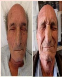
Objectives: Hypothenar hammer syndrome is a rare clinical entity, typically presenting unilaterally, in middle-aged men, with a history of persistent trauma to their hypothenar eminence. We report a case of bilateral hypothenar hammer syndrome in a 77-year-old man with no recent occupational exposure.

Methods: We performed an extensive literature search using the Pubmed database and identified less than 175 results on hypothenar hammer syndrome. Only 12 articles referenced bilateral hypothenar hammer syndrome. Additionally, only 16 articles included patients 65 and older. The following search terms were used: hypothenar hammer syndrome, bilateral, “two aneurysms,” “multiple aneurysms,” and symptoms.

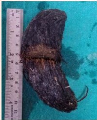
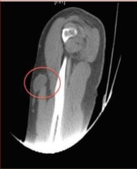
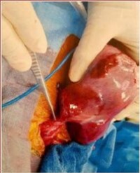
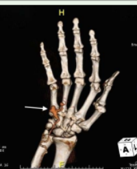
Results: A 77-year-old man who presented with pulsatile hypothenar masses bilaterally and was diagnosed with hypothenar hammer syndrome. He worked as a carpenter 15 years before presentation. He currently uses a rolling walker. CT angiography confirmed the presence of two aneurysms in his dominant right hand, the largest measuring 1.8 cm in diameter. The patient had a motor function deficit along the distribution of the ulnar nerve on the right hand. He underwent resection of the aneurysms in the right hand and reconstruction of the ulnar artery.

Conclusion: Unusual aspects of this case include age of presentation, bilateral aneurysms, and lack of recent occupational exposure. We assert that the use of a rolling walker may have contributed to the progressive aneurysmal degeneration of his ulnar artery.

Charles DeCarlo¹, John Phair², Katie Weichman³, Evan Lipsitz², and Karan Garg²*


Article Image 1

Prenatally Diagnosed Malignant Intrapericardial Teratoma with Yolk Sac Element: Importance of Early Diagnosis and Its Impact on Patient Management

Primary cardiac tumors are rare, less than 0.2% of all childhood tumors. They can be diagnosed prenatally. Although they are usually benign, they can be fatal because of the mass effects. We present a case of a prenatally diagnosed cardiac teratoma in a female fetus at 29 weeks gestation. The mass was large and the fetus showed hydrops. She was delivered at 32 weeks and underwent emergency surgery. Histologically, the tumor showed malignant yolk sac elements. One year follow up showed no recurrence. This case represents the importance of prenatal diagnosis and a multi-disciplinary approach to ensure favorable outcome.

Laura Thompson¹, Adina Alazraki², James W Parks³, Shri Deshpande³, Saneeha Shahid¹, Nasim Khoshnam¹, Sydney R Willis¹, and Bahig M Shehata¹*


Article Image 1

Surgical Reconstruction of Microtia with

Microstomia refers to limitation in mouth opening. It is usually seen as an acquired situation but it can also be seen congenitally. As a result, it leads to aesthetic and functional deformities. Narrowing of mouth opening causes restriction in intake of nutrients, implantation of prostheses, ensuring dental hygiene and dental treatment, making intubation difficult or even impossible. In this article, we present the result obtained by the “fishtail flap” method with commissuroplasty in a case referred to the microstomia seen after a surgical procedure.

Melis Demirag Evman*, Sedat Aydin, Hakan Avci, and Nazmiye Yogurtcu


Article Image 1

Stiff Person Syndrome

Stiff Person Syndrome is a rare neurologic disorder characterized by progressive rigidity and stiffness and linked with features of an autoimmune disease. Stiffness, which primarily influences the truncal muscles, eventually leads to postural deformities. Here, I report a case of Stiff Person Syndrome in association with thyroiditis, epilepsy and vitiligo.

Norah A Alharbi*


Article Image 1

Shoulder Pain as the First Symptom of Advanced Breast Cancer

Shoulder pain is one of the most common orthopaedic complaints in clinical practise. Its high prevalence makes it difficult to distinguish between potentially harmful disorders and other more benign pathologies, so clinicians must pay attention to the well-known red flags. The authors present an atypical case of shoulder pain in which the adequate knowledge of red flags allowed the diagnosis of a systemic disease. Thirty-one-year-old female suffering with severe shoulder pain on physical examination and an axillary mass. Imaging studies suggested it to be a metastatic lesion. Further exams and biopsies were obtained, revealing a poorly-differentiated malignancy, positive for vimentin and cytokeratin, confirmed to be a breast malignancy. This clinical report alerts orthopaedists to the need of valuing red flags in order to adequately advise their patients.

Rita Grazina*, Henrique Sousa, Renato Ramos, Moisés Ventura, André Costa, and Rui Lemos


Article Image 1

Sternal Horns: A Thoracic Cage Variant

Abnormalities of the thoracic ribs are rare (thought to be less than 0.5% of the population). We present a case of a middle-aged man who, presenting with chest discomfort and mild shortness of breath was found to have an abnormal thoracic cage on imaging. His chest radiograph showed bony outgrowths from the manubrium sterni, giving the appearance of what we have termed ‘sternal horns’. The abnormality was further evaluated with computed tomography 3-D volume rendered imaging.

Peter Creber*, David Smith, and Ladli Chandratreya


Article Image 1

Severe Course of Morbus Lemierre Necessitating Extracorporeal Life Support

Lemierre’s disease, also known as Post-Angina-Sepsis, describes an acute oropharyngeal infection caused by Fusobacterium necrophorum that is complicated by thrombophlebitis of the V. jugularis interna and septic pulmonary emboli. Sepsis and pulmonary embolism may result in life-threatening hemodynamic instability affording interdisciplinary intensive care. Here we report about the life-threatening course of M. lemierre in a young female patient with successful surgical and anti-infective treatment supported by veno-arterial ECMO therapy.

Stephanie Kraus¹, Andreas Kremser¹, Marcus Creutzenberg¹, Hermann Hilber², Reiner Neu³, Alois Philipp³, Stephan Schroll⁴, Bernhard Graf¹, Thomas Bein¹, and Andreas Redel¹*


Article Image 1

Ectodermal Dysplasia-A Case Report

Hypohidrotic ectodermal dysplasia is a congenital, non-progressive disorder characterized by hypodontia, hypohidrosis and hypotrichosis. It is inherited in an autosomal dominant, autosomal recessive, or X-linked patterns. The diagnosis is established by genetic tests or after infancy, based on physical features. In some patients, the pattern of inheritance is determined by family history and in others by molecular genetic testing. Characteristic changes in teeth in these patients are: both deciduous and permanent teeth are affected, the alveolar ridges are hypoplastic, missing teeth or retarded growth of teeth, peg-shaped, tooth enamel is also defective. Dental treatment is necessary and children as young as 2 years may need dentures. Through this manuscript, we report a case of hypohidrotic ectodermal dysplasia.

Ambarkova V¹*, Jovanovska M², Bajraktarova E³, Batra M⁴, and Popovski V⁵


Article Image 1

Surgical Management with a Single Operation of Intrathoracic Appendicular Abscess: A Clinical Observation and Literature Review

The authors describe the case of intrathoracic appendiceal abscess associated with right diaphragmatic hernia (hiatal hernia) discovered in a 52 years old woman. The surgical treatment consisted of conventional laparotomy appendectomy after reduction of paraoesophagal hernia into the abdominal cavity in the first time. In second time, we realised a pleural decortication using esophageal hiatus like an uniportal video-assisted thoracic surgery and at the end, reparation of paraoesophagal hernia. The clinical course was satisfactory. A review of the literature allowed us to understand and discuss the diagnostic and surgical approches of this association of two pathologicals entities, benign and anodyne in its isolated and uncomplicated clinical forms. The available literature on intrathoracic appendicitis is reviewed.

Razafimanjato NNM¹*, Ravoatrarilandy M¹, Hunald FA², Samison LH², and Rakotovao HJL¹


Article Image 1

Topical Treatment of Vaginal Melanoma in situ with Imiquimod (Aldara

Melanomas of the vulva and vagina comprise less than 2% of melanomas in women and the overall prognosis is poor. Imiquimod (Aldara®) cream has shown to be effective in non-gynecologic cases of melanoma in situ but is less frequently used in gynecologic cases. We report a case of vaginal melanoma in situ treated with imiquimod. The usual treatment of vaginal melanoma in situ and vaginal melanoma is surgery. There are only a few case reports in the literature that show the effect of treatment of vaginal and vulvar melanoma in situ with imiquimod.

Charles RBW¹*, Van de Vijver KK², Hermans RHM³, and Lok CAR¹


Article Image 1

Endocervical Adenocarcinoma in situ with Replacement of the Entire Corpus Endometrium

Adenocarcinoma in situ (AIS) of the uterine cervix is a premalignant glandular condition and is the precursor to cervical adenocarcinoma. Extension of endocervical AIS proximal to the endocervical canal up to the endometrium of the corpus uteri, fallopian tubes and even the ovaries is rare. In the present case we describe a very unique pattern in which AIS has spread, starting from within the endocervical canal all the way up covering the entire inner lining of the uterus and into the salpinges but not in the ovaries.

Charles RBW¹*, Van de Vijver KK², and Lok CAR¹


Article Image 1

Idiopathic Pulmonary Fibrosis Associated with Alpha1-Antitrypsin Deficiency: Concomitant Finding or Real Association?

Background: Idiopathic Pulmonary Fibrosis (IPF) is a chronic and progressive fibrotic interstitial lung disease of unknown etiology. It is unrelated to Alpha1-Antitrypsin Deficiency (AATD). Despite the progress in the pathogenetic knowledge, many aspects are still unclear. Lung fibrosis is actually regarded as a consequence of a chronic epithelial lung injury characterized by irreversible fibroblast activation and abundant amounts of collagens and other extracellular matrix substances deposition. Several proteins involved in oxidant/antioxidant balance and protease/antiprotease equilibrium have been associated with lung fibrogenesis, although AATD has never been clearly correlated with IPF development.

Case presentation: In this paper, we describe the history of twin homozygous sisters with a familiar homozygous Z type deficiency for Alpha1-Antitrypsin (AAT). One presented liver cirrhosis and the other twin sister developed IPF. This IPF patient with Z/Z mutation and very low AAT serum concentration had no signs of pulmonary emphysema, asthma or liver cirrhosis but she showed only radiological findings of IPF, and she started antifibrotic therapy.

Conclusion: An unusual association between the most common mutation in AAT (Z/Z mutation) and IPF development was reported in this letter supporting the hypothesis that antiprotease AAT maybe involved in IPF pathogenesis.

Alessandro Giuseppe Calabrò¹, Elena Torricelli¹, Elisabetta Rosi¹, Chiara Cresci¹, Anna Maria Grosso¹, Moroni Chiara², Katia Ferrari¹, Massimo Pistolesi¹, Luca Voltolini³, and Elena Bargagli¹*


Article Image 1

Laparoscopic Removal of Intrauterine Device from the Wall of Sigmoid Colon

Laparoscopy is a suitable method for the diagnosis and surgical retrieval of the perforated IUD. A case of uterine perforation caused by an IUD is presented. The IUD was located by using x-ray

Ashraf A Foda*


Article Image 1

Cytomegalovirus Infection and Multiple Venous Thrombosis: A Rare Complication of a Common Infection

Background: Cytomegalovirus (CMV) is a common cause of viral infection. In immunocompetent patients, CMV infection is usually mild or asymptomatic and does not require any treatment. However, in some patients systemic manifestations can occur.

Case presentation: We report the case of a 40-year-old woman with a medical history of Graves’ disease and papilloma virus infection presenting with swollen ankles associated with fever and pain in the lower limbs for several weeks. Clinical examination revealed bilateral in duration of the calf. Laboratory tests showed elevated liver enzymes and systemic inflammation. Exploration by ultrasound of lower limbs and CT scan of the abdomen demonstrated superficial venous thrombosis of the lower limbs and thrombosis of the left hepatic vein. A thorough evaluation for hypercoagulability and septic states were negative. Finally, serological conversion for CMV was demonstrated with positive IgM and IgG with negative prior testing done seven years earlier. The patient was given an anticoagulation treatment and oral contraception was discontinued. The patient improved clinically and biologically after few weeks.

Conclusion: We describe a rare case of sub-acute CMV primary infection in an immunocompetent patient complicated with multiple thromboses. This case illustrates a rare complication of a common infection in order to raise awareness of this entity.

Gauthier Molls*, Valérie Gangji, and Muhammad S Soyfoo