SM Journal of Clinical Anatomy

Archive Articles

Article Image 1

Assessment of an Anatomic Variant That May Mimic Prefracture Findings of Drug-Associated Atypical Femoral Fractures on Conventional Radiographs: The Third Trochanter

Introduction: The study objective was to assess lateral femoral cortex variants that may mimic prefracture findings of drug-associated atypical femoral fractures (AFF) among hip radiographs.

Materials and Methods: Bilateral hip radiographs of 1493 consecutive patients (mean age 67.7, 804 women) were reviewed. Hips were positive if localized lateral subtrochanteric femoral cortical thickening (LSFCT) was present. Positive studies were divided into a medication group if history of bisphosphonate or denosumab use was present or a variant group. The medication group was subcategorized into a prefracture group if classing beaking LSFCT or a contralateral AFF was presentor a non-prefracture group. The LSFCT width, femoral head and lesser subtrochanteric distances were measured. Analysis of Variance (ANOVA) was performed (p <0.01) to compare the three groups, with post hoc Tukey HSD evaluation. Cross-sectional imaging for each group was reviewed.

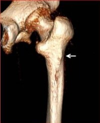
Results: Of the1493 exams, 1079 were included. In the 24 patients with LSFCT, 8 patients were assigned to the medication group and 16 to the variant group. Of the 8, 3 met criteria for the prefracture group and 5 were subcategorized to the non-prefracture group. Differences among the prefracture versus the non-prefracture and variant groups were statistically significant (p<0.01). Cross-sectional imaging of the correlated LSFCT with the third Trochanter posterolaterally in the variant group and laterally for the prefracture group

Conclusions: The third trochanter is an anatomic variant that may mimic prefracture findings of drugassociated AFF. The third trochanter can be differentiated radiographically from prefracture AFF findings with a < 3 mm width and < 3mm lesser subtrochanteric distance.


Troy H Maetani1 *, Stacy E Smith2 and Barbara N Weissman2


Article Image 1

Branching Pattern of Collateral Branches of the Subclavian Artery

Introduction: The arteries of the posterior cervical triangle (lateral cervical region) are frequently used for flap design in plastic and reconstructive surgery. In particular, the musculocutaneous flaps, such as the flap of the lower trapezius and the dorsal scapular flap, are based on the integrity and functional availability of the dorsal scapular artery. Likewise, the cervico-dorsal and cervico-scapular flaps are based on the superficial cervical artery. We can assume that the mode of birth of these arteries - and the possibility of their origin directly from the subclavian artery - will have consequences on their vascular dynamics. These consequences can result in good or bad viability of the irrigated flaps. The objective of the present study was to highlight the birth modalities of the subclavian artery branches

Material and Methods: Dissection was performed in 58 anatomical regions from 32 non-formalin fixed cadavers. The cadavers had no history of surgery or deformity in the areas targeted for dissection (supraclavicular and dorsal regions). They were embalmed using a glycerin-rich, formalin-free solution to preserve tissue suppleness.

Results: The average age of the donors was 72.32 years (range, 34–90) and there were 21 males and 11 females. We find some similarities, with a predominance for types D (Cervico-scapular trunk from the thyrocervical trunk: 28%), A (Separate origin of superficial cervical, dorsal scapular, and suprascapular arteries: 19%), and C (Cervico-dorsal trunk from the subclavian artery: 15%).

Conclusion: There are many anatomical variations concerning the birth modalities of the collateral branches of the subclavian artery. Surgical teams should consider these variations when exploring the region of the lateral triangle of the neck.

Philippe Manyacka Ma Nyemb1,2*, Christian Fontaine3, Xavier Demondion3, Maurice Demeulaere3, Fabien Descamps3 and Jean-Marc Ndoye4


Article Image 1

Anatomical Apps and Smartphones: A Pilot Study with 100 Graduation Students

Background: Anatomical teaching is going through several changes. Several studies observed that the students are deeming anatomy as outdated and irrelevant. The rates of lectures attendance and interest are declining. In this context, tools such as body painting, board games and team quizzes are appearing in the classroom to aid and motivate the student. Often, these complementary tools showed positive results towards their goals in the learning process of several branches of medicine. The smartphone has also been used for these purposes, although the literature lacks studies regarding anatomy. The study conducted herein aimed to create a pilot version of an anatomical application for the smartphone and disseminate its usage in nursing students

Methods: A smartphone application was self-produced with the tool known as Fábrica de Aplicativos© (https://fabricadeaplicativos.com.br) and consisted of anatomical terms and their meaning (Dictionary of Anatomy). The students had the opportunity to use this application during the semester and by the end of the period they answered a questionnaire with questions about the quality and efficacy of the app, with the option of submitting suggestions for its improvement.

Results: 100 graduation students were enrolled in this study. The overall evaluation of the dictionary was excellent/great (65%), while critics such as the lack of better images, the lack of a search resource and the lack of sections were pointed out.

Conclusion: As such, it was observed that the smartphone may have a positive effect in the learning process of anatomy, as demonstrated by our study

Lucas Alves Sarmento Pires1 , Tulio Fabiano de Oliveira Leite2 , Albino Fonseca Junior1 , Marcio Antonio Babinski1 and Carlos Alberto Araujo Chagas1


Article Image 1

A Symptomatic Multiseptate Gallbladder without Gallstones

Multiseptate Gallbladder is a rare congenital abnormality, which can be totally asymptomatic or responsible for biliary pain in the absence of cholelithiasis or cholecystitis. In case of persistent symptomatology, cholecystectomy represents the treatment of choice

Damien Dousse1 *, Laura Marcu2 and Francesco Martini1


Article Image 1

Evidence for Endogenous Neurosteroid Production in the Mammalian Olfactory Mucosa: Immunocytochemical Localization of Cytochrome P450 SideChain Cleavage Enzyme

Steroid hormones and their metabolizing enzymes have previously been identified in the olfactory mucosa. Enzymes of the cytochrome P450 family that are thought to play a role in the metabolism or activation of airborne toxins have also been identified in mammalian olfactory mucosae. In the synthesis of steroid hormone, cholesterol is transported to the mitochondria where the side-chain cleavage enzyme (cytochrome P450scc) converts cholesterol into pregnenolone. Conversion of cholesterol to pregnenolone is an obligate step in steroid hormone production. The specific aim of this study is to identify cytochrome P450scc in the mammalian olfactory mucosae. Using polyclonal antibodies to cytochrome P450scc, we found immunoreactivity for cytochrome P450scc in the rat olfactory mucosa. Within the olfactory epithelium, the supranuclear region of sustentacular cells was immunoreactive for cytochrome P450scc. Olfactory neurons, basal cells, olfactory nerve axons and acinar cells of Bowman’s glands were unstained. In positive control tissue (adrenal glands), staining for cytochrome P450scc was seen in all layers of the adrenal cortex. The localization of cytochrome P450scc to sustentacular cells is consistent with a functional role for mitochondrial cytochrome P450scc in the production of olfactory mucosa-specific neurosteroids. These endogenous neurosteroids are most likely involved in the modulation of olfactory function

James D Foster*


Article Image 1

Anatomical and Anthropological Investigation of the Articular Surface of the Human Glenoid Cavity in Brazilian Corpses

The glenoid cavity of the scapula shows high morphological variability that has not been clearly elucidated in the literature. Its morphological aspects knowledge can be used in the sex determination, to improve the development of more functional prostheses and in the accurate image diagnostic. The purpose of the present study was to analyze and compare sex and dominance differences in the articular surface of the human glenoid cavity. Anthropometric measurements of the glenoid cavities of scapula were taken of 200 specimens, besides we developed a morphological classification system according the shape of inferior and superior poles and the presence of the glenoid notch in the anterior margin. For all anthropometric parameters, the male scapula showed higher values than the measures in female scapula both in right and left sides, but we did not find a significant difference between the left and right scapula in either sex. On the other hand, taking into consideration the anthroposcopic aspects, 50% of male and 58% of female were classified in a different morphological group when compared to the contralateral glenoid in the same individual. The present study revealed an evident sexual dimorphism and provides information about the articular surface of normal glenoid cavities to improve the diagnosis of orthopedic lesions, to produce better prosthesis and to improve the medico-legal identification

Jefferson M Homem1 , Aline S DeMaman3 , Denise Lachat1 , Ariane Zamarioli2 , José A Thomazini1 and João-José Lachat1 *


Article Image 1

Decoupling Interrupts from Red-Black Trees in Model Checking

Congestion control and red-black trees, while technical in theory, have not until recently been considered appropriate. In fact, few security experts would disagree with the construction of DHCP. Though it is regularly a practical goal, it is buffeted by previous work in the field. We introduce an analysis of IPv7, which we call Lakin.

Candela Bro de Velas, Aitor Tilla, Borja Mon de York and Josetxu Leton*


Article Image 1

Learning Strategies of Anatomy in First and Second Years of Medical Studies

Introduction: For many years, one of the current trends has been to give to medical students the tools they need to benefit from the best possible strategic skills for the learning of medicine. Anatomy is one of the essential disciplines for the implementation of this change. This process must involve identifying the learning styles used by the students. The purpose of this work was to describe the learning styles developed by the students of the Saint-Louis Faculty of Medicine during the anatomy course

Materials and methods: A descriptive and cross-sectional study was conducted among the first and second year medical students. An anonymous questionnaire was used to gather the opinion of the students.

Results: All students answered the questionnaire. The sample of 133 students was made of 98 men and 35 women. The learning styles of our students were varied, but they had a preference for intuitive methods

Conclusion: It is important to adapt the teaching of anatomy to the learning styles of our medical students. An effort must also be made to help students diversify their learning strategies.

Philippe Manyacka Ma Nyemb*


Article Image 1

Organization of a Student Peer Tutoring For the Learning of Anatomy

Introduction: In the first and second years of medical studies at the Saint-Louis Faculty of Medicine in Senegal, students receive a complete teaching in anatomy. To better follow this teaching, it has been introduced tutoring in anatomy, managed by peer students. The aim of our study was to assess the global opinion of medical students on this pedagogical experience

Materials and Methods: An anonymous questionnaire combining open and closed questions was designed and administered to 133 first and second year medical students

Results: The tutored students were satisfied with the principles and objectives of tutoring, the planning, organization and running of tutoring, as well as the pedagogical relationship and the communication around tutoring

Conclusion: In anatomy, student peer tutoring is an interesting experience, as tutors act directly in the initial training of first and second year medical students.

Philippe Manyacka Ma Nyemb*


Article Image 1

Apigenin and Losartan Combinations Attenuate Monosodium GlutamateInduced Changes in the Rat Exocrine Pancreas: Histological and Immunohistochemical Study

Background: Monosodium glutamate is an important food additive used as a flavor enhancer. In spite, many investigations showed its toxicity to the human and animals. Apigenin is a natural flavonoid with antioxidant, anti-inflammatory, and anti-proliferation properties. Losartan is Ang II (AT1) receptor blocker, used as an antihypertension and antifibrotic agent. The aim is to investigate apigenin and losartan protection separately and in combinations against monosodium glutamate-induced changes in the rat exocrine pancreas using histological and immunohistochemical studies.

Materials & Methods: 70 Wistar rats used, divided into 7 groups (10 rats each); Group 1: served as control, Group 2 (MSG): rats received monosodium glutamate 2 mg/g body weight/day in drinking water, Group 3 (Api): rats received apigenin 50 µg/rat/day, Group 4 (LST): rats received losartan 10 mg/kg/day, Group 5 (MSG+Api): rats treated with Api 50 µg/rat/day 30 min before MSG, Group 6 (MSG+LST): rats treated with LST 10 mg/kg/ day 30 min before MSG and Group 7 (MSG+Api+LST): rats treated with Api and LST 50 µg/rat/day and 10 mg/ kg/day respectively 30 min before MSG.

Results: MSG group showed marked acinar cell damage, apparently dilated ducts, inflammatory cellular infiltration, congested blood vessels with intimal loss and dissolution of the internal elastic lamina. These besides, significantly increased collagen fibers, significant decrease in PCNA positive acinar cell nuclei and lost E-cadherin expression. Conclusion: Apigenin and losartan combination attenuate MSG damaging effects on the exocrine pancreas. This is through their antioxidant, anti-inflammatory as well as antifibrotic effects.

Samah Kandeel1 * and Mohamed Balaha2,3


Article Image 1

A Detailed Review on the Clinical Anatomy of the Pectoralis Major Muscle

The pectoralis major is a muscle of the upper limb girdle. This muscle has a unique morphological architectonic and a high rate of clinical applications. However, there is lack of data regarding the morphological and functional interactions of the pectoralis major with other muscle and fascial compartments. According to the applied knowledge, the “Humero-pectoral” morpho-functional concept has been postulated. The purpose of this review was the dissectible investigation of the muscle anatomy and literature review of surgical applications.

Alexey Larionov, Peter Yotovski and Luis Filgueira*


Article Image 1

Perception of Medical Students in the Front of an Active Evaluation Model of Cardiovascular Anatomy

The learning model based on active methodologies has been increasingly explored in undergraduate programs, especially in the health area. Many authors support this preference in a better consolidation of knowledge and in a more dynamic learning process. In this context, the present study aims to evaluate the students' perception of medicine against a practical evaluation model of human anatomy. An analytical, descriptive study with quantitative approach was carried out 38 students of the medical course. The students were submitted to a simulation of practical evaluation, in which the students should point out in the anatomical piece questioned structures. They then answered a structured questionnaire. It was observed that 92.10% of the students stated that they had no contact with an active teaching methodology previously and 94.73% reported that they had no contact with this methodology in the area of morphological sciences. When questioned about the active methodology, 92.10% of the students answered that this method would be better for learning consolidation, and 97.36% of the interviewers think there should be more moments with this type of methodology during monitoring activities. It was found that there was a good acceptance of the active evaluation method, thus being an active tool for the discipline of human anatomy

João Erivan Façanha Barreto2 , João Victor Souza Sanders1 *, Hudson Martins de Brito1 , Michelly Carneiro Collyer1 , Francisco Ewerton de Paula Uchôa1 , Jonathan Barros Cavalcante1 , José Ricardo Souza Ayres de Moura3 , Osvaldo Pereira da Costa Sobrinho1 , Jalles Dantas de Lucena2 , Ivan do Nascimento da Silva4 and Gilberto Santos Cerqueira2


Article Image 1

Lantana Trifolium Ethanolic Extract has a Protective Potential against Ethambutol Induced Changes in Visual Acuity

Introduction: Ethambutol (EMB), an important drug in treatment of multidrug resistant tuberculosis, has been associated with severe side effects including visual impairment.

Purpose: To establish the protective potential of Lantana trifolium ethanolic extract against EMB induced changes in visual acuity.

Materials and methods: Experimental design involving 25 male adult Wistar rats of 110-130g average weight, divided into five groups each comprising five animals. Group A, the negative control received distilled water. Group B, the positive control was treated with EMB 100 mg/kg/day. Test groups C, D, and E were treated with 25, 50, and 100 mg/kg/day of Trifolium Extract (TE) respectively, one hour before administering 100 mg/ kg/day of EMB for five weeks. Visual acuity was determined by the mean escape latencies obtained using a modified Morris water maze

Results: Lantana trifolium ethanolic extract had a dose dependent protective potential against EMB induced changes in visual acuity. This was shown by the significant increase in the escape latencies of positive control group when compared with those of the group A (4.35±0.50), D (4.85±0.65), and E (3.6±0.38). This effect is likely due to the ability of Latana trifolium to prevent inflammation and accumulation of anti-oxidants in the optic nerve.

Conclusion: Lantana trifolium ethanolic extract has a dose dependent protective potential against EMB induced changes in visual acuity. Studies to determine the exact phytochemical component and mechanism of action responsible for this effect should be conducted.

Owembabazi Elna1 *, Ninsiima Herbert Izo2 , Keneth Iceland Kasozi2 , Abili Sadrax3 , Ssempijja Fred1 , Echoru Isaac1 and Bukenya Edmund1