Back to Journal

Journal of Radiation Oncology & Research

Primary Diffuse Large B cell Lymphoma of the Uterus: A Case Report and Literature Review

[ ISSN : 3068-0832 ]

Abstract Citation Introduction Case Report Discussion References
Details

Received: 01-Oct-2018

Accepted: 22-Oct-2018

Published: 31-Oct-2018

Qianqian Wang1, Wenjuan Wang2, Wanfeng Fan1, Qian zhou1, Liguang Yang1 and Xinjiang Liu1*

1Department of Radiology, Binzhou Medical University Hospital, China

2Binzhou Medical University, China

Corresponding Author:

Xinjiang Liu, Department of Radiology, Binzhou Medical University Hospital, Binzhou 256603, China, Tel: 13356286368

Keywords

Diffuse Large B-Cell Lymphoma (DLBCL); Primary Female Genital System Lymphoma (PFGSL); Extranodal lymphoma; Diagnosis

Abstract

A 64-year-old woman who was found to have an abdominal mass on ultrasound was misdiagnosed with a uterine fibroid/uterine sarcoma. Pelvic MRI showed multiple soft tissues mass shadows in the pelvic cavity, and the larger shadows were approximately 14.2 cm × 9.7 cm × 15.6 cm, with clear boundaries dominated by an iso long T1 and slightly longer T2, locally visible nodular long T2 and short T2 signal foci, and obvious high signals on DWI and decreased ADC signals. The enhancement of the densification was obviously non-uniform. The immunohistochemical results were positive for CD20, LCA, PAX-5+, CD43 +, c-myc, bcl2 and bcl6 and negative for SAM, Desmin, Myo D1, MPO, CD99, CD30, CD23, S-100, and CD10. The majority (90%) of tumor cells were positive for Ki-67. The diagnosis was a diffuse large b-cell lymphoma. Preoperative diagnosis of primary lymphoma of the uterus is difficult in clinical situations. Primary Female Genital System Lymphoma (PFGSL) is a rare disease that is mostly associated with non-Hodgkin’s lymphoma, usually Diffuse Large B-Cell Lymphoma (DLBCL). The majority of primary uterine DLBCL originates from endometrial stroma

Citation

Wang Q, Wang W, Fan W, Zhou Q, Yang L and Liu X. Primary Diffuse Large B cell Lymphoma of the Uterus: A Case Report and Literature Review. J Radiat Oncol Res. 2018; 2(1): 1007.

Introduction

The global incidence of Non-Hodgkin’s Lymphoma (NHL) is 3%, of which 1% comes from extranodal lymphoma [1]. Diffuse Large B-Cell Lymphoma (DLBCL) is the most common histologic subtype in extranodal lymphoma, accounting for approximately 50% of cases [2]. Primary Female Genital System Lymphoma (PFGSL) of the reproductive system is a rare disease, accounting for only 0.21-1.1% of extranodal lymphoma [2,3]. Frequently occurring parts include the ovary, uterus, vagina and vulva. This paper reports a case of primary lymphoma of the uterus that was misdiagnosed as uterine fibroids.

Case Report

Clinical evidence

The female patient, who was 64 years old, was found to have uterine fibroids by a physical examination 23 years ago. Natural menopause occurred 11 years ago, with no abnormal vaginal bleeding after menopause. Over the past 2 months, the tumor in the lower abdomen was spontaneously palpable and hard, and a color ultrasound examination performed 4 days ago suggested a hypoecho in the pelvic cavity, possibly fibroids with diameters of approximately 10 cm. The patient had no nausea and vomiting, occasional abdominal distension, no diarrhea, no abdominal pain, discomfort in the lumbosacral region, anorectal heaving and distension in January, as well as no frequent urination, urgent urination or pain; no insensitivity to urination; and no abnormal leucorrhea.

Gynecological examination

Vulva development is normal; married and delivered; vagina unobstructed, smooth wall; cervical atrophy, incomplete exposure due to tumor compression, smooth palpation surface, no lift pain and sway pain; palpation of the uterus is not clear; the pelvic cavity touches a hard surface; irregular, movement is acceptable; the upper edge reaches the level of the umbilicus, no obvious tenderness; mild tenderness in the left adnexal region. No obvious abnormalities were found in the right accessory area.

Intraoperative observations

The uterus increases with size, such as during pregnancy, and congestion, with a brittle, anterior wall only exposed at the bottom of the palace; most of the anterior wall had edema thickening and hardening of the urinary bladder as well as invasion adhesion coverage; double accessories were not observed, but there was an obvious abnormal appearance; the rectum anterior wall had bumps that were approximately 5cm in diameter; the oppressed rectum was larger than the other small intestine segments and was mesenteric was the greater omentum 0.88cm in size with multiple lesions; most of the lines indicate a uterus excision and double appendix resection.

Imaging findings

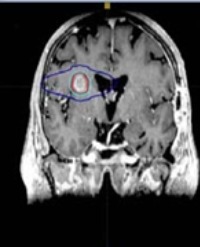
There were multiple large soft tissue shadows in the pelvic cavity, and the larger ones were approximately 14.2cm × 9.7cm × 15.6cm in size; the boundary was still clear. The lesions were characterized by an equal length T1 and slightly longer T2, with locally visible nodular long T2 and short T2 signal foci. The DWI signal was obviously high and the ADC signal was reduced. The part of the enhancement showed obvious non-uniform reinforcement. The uterus was imbedded, the normal musculoskeletal structure was unclear, and the posterior part of the lesion was closely related to the rectum, as shown in Figure 1.

Figure 1: A 64-year-old woman, Primary female genital system lymphoma. Pelvic MRI showed multiple soft tissue mass shadows in the pelvic, T2 coronal position, T2-fs sagittal position, T2-fs axis position, Obviously high signal on DWI, Low Signal on ADC. The part of the enhancement showed obvious non-uniform reinforcement.

Pathology findings

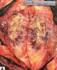
The uterine volume was approximately 6×6×5.5 cm, and the endometrium was smooth with a mass under the endometrium, with a volume of 14×13×10.5 cm. There were numerous white nodules in the left mesosalpinx, with a diameter of 0.1-1.3 cm, and the volume of the left ovary was 1.7×0.8×0.6 cm. There was one gray-white nodule in the mesosalpinx and the right ovary area each, a volumes of 2.7×1.7×0.8 cm and 3.5×3.5×2.5cm, respectively. The cut surface was all soft and gray-red, and there was 1 white fish-like nodule (mesenteric metastasis), with a volume of 3×2.5×1.2 cm, as shown in Figure 2.

Figure 2: Diffuse large b-cell lymphoma, the Pathological gross specimen. The microscopic 10 x10.

We concluded that the masses represented diffuse large b-cell lymphoma.

Immunohistochemical

CD20+, PAX-5+, CD3 -, CD5 -, CD43+, CD10-, the Bcl- 6part+, scattered in weak MUM - 1+, the Bcl - 2+, cyclin D1 scattered +, CD23 -, CD30 -, c - myc part weak +, LCA +, CK-, Vimentin part +, SMA -, Desmin-, MPO -, Myo D1-, Myogenin -, CD99 -, S-100 -, ki - 67 approximately 90% (Table 1).

Table 1: Description of the immunohistochemistry and its significance.

Marker Description Status in our Significance
patient
CD20 Pan-B-cell antigen + Indicates B-cell origin
CD43 Pan-T-cell antigen + Indicates T-cell origin
CD30 Tumor necrosis factor receptor; - Used in diagnosis of Hodgkin's lymphoma
lymphocyte activation antigen
CD3 pan-T-cell antigen - Indicates T cell origin of lymphocytes
BCL6 Germinal center marker + positive in T-cell, DLBCL, Burkitt’s, Follicular, and Hodgkin's lymphomas
BCL2 Proto-oncogene; prevents cells from + positive in Follicular, Burkitt’s, DLBCL, Hodgkin's, Mantle Cell, and
Marginal Zone lymphomas
Multiple Myeloma 1 Intra- and post-germinal center B-cell + Helps distinguish between germinal center and non-germinal center DLBCL
(MUM1) marker
Ki-67 Labile, non-histone nuclear protein. Marker 0.9 Higher nuclear staining indicates more aggressive tumor
of cell proliferation
CD10 Cell membrane metallopeptidase; germinal - positive in germinal center DLBCL, Burkitt’s, Follicular, Hairy cell
center marker lymphomas. Occasionally expressed in uterine smooth muscle tumors
Smooth muscle actin Expressed by smooth muscle cells - Indicates smooth muscle origin
(SMA)
Desmin Expressed by smooth muscle cells - Indicates smooth muscle origin
S100 Cytoplasmic EF-hand Ca2+-binding - Marker of neural tissue and melanocytic differentiation
protein

Discussion

PFGSL is a rare disease that is mostly associated with non-Hodgkin’s lymphoma, usually diffuse large b-cell lymphoma [4]. Nasioudis et al. [3] found that 667 cases in the SEER database were primary tumors in female genital lymphoma from 1988 to 2012. The most common histological subtypes were diffuse large B cells (59.8%) and follicular lymphoma (11.9%), which occurred in the ovary (37%), cervix (21.4%) and uterus (16.5%). Of these, 62.8% underwent surgery. The majority of primary uterine DLBCL originated from endometrial stroma. The discovery of DLBCL in uterine fibroids has been reported only twice before. Zhao [5] reported a 73-year-old female with an endometrial mass of 17 cm, which was diagnosed as a primary DLBCL. The DLBCL originated from uterine fibroids, and the tumor extended to the adjacent myometrium, including intravascular tumor thrombosis. Alexandra Martin [2] reported a case with a uterine mass of 10×8×5.8 cm, with white-brown orbicular nodules. The biopsy showed benign hysteromyoma accompanied by mucinous changes, and hysteromyoma showed abnormal nodules infiltrated by local lymphocytes. The tumor was composed of large atypical lymphocytes with an obvious enhancement of mitosis and active apoptosis in large nuclei and diverse nucleoli. Ana Regalo [6] reported a large b-cell lymphoma of the cervix. Pelvic MRI showed an uneven mass of the cervix, the size of which was 7.9×7.6×6.9 cm; interstitial destruction; and obvious involvement of the bladder. Patro [7] reported a case of uterine primary diffuse large b-cell lymphoma with a specimen size of 13.5 ×10× 8 cm. The myometrium was diffusely thickened, with a fish-like appearance. On immuno-histochemical evaluation, tumor cells were found to be immuno-positive for CD 20, and LCA and were immuno-negative for CD 3, CD 5, MPO, CD 23 and CD 10. p53 showed a positive response, and 80% of tumor cells were positive for Ki-67.

A preoperative diagnosis of primary lymphoma of the uterus is difficult in clinical situations. Patients with a primary lymphoma of the uterus often have no symptoms or non-specific abdominal symptoms, including abdominal pelvic pain, abdominal distension, vaginal bleeding or bleeding in their urine, or gastrointestinal symptoms [8,9]; only 17% of non-Hodgkin’s lymphoma patients reported physical symptoms, such as fever, night sweats or weight loss. Zhu Yilu [10] believed that the significant high signal of diffusion weighted imaging (DWI)was characteristic of uterine lymphoma on MRI, but the final diagnosis still required pathology. Fox and Vang [11] et al. developed the following diagnostic criteria [2,7,9]: 1) the reproductive system is the only affected organ; 2) no bone marrow or peripheral blood abnormalities; 3) no history of lymphoma; and 4) the presence of lymphoma at a distant site should be separated from the primary lymphoma for several months.

Genital lymphoma is rare, without standard treatment guidelines, and is often treated with surgery and chemotherapy combined with radiotherapy according to the patient’s situation. Diffuse large b-cell lymphoma chemotherapy usually adopts the r-chop regimen, namely, rituximab combined with cyclophosphamide, doxorubicin and prednisone [12,13]. Tarec [14] believes that DLBCL involvement in the uterus is associated with a high risk of secondary central nervous system involvement, and these patients should be considered for central nervous system staging and prevention.

References

1. Cubo AM, Soto ZM, Cruz MA. Doyague MJ, Sancho V, Fraino A, et al. Primary diffuse large B cell lymphoma of the uterine cervix successfully treated by combined chemotherapy alone: A case report. Medicine (Baltimore). 2017; 96: 6846.

2. Martin A, Medlin E. Primary, non-germinal center, double-expressor diffuse large B cell lymphoma confined to a uterine leiomyoma: A case report. Gynecol Oncol Rep. 2017; 22: 78-81.

3. Nasioudis D, Kampaktsis PN, Frey M. Witkin SS, Holcomb K. Primary lymphoma of the female genital tract: An analysis of 697 cases. Gynecol Oncol. 2017;145: 305-309.

4. Samama M, van Poelgeest M. Primary malignant lymphoma of the uterus: a case report and review of the literature. Case Rep Oncol. 2011; 4: 560-563.

5. Zhao L, Ma Q, Wang Q, Ying Zeng, Qingya Luo, Hualiang Xiao. Primary diffuse large B cell lymphoma arising from a leiomyoma of the uterine corpus. Diagn Pathol. 2016; 11: 9.

6. Regalo A, Caseiro L, Pereira E, José Cortes. Primary lymphoma of the uterine cervix: a rare constellation of symptoms. BMJ Case Rep. 2016; 2016.

7. Patro MK, Bal AK, Santosh T, Mishra B. Primary Non-Hodgkin’s Lymphoma of Diffuse Large B-Cell Phenotype [DLBCL] of Uterine Corpus: A Rare Case Report with Brief Review of the Literature. J Obstet Gynaecol India. 2016; 66: 675-678.

8. Chen R, Yu Z, Zhang H, Ding J, Chen B. Primary malignant lymphoma of the uterus and broad ligament: a case report and review of literature. Onco Targets Ther. 2015; 8: 265-268.

9. Nasioudis D, Frey MK, Chapman-Davis E, Caputo TA, Holcomb K. Fertility preserving surgery for advanced stage ovarian germ cell tumors. Gynecol Oncol. 2017; 147: 493-496.

10. Zhu YL, Qian HF, Shen J. MRI diagnosis of primary lymphoma of uterus. Modern practical medicine. 2012; 1052: 1038-1039.

11. Vang R, Medeiros LJ, Deavers MT. Current problems with staging lymphomas involving the ovary. Am J Surg Pathol. 2006; 30: 1202-1203.

12. Shen H, Wei Z, Zhou D, Zhang Y, Han X, Wang W, et al. Primary extra-nodal diffuse large B-cell lymphoma: A prognostic analysis of 141 patients. Oncol Lett. 2018; 16: 1602-1614.

13. Upanal N, Enjeti A. Primary lymphoma of the uterus and cervix: two case reports and review of the literature. Aust N Z J Obstet Gynaecol. 2011; 51: 559-562.

14. El-Galaly TC, Cheah CY, Hutchings M, Mikhaeel NG, Savage KJ, Sehn LH, et al. Uterine, but not ovarian, female reproductive organ involvement at presentation by diffuse large B-cell lymphoma is associated with poor outcomes and a high frequency of secondary CNS involvement. Br J Haematol. 2016; 175: 876-883.

Other Articles

Article Image 1

Volumetric Modulated Arc Therapy Versus 3D Conformal Planning Technique for Esophageal Cancer: Should Field Based Planning Be the Universal Standard?

Carcinoma of the esophagus is among the most rapidly increasing cancers in incidence. With the use of aggressive bimodality and trimodality treatment strategies, the reduction of treatment toxicity is of prime importance [1]. Radiotherapy plays a key role in definitive, adjuvant and neoadjuvant treatment for carcinoma of the esophagus. Due to extensive vascular and lymphatic drainage, and therefore tendency to present at an advanced stage, the volumes required to adequately cover gross disease are substantial. Other critical organs in close proximity are therefore at risk for radiotherapy-induced toxicity, including the lung parenchyma, heart, spinal cord, stomach and others. Conventional 3D conformal radiotherapy techniques (3 field or 4 field) have traditionally been used at our center to provide adequate coverage to the target volume of the esophageal tumor and lymph nodes; however as a consequence doses delivered to these Organs At Risk (OARs) may be high. Intensity Modulated Radiotherapy (IMRT) and Volumetric Arc Radiotherapy (VMAT) has also been considered in the past, however comparisons of these plans have shown variable results in normal tissue sparing. Furthermore these techniques may impart a higher volume of low dose radiotherapy to substantial amounts of normal tissue. We compared conventional 3D CRT plans with IMRT or VMAT plans for a series of esophageal cancer patients with tumors of varying location at our center to determine the optimal treatment planning strategy.

Elysia Donovan1*, Tom Chow2, Jack Skoczny1 and Ranjan Sur1


Article Image 1

Definitive Radiotherapy for the Treatment of Non-Small Cell Lung Cancer in Patients Aged 70 Years and Older

Background: The factors affecting survival were evaluated in patients aged ≥ 70 years with Non-Small Cell Lung Cancer (NSCLC) treated with definitive Radiotherapy (RT).

Methods: Between January 1996 and April 2012, 52 patients were treated. The median age was 73 (range 70-80) and 73% and 75% of patients with stage III according to AJCC 2002 and AJCC 2010 staging, respectively. Radiotherapy was performed median 6160 cGy (range: 3600-6660 cGy) and CHE were given 75% of the patients.

Results: Median follow-up was 12.5 (range: 2.5-103) months. Median overall (OS) and Disease-Free (DFS) Survival were 22 and 18.5 months, respectively. Radiotherapy related death was not observed. In Univariate analysis; AJCC 2002 stage I-II, RT dose, RT duration, for OS and RT dose, RT duration, neoadjuvant CHE ≤ 3 cycles, complete response, ≥ 4 cycles of CHE for DFS were significant (p <0.05). In multivariate analysis, RT duration > 49 days related with higher RT dose (≥ 60 Gy) were found a positive impact on OS (HR: 3.235, 95% CI: 1:25 to 8:32 p = 0.01).

Conclusion: In our study, elderly patients with NCSLC can be given ≥ 60 Gy without complications and was seen positively impact on survival.

Süreyya Sarıhan*1, Türkkan Evrensel2 and Deniz Sığırlı3


Article Image 1

Fractionated Stereotactic Radiotherapy and Stereotactic Radiosurgery as Salvage Treatment for Recurrent Malignant High Grade Gliomas

High-Grade Gliomas (HGG) are the most common and aggressive primary brain malignancies in adults. Fractionated Stereotactic Radiotherapy (fSRT) and Stereotactic Radiotherapy (SRS) have emerged as a novel technique to deliver high doses of RT in the recurrent setting with aim for durable local control and potential overall survival benefit. This study evaluated the patterns of recurrence, prognostic indicators as well as the efficacy of treatment in patients with recurrent HGG treated by fSRT or SRS. Favorable outcomes were observed in patients treated with fSRT and SRS as salvage therapy. The predominant mode of failure post salvage SRS and fSRT remains in-field. Prospective trials are required to better define patient selection, tumor recurrence size, and effective dose regimen.

Kevin King*, Philip Blumenfeld, Jacob Y Shin, George Tolekidis and Aidnag Diaz


Article Image 1

Radiotherapy of Malign Melanoma: A Single-Centre Experience from the Eastern Black Sea Region of Turkey

Radiation therapy is used for adjuvant therapy on patients with recurrence or lymphadenectomy in the malignant melanomas. Treatment is used for palliative on metastatic patients. Between 1996 and 2017, 35 patients who received adjuvant radiotherapy and 55 patients who received palliative radiotherapy were retrospectively analyzed. Of the 90 patients included in the study, 56 (62%) were men and 34 (38%) were women. Mean age of patients was 63 years (age range: 22–95 years). For all patients, the overall survival time was 32.23 months (95% CI, 16.8–47.6). As an adjuvant therapy, 48 Gy (hypofractionated) radiotherapy was used in 4 patients and 50-66 Gy radiotherapy was used in 31 patients. Overall survival in these patients was 66.14 (95% CI 36.6-95.6) months. Mean metastases that occur in patients is 37.19 months (range: 2-138 months), which are 37 (41%) patients in the brain, 12 (13%) patients in the bone and 6 (7%) patients in the non-regional lymph node metastasis. 20-30 Gy radiotherapy was used for metastasis treatment. Overall survival in brain metastasis, bone metastasis and lymph nodes metastatic patients were 4.22 (95% CI 2.62-5.82), 7.6 (95% CI 1.14-14.05) and 7.33 (95% CI 0-15.03) months respectively. Although, the regional nodal disease can be favorably controlled with lymphadenectomy and radiotherapy, the risk of distant metastases and the risk of disease-related death are high. In addition, metastasis-related symptoms (particularly pain) can be controlled by radiotherapy.

Mustafa Kandaz1*, Özlem Aynacı2, Alperen Tüysüz3, Emine Canyılmaz1 and Adnan Yöney1


Article Image 1

Examining the Survival benefit of Radiation Therapy on Leptomeningeal Carcinomatosis and Identifying factors Associated with Survival Benefit of WBRT

Purpose: To report the survival benefit of Radiation Therapy (RT) in Leptomeningeal Carcinomatosis (LMC) patients with different primary cancers and to identify the factors of LMC patients that had survival benefit from Whole Brain Radiation Therapy (WBRT).

Methods and Materials: Systematic literature review was performed. Search terms included ‘Leptomeningeal’, ‘prognostic factors’ and ‘lung’ or ‘breast’. Literatures were included if patient profile was reported. Specifically, age, median survival, diagnosis time line, type of cancer, received treatments, performance score, and systemic disease were sought after. Literatures were excluded if they were a) case reports, b) written in language other than English, or c) not including patient profile.

Results: RT treatment has survival benefit for LMC patients with CSF obstruction. For LMC patients with primary breast cancer, 7 studies were found since 1991 that met searching criteria. Factors which would allow breast cancer-caused LMC patients to benefit from WBRT with survival are identified with statistical significance. They include KPS>60, Intrathecal (IT) chemotherapy, systemic therapy, hormonal receptor status and severity of systemic disease. Patients who met aforementioned criteria and received WBRT had longer median survival (17 weeks vs. 11.9weeks, p=0.015). Similarly, 10 studies were found that met searching criteria for Non-Small Cell Lung Cancer (NSCLC) caused LMC. Beneficial factors for NSCLC-caused LMC are identified. They include IT chemotherapy, KPS > 60, and EGFR TKI treatment. Patient who met these criteria had longer median survival when received WBRT (17.6 weeks vs 12.2 weeks, p = 0.0412).

Conclusion: Profiles of LMC patients that can benefit with longer survival by receiving WBRT are identified for breast cancer and NSCLC. Algorithms in identifying such patients are provided.

Jae Kim*


Article Image 1

Anal Canal Squamous Cell Cancer; Pattern of Recurrence and Survival, 25 Years Experience

Background and objectives: Combined chemo radiation is the standard of care for treatment of squamous cell carcinoma of the anal canal. Our objective was to analyze the treatment results of patients with squamous cell carcinoma of the anal canal treated at our institution.

Methods: We identify patients with confirmed diagnoses of anal canal squamous cell carcinoma treated in our institution (KFSHRC), Riyadh between1994-2017. We retrospectively reviewed their pattern of recurrence and survival rate.

Results: 35 males (70%) and 15 females (30%) were identified. Median age at diagnosis was 58 years. 5(10%) patients underwent initial Abdomino Perineal Resection (APR) while 45 patients (90%) received definitive concurrent chemo radiation (30 using 2D/3DCRT and 15 patients using IMRT). All patients completed their planned treatment course except 4 patients (in 2D/3DCRT arm). IMRT resulted in significant decrease in all toxicity grades in comparison to 2D/3DCRT arm (p value 0.035). After median follow up of 13months, two out of the 5 patients (40%) who underwent initial APR had local recurrence; while after 18 months median follow up 8(19.6%) patients who underwent definitive chemo radiation had local recurrence. The 2 and 5 years Disease Free Survival (DFS) were 79.4% and 53% respectively in IMRT group vs 64% and 55% respectively in 2D/3DCRT group (p value 0.79). Regarding Overall survival (OS), the 2 and 5 years OS were 82% and 41% respectively in IMRT vs 66% and 44% respectively in the 2D/3DCRT group(p value 0.36). In Univariate analysis, only number of chemotherapy cycles was statistically correlated with DFS and OS with (p value of 0.02 and <0.0001) respectively.

Conclusion: Combined chemo radiation therapy for anal canal squamous cell carcinoma is effective treatment in term of local control and survival. The recurrence and survival pattern of our patients’ cohort compare favorably to the international results. Radiation therapy using IMRT resulted in significant decrease of all toxicity grades over 2D/3DCRT, with improvement of the 2 years DFS and OS in comparison to the 2D/3DCRT although it was not statistically significant. In Univariate analysis initial Hb level was not significantly correlated with DFS or OS at 5 years, only number of chemotherapy cycles was statistically correlated with survivals at 5 years

Ahmed Elashwah1,3*, Abdullah Alsuhaibani2, Alaa Abduljabbar, Samar Alhomoud4, Luai Ashari4, Shouki Bazarbashi5, Ali Aljubran5, Ahmed Alzahrani5 and Nasser AlSanea4


Article Image 1

Adjuvant and Definitive Radiotherapy in Cervical Cancer

The objective of this study is to evaluate early side effects, local control and early response rates of adjuvant or definitive radiotherapy and/or chemo radiotherapy in patients diagnosed with cervical cancer and treated in our clinic. Twenty-one patients who were admitted to our clinic with a diagnosis of stage IB-IVA cervical cancer between January 2017 and November 2018 were included in the study. Adjuvant radiotherapy was performed in 1 patient (5%), adjuvant chemo radiotherapy in 3 patients (14%) and definitive chemo radiotherapy in 17 patients (81%). All patients received intracavitary brachytherapy after radiotherapy. Median follow-up period is 13 months (range: 5-23 months). 12 patients (57%) had a complete response, in 7 patients (33%) had the partial response and in 2 patients (10%) had no response to treatment. 1 (5%) patients who did not respond to the treatment had died. One patient (5%) had lung metastasis, 1 (5%) patients had breast cancer as the second primary. Cystitis and diarrhea were the most common early side effects of radiotherapy. 8 patients (38%) had grade II diarrhea and cystitis and 2 patients (10%) had grade III diarrhea. Nine patients (43%) who received concurrent chemotherapy had grade II-III neutropenia as hematological toxicity. None of the cases had grade IV toxicity. In conclusion, concurrent radio chemotherapy or radiotherapy alone is the preferred treatment modalities in patients with adverse risk factors in stage ≥ IB cervical cancers because of high response rates and acceptable toxicity.

Elif Eda Tiken* and Gülşen Pınar Soydemir


Article Image 1

Clinical outcomes in Nasal Cavity and Paranasal Sinus Tumors: A Single Institution Experience from the Eastern Black Sea Region of Turkey

Nasal cavity and paranasal sinus tumors constitute 3-5% of the upper respiratory tract tumors, 0.2-0.8% of all tumors. Tumors are located approximately 40-50% in the nasal cavity, 30-40% in the maxillary sinus, 10% in the ethmoid and frontal sinuses and 5% in the sphenoid sinuses. Sixty Two patients who were admitted to our clinic with a diagnosis Nasal cavity and paranasal sinus tumors between January 1997 and December 2018 were included in the study. There were 28 (44%) females and 34 (55%) males, for a ratio of 1/1.2. Median age at presentation was 59.4 (range 32 to 87) years. The mean overall survival 59.7± 7 (95% Confidence Interval (CI), 41.3-79.6) months and the median overall survival 38 ± 7 (95% CI, 24.4-49.6) months. The -1, -3 and -5 year overall survival rates were 72.2 %, 51.4 % and 34.6% respectively. The mean disease-free survival 77.4± 8 (95% CI, 51.4-86.1) months and the median disease-free survival 36± 8 (95% CI, 7.65-78.5) months. The -1,-3 and-5 year DFS rates were 69.8%, 49.1% and 39.9% respectively. On Univariate analysis, the OS was significantly better with female sex (p<0.005), < 50 age (p<0.021), T stage (p<0.0001), the absence of lymph node involvement (p<0.0001). In conclusion, PNS/NC is a rare group of cancers. Surgical resection followed by PORT is an effective treatment. RT increases tumor control rate. Local recurrence was the main common cause of failure followed by distant metastasis and regional relapse of the lesions.

Gülşen Pınar Soydemir1 and Mustafa Kandaz2*


Article Image 1

Recurrent Radiation-Associated Breast Angiosarcoma. Report of a Case with Uncommon Clinical Presentation and Brief Review of the Literature

Though rare, angiosarcomas are one of the most common tumors to arise following radiation therapy for the treatment of malignancies, such as breast cancer and Hodgkin’s lymphoma. Investigation of radiation-associated angiosarcomas has always proven difficult due to the extremely low incidence. This means that case studies are the predominant type of investigation found in the literature. Breast cancer treatment can lead to chronic lymphedema due to the dissection of axillary lymph nodes, as well as radiation treatment, often following conservative breast surgery. Due to these risk factors, breast cancer patients are at an increased risk of development of angiosarcoma. We present a case of breast angiosarcoma that developed after radiation therapy for breast ductal carcinoma. In this case, there were multiple metastases and recurrences in the involved breast, as well as development of angiosarcoma in the opposite breast. We also provide a brief review of the literature.

Samantha Webking1*, Mary Morse1, Nevill Duncan4, Daniel Hildebrandt1, Sherif Nasr3, Stephanie Bernik2, Mohamed Aziz3


Article Image 1

Low Frequency Magnetic Field in a CT Area

Introduction : Throughout history it has been proven that electromagnetic fields at uncontrolled levels can be harmful to health. The Computerized Axial Tomography (CT) is a high electromagnetic field generator. In TAC areas where workers are exposed to these radiations for extended periods of time, it is necessary to keep not only ionizing radiation under control, but also non-ionizing radiation.

Goals: Carry out magnetic field measurements in a CT area in a hospital in the city of Havana, compare them with international standards and check the state of the electromagnetic environment.

Methods: The magnetic field measurements were carried out using a gaussmeter located one meter above the floor level and punctually meter by meter. To verify the state of the environment, the recommendations of the International Commission on Non-Ionizing Radiation Protection (ICNIRP) in 2010 were used as a point of comparison.

Results: The results showed values below those recommended by the ICNIRP, but very close to those provided by some authors as causing leukemia.

Conclusions: It was concluded that the non-ionizing radiation values obtained in µT do not exceed those recommended by this commission, with respect to occupational exposure, but strict surveillance must be maintained.

Yenisel Díaz Roller1, Janet Carvajal de la Osa1*, Ignat Pérez Almirall1, Alejandro Rivas Gamallo1, Miguel Castro Fernández1 and Iosvany Aguiar Ramos2