JSMC Nanotechnology and Nanomedicine
-
Review ArticleThe Novel Applications of Chitosan Nanoparticles for Photodynamic Therapy TechniqueMohammadreza Saboktakin*1Department of Nanomedicine, NanoBMat Company, Germany*Corresponding author: Mohammadreza Saboktakin, NanoBMat Company, GmbH, Hamburg, Germany, Email: saboktakin123@gmail.comSubmitted: 13 May 2019; Accepted: 28 May 2019; Published: 30 May 2019
-
Nanoparticles formulated from biodegradable polymers such as Chitosan/Dextran Sulfate are being extensively investigated as drug delivery system due to their controlled release characteristics and biocompatibility. Chitosan/Dextran Sulfate nanoparticles for drug delivery are mainly formulated by a routine technique using PVA as a stabilizer generating negatively charged particles and heterogeneous size distribution. The objective of the present study is to formulate cationic Chitosan/Dextran Sulfate nanoparticles with defined size and shape that can efficiently bind drug. This technique to make cationic nanoparticles with very low size composed of biodegradable and biocompatible. PVA-chitosan blend was used to stabilize the Chitosan/Dextran Sulfate nanoparticles.Keywords: Chitosan; Drug; Indocyanine green (ICG); ASA; Photodynamic therapy (PDT); Cancer treatment
-
Nanoparticles present great potential in drug delivery applications, yet there are some issues regarding their stability. In this study was conducted to define the conditions to stabilize polysaccharide (chitosan/dextran sulfate, CS/DS) nanoparticles by a process of freeze-drying, assessing the cryoprotectant capacity of two sugars (sucrose and glucose). Additionally, it was also intended to find if the solubilisation of chitosan in different acids affected nanoparticle preparation and characteristics. Chitosan is obtained by the N-deacetylation of chitin, a polymer that can be extracted from various sources (crustacean’s shells, exoskeletons of certain invertebrates – ladybugs – and the cell walls of fungi, for example). It is considered the second most abundant polymer on Earth [1], after cellulose [2]. However, chitin is not very versatile due to its structure and poor solubility in many solvents. Chitin plays a role in the protection of certain animals in nature, being organized in semi-crystalline microfibrils to provide the said protection [3]. The deacetylated form of chitin is chitosan, which structure can be seen on Figure 1.
-
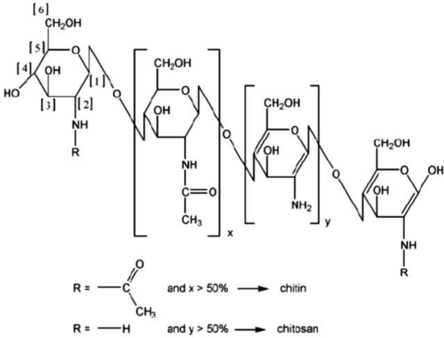 Figure 1: Chemical structure of Chitin and Chitosan. View Figure
Chitosan (MW ~30-190 kDa) is the polymer obtained when deacetylation surpasses 50% and is comprised of β (1-4)-links of D-glucosamine and N-acetyl-D- glucosamine monomers that are distributed randomly throughout the chain. This is the only natural polymer exhibiting a cationic character [4]. Due to the fact that the amine groups are present throughout the structure of chitosan, the more deacetylated the polymer is, the more susceptible it is to protonation because nitrogen has an unused pair of electrons that can easily interact with electrophilic groups. When chitosan undergoes protonation, which occurs at low pH levels, it acquires a positive charge, thus providing the possibility to interact with negatively charged groups. This ability has been widely explored in drug delivery, with the preparation of nanoparticles by electrostatic interaction, as reported using carrageenan [5], tripolyphosphate (TPP) or dextran sulfate [6] as counter ions. Chitosan is not soluble in water. Instead, and because the amine groups have a logarithmic acidity constant (pKa) of ~6.5, chitosan dissolves easily in acidic media. The most usual solvent for its dissolution is 1% (v/v) acetic acid. Due to the fact that the polymer is easily protonated, it can also be dissolved in formic and lactic acids and in hydrochloric acid solutions [7]. In a pH of 7 or higher, as the pH is higher than the pKa of amine groups, chitosan becomes insoluble, rendering its biologically applications are scarce or non-existent. As mentioned earlier, chitosan presents adequate characteristics regarding biological applications [8], including biocompatibility, biodegradability and low toxicity. Its biodegradability is due to a metabolism by lysozyme [9]. Moreover, due to its protonation in acidic media, it presents a favourable interaction with mucus [10], which is particularly relevant in the intestine, where there is a high amount of mucus. This is actually one of the reasons justifying its application in specific disease conditions, such as colonic inflammation. Nevertheless, the description of chitosan applications in literature is endless, including pulmonary and oral insulin delivery as well as the delivery of antibiotics and small genetic fragments [11-13], just to mention some examples. Finally, chitosan can also be found in a variety of dietary supplements (Lipoforte® and EasySlim® Blocker, just to name a few) for weight loss. However, the European Food Safety Authority (EFSA) considers that there is not enough evidence that supports this indication. There are other studies that report its beneficial use on lowering LDL-cholesterol [14,15], a claim that EFSA found to have enough evidence that supports it. However, some problems have also been described for supplements, reporting chitosan interference with certain treatments, for example, anticoagulation therapy with warfarin [16], and anti-epilepsy therapy with valproate [17]. Dextran sulfate was the other polymer used in this experimental work. It is also a natural polymer, obtained from bacteria of the Leuconostoc genus. It is comprised of a branched an hydroglucose backbone with attached sulphur groups, the latter believed to be 17% of the total mass of the polymer. This means that, 2.3 sulphur groups are present for each glucosyl residue, thus the polymer being expected to be negatively charged. Its chemical structure can be seen in Figure 2. Dextran sulfate (MW ~5-500 kDa [18-20] is also reported as biocompatible and biodegradable [21], thus theoretically complying with mandatory requisites for biomedical applications. Studies regarding the use of dextran sulfate as a component of DDS are scarce, even though it was widely studied in the eighties for its anti-HIV activity, against HIV-1. Its mechanism of action, in this context, involves inhibiting the reverse transcriptase, a viral enzyme responsible for turning the viral RNA in viral DNA that will, ultimately, be part of the host cell DNA resulting in the production of viral particles [22]. Additionally, dextran sulfate has also been shown to activate macrophages by establishing an interaction with the scavenger receptor present in these cells [23]. Moreover, regarding the immune system, it is believed that dextran sulfate induces activation of B lymphocytes even though that mechanism is mediated by macrophages [24]. Another aspect that is widely mentioned in studies is an anticoagulant capacity similar to heparin, even though it wasn’t studied further [25]. Finally, dextran sulfate is used, nowadays, to induce colitis in mouse models [26,27]. Approved formulations containing this polymer include two EMA-approved orphan medicines: i) a medicine for the prevention of graft rejection and its further damage on pancreatic transplantation by inhibiting the activation of complement system and clotting [28] and ii) a medicine to promote mobilisation of progenitor cells, prior to stem cell transplantation, through a cytokine released by bone marrow that can guide these important cells through blood stream [29]. Dextran sulfate is a very interesting polymer with many characteristics that can be used in drug delivery. However, there is still much work to do and, more importantly, study possible associations with other polymers to produce nanocarriers with a potential use in therapeutics [30-34]. Chitosan extracted from Metacarcinus magister shells using demineralization with 1.5M and 2M HCl showed the characteristic functional groups of CS. The CS-CMC nanoparticle were chelated with five different concentrations of TPP and BaCl2 (0.1%, 0.2%, 0.3%, 0.4% and 0.5%) and embodied with CUR. The SEM analysis indicated the size of the nanoparticles to below 500 nm which was also confirmed by AFM. The encapsulation efficiency was absolutely time dependent. The drug release was found to be best with higher concentration of chelating agents used and the drug release was prolonging to 80th minute for all the samples. In vitro drug release showed antibacterial activity when ethanol and acetic acid was used as solvent system [35]. A new granule hydrogel composite of natural biocompatible polymeric chitosan and gelatin (C-G) have prepared. The gelatin side groups (hydroxyl and carboxyl) can attach with amine groups of chitosan, which improve the interlink cross linking in the final nanocomposites. The diameter of the granules is around 3mm to 6 mm. Furthermore, in order to reinforce the mechanical properties of granules, for the first time, octa (ammonium chloride) substituted Polyhedral Oligomeric Silsesquioxane C-G/ POSS-(NH3+ Cl-)8 has been used as a nanofiller in the structure of this nanocomposite. The effect of C-G/POSS-(NH3+ Cl-)8 on mechanical stability and swelling behavior of hydrogels has been investigated in three pH of 1.2, 7.4 and 9.5, respectively. Results demonstrate that the C-G/POSS- (NH3+ Cl-)8 granules in acidic pH has the maximum swelling percent. The encapsulation efficiency (EE) values for C-G granules in three pH of 1.2, 7.4 and 9.5 are obtained 65%, 58% and 53% and for C-G/POSS-(NH3+ Cl-)8 sample are 59%, 52% and 48%, respectively, which conform with the swelling behavior of these hydrogels. According to the obtained results from Metronidazole (MTZ) release, the assynthesized hydrogels may have intrinsic ability in the controlled release of drugs. The as-prepared hydrogels are characterized via FT-IR, SEM, TGA and XRD analysis [36]. Chitosan nanoparticles (CS NPs) exhibit good physicochemical properties as drug delivery systems. Haliza Katas and et al. [37], have determined the modulation of preparative parameters on the physical characteristics and colloidal stability of CS NPs. CS NPs were fabricated by ionic interaction with dextran sulphate (DS) prior to determination of their storage stability. The smallest CS NPs of 353 ± 23nm with a surface charge of +56.2 ± 1.5mV were produced when CS and DS were mixed at pH 4 and with a DS: CS mass ratio of 0.5 : 1. An entrapment efficiency of 98% was achieved when BSA/siRNA was loaded into the nanoparticles. The results also showed that particle size and surface charge of CS NPs were slightly changed up to 2 weeks when stored at 4°C. Greater particle size and surface charge were obtained with increasing the concentration of DS. In conclusion, NPs were sufficiently stable when kept at 4°C and able to carry and protect protein.-
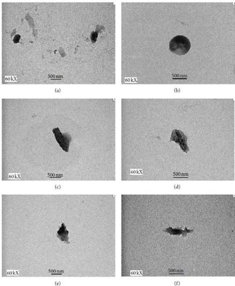 Figure 2: TEM images of CS NPs. (a) and (b) Unloaded CS NPs at 0.5: 1
and 1: 1, (c) and (d) BSA loaded CS NPs at 0.5: 1 and 1: 1, and (e) and
(f) siRNA loaded CS NPs at 0.5 : 1 and 1 : 1, respectively. All the images
were taken at 60 kX magnification. View Figure
Figure 2: TEM images of CS NPs. (a) and (b) Unloaded CS NPs at 0.5: 1
and 1: 1, (c) and (d) BSA loaded CS NPs at 0.5: 1 and 1: 1, and (e) and
(f) siRNA loaded CS NPs at 0.5 : 1 and 1 : 1, respectively. All the images
were taken at 60 kX magnification. View Figure
Ji Woon Suh and et al. [38], have formulated buccal mucoadhesive nanoparticles (NPs) using the natural mucoadhesive polymers. The natural mucoadhesive polymers chitosan (CS) and dextran sulfate sodium salt (DS) were used to prepare mucoadhesive NPs using the ionic gelation method. As the molecular weight of DS decreased, the amount of mucin and the number of buccal cells adsorbed on DS increased. The CS/DS NPs ranged from 100 to 200nm in diameter. The adhesive interactions of CS/DS NPs with mucin were not significantly different from those of CS/sodium triphosphate pentabasic (TPP) NPs; however, CS/DS NPs exhibited 5 times greater mucoadhesive activity to buccal cells compared to control CS/ TPP NPs in ex vivo adhesion tests. These results indicate that the buccal mucoadhesive properties of NPs can be improved using natural mucoadhesive polymers. A novel nanoparticle delivery system has been developed by employing the oppositely charged polymers chitosan (CS) and dextran sulfate (DS), and a simple coacervation process. Under the conditions investigated, the weight ratio of the two polymers is identified as a determining factor controlling particle size, surface charge, entrapment efficiency and release characteristics of the nanoparticles produced. Particles of 223 nm mean diameter were produced under optimal conditions with a zeta potential of approximately -32.6 mV. A maximum of 75% anti-angiogenesis peptide entrapment efficiency was achieved with a CS: DS weight ratio of 0.59:1. The same nanoparticle formulation also showed slow and sustained peptide release over a period of 6 days. In contrast, the formulation containing a lower ratio of CS: DS (0.5:1), was found to have reduced entrapment efficiency and more rapid peptide release characteristics. The results suggest that physicochemical and release characteristics of the CS-DS nanoparticles can be modulated by changing ratios of two ionic polymers. The novel CS–DS nanoparticles prepared by the coacervation process have potential as a carrier for small peptides [39]. Fwu-Long Mi [40], have prepared chitosan-based nanoparticles containing indocyanine green (ICG) for hyperthermia therapy and imaging. ICG was incorporated into chitosan and poly-gamma-glutamic acid self assembled nanoparticles using the polyelectrolyte complex method. The ICG encapsulation efficiency of the nanoparticles was higher than 95% and the ICG-loaded nanoparticles showed effective temperature elevation upon NIR laser irradiation (808 nm, 1.5 W/cm2). The cytotoxicity results from NIR laserinduced hyperthermia effect showed an enhanced toxicity in overcoming doxorubicin (Dox) resistance of MCF-7/ADR cells. Pei-Ru Wei et al. [41], have designed a study for photodynamic therapy (PDT) using chitosan coated Mg-Al layered double hydroxide (LDH) nanoparticles as the delivery system. A Food and Drug Administration (FDA) approved near-infrared (NIR) fluorescent dye, indocyanine green (ICG) with photoactive properties was intercalated into amine modified LDH interlayers by ion-exchange. The efficient positively charged polymer (chitosan (CS)) coating was achieved by the cross linkage using surface amine groups modified on the LDH nanoparticle surface with glutaraldehyde as a spacer. The unique hybridization of organic-inorganic nanocomposites rendered more effective and successful photodynamic therapy due to the photosensitizer stabilization in the interlayer of LDH, which prevents the leaching and metabolization of the photosensitizer in the physiological conditions. The results indicated that the polymer coating and the number of polymer coats have a significant impact on the photo-toxicity of the nano-composites. The double layer chitosan coated LDH–NH2–ICG nanoparticles exhibited enhanced photo therapeutic effect compared with uncoated LDH–NH2– ICG and single layer chitosan-coated LDH–NH2–ICG due to the enhanced protection to photosensitizers against photo and thermal degradations. This new class of organic-inorganic hybrid nanocomposites can potentially serve as a platform for future non-invasive cancer diagnosis and therapy. A new system have examined by chitosan (CS)-dextran sulfate (DS) nanoparticles coated iron oxide as drug carriers detectable using magnetic resonance imaging (MRI) technique. The 5- aminosalicylic acid (5-ASA) was chosen as model drug molecule. CS-DS hydrogels were formulated by a complex coacervation process under mild conditions. The influence of process variables, including the two ionic polymers, on particle size, and hydrogel entrapment of 5-ASA was studied. The in vitro release of 5-ASA were also evaluated, and the integrity of 5-ASA in the release fraction was assessed using sodium dodecyl sulfate-polyacrylamide gel electrophoresis. The release of 5-ASA from hydrogel was based on the ion-exchange mechanism. The CS–DS hydrogel developed based on the modulation of ratio show promise as a system for controlled delivery of drug detectable using magnetic resonance imaging (MRI) technique. Results of release studies indicate that super paramagnetic chitosan– dextran sulfate hydrogel offer a high degree of protection from premature drug release in simulated upper conditions. These hydrogels deliver most of the drug load in the colon, an environment rich in bacterial enzymes that degrade the chitosan–dextran sulfate and allow drug release to occur at the desired site. Thus, spherical super paramagnetic hydrogel is a potential system for colon delivery of 5- ASA. Also, this hydrogel can be detected by magnetic resonance imagining (MRI) technique [42] (Figure 3).-
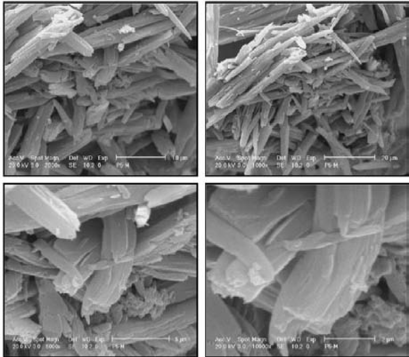 Figure 3: Morphology of Chitosan-dextran sulfate(CS-DS) hydrogel by
Emission Scanning Microscopy field. View Figure
Figure 3: Morphology of Chitosan-dextran sulfate(CS-DS) hydrogel by
Emission Scanning Microscopy field. View Figure
Antony V. Samrot et al. [43], have used chitosan derived from crab shell using two concentrations of hydrochloric acid i.e.1.5M and 2M HCl during demineralisation step. The obtained chitosan was utilized for synthesis of polymeric nanoparticles using sodium tripolyphosphate and barium chloride as chelators. The nanoparticles were encapsulated with hydrophobic curcumin and analysed for drug delivery in vitro. Also, the synthesized nanoparticles were characterized by FTIR, SEM and AFM analysis and subjected for drug encapsulation efficiency, in vitro drug release kinetics and controlled drug delivery studies in vitro against Pseudomonas aeruginosa. Barium chloride was found to produce spherical shaped drug loaded (Figure 4), nanoparticles of size below 500nm.-
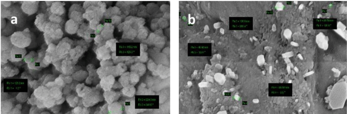 Figure 4: Scanning Electron Microscopy analysis of 1.5M demineralized
CS-CMC Nanoparticles and chelated with different concentrations of
BaCl2. View Figure
Figure 4: Scanning Electron Microscopy analysis of 1.5M demineralized
CS-CMC Nanoparticles and chelated with different concentrations of
BaCl2. View Figure
Multifunctional biodegradable and biocompatible chitosan nanoparticles loaded with drug as an optical-imaging contrast agent for cancer imaging and as a photothermal therapy agent for cancer treatment. To improve the efficacy of PTT, chitosan nanoparticles (NPs) encapsulating super paramagnetic iron oxide (Fe3O4), drug, and perfluoropentane (PFP) as synergistic agents for NIR laser-induced PTT. -
-
- Cui Z, Nair LS. Chitosan: A Versatile Biomedical Polymer. Recent Pat Biomed Eng. 2010; 3: 129-137.
- Yang J, Han S, Zheng H, Dong H, Liu J. Preparation and application of micro/nanoparticles based on natural polysaccharides. Carbohydr Polym. 2015; 123: 53-66.
- Pillai CKS, Paul W, Sharma CP. Chitin and chitosan polymers: Chemistry, solubility and fiber formation. Prog Polym Sci. 2009; 34: 641-678.
- Tiyaboonchai W, Limpeanchob N. Formulation and characterization of amphotericin B-chitosan-dextran sulfate nanoparticles. Int J Pharm. 2007; 329: 142-149.
- Sonaje K, Chen YJ, Chen HL, Wey SP, Juang JH, Nguyen HN, et al. Enteric- coated capsules filled with freeze-dried chitosan/poly(γ-glutamic acid) nanoparticles for oral insulin delivery. Biomaterials. 2010; 31: 3384-3394.
- Luo Y, Teng Z, Li Y, Wang Q. Solid lipid nanoparticles for oral drug delivery: Chitosan coating improves stability, controlled delivery, mucoadhesion and cellular uptake. Carbohydr Polym. 2015; 122: 221-229.
- Duttagupta DS, Jadhav VM, Kadam VJ. Chitosan: A propitious biopolymer for drug delivery. Curr Drug Deliv. 2015; 12: 369-381.
- Rodrigues S, Costa AMR Da, Grenha A. Chitosan/carrageenan nanoparticles: Effect of cross-linking with tripolyphosphate and charge ratios. Carbohydr Polym. 2012; 89: 282-289.
- Chen Y, Mohanraj VJ, Parkin JE. Chitosan-dextran sulfate nanoparticles for delivery of an anti-angiogenesis peptide. Lett Pept Sci. 2003; 10: 621-629.
- Rinaudo M, Pavlov G, Desbrières J. Influence of acetic acid concentration on the solubilization of chitosan. Polymer (Guildf). 1999; 40: 7029-7032.
- Saboktakin MR, Tabatabaie RM, Maharramov A, Ramazanov MA. Synthesis and characterization of pH-dependent glycol chitosan and dextran sulfate nanoparticles for effective brain cancer treatment. Int J Biol Macromol. 2011; 49: 747-751.
- Vårum KM, Myhr MM, Hjerde RJN, Smidsrød O. In vitro degradation rates of partially N-acetylated chitosans in human serum. Carbohydr Res. 1997; 299: 99-101.
- Dhawan S, Singla AK, Sinha VR. Evaluation of mucoadhesive properties of chitosan microspheres prepared by different methods. AAPS PharmSciTech. 2004; 5: e67.
- Janes KA, Fresneau MP, Marazuela A, Fabra A, Alonso MJ. Chitosan nanoparticles as delivery systems for doxorubicin. J Control Release. 2001; 73: 255-267.
- Xiao B, Laroui H, Viennois E, Ayyadurai S, Charania MA, Zhang Y, et al. Nanoparticles with surface antibody against CD98 and carrying CD98 small interfering RNA reduce colitis in mice. Gastroenterology. 2014; 146: 1289-1300.
- Tekie FSM, Atyabi F, Soleimani M, Arefian E, Atashi A, Kiani M, et al. Chitosan polyplex nanoparticle vector for miR-145 expression in MCF-7: Optimization by design of experiment. Int J Biol Macromol. 2015; 81: 828-837.
- European Food Safety Authority. Scientific opinion on the substantiation of health claims related to chitosan. EFSA J. 2011; 9: 1-21.
- Shields KM, Smock N, McQueen CE, Bryant PJ. Chitosan for weight loss and cholesterol management. Am J Health Syst Pharm. 2003; 60: 1310-1313.
- Patti AM, Katsiki N, Nikolic D, Al-Rasadi K, Rizzo M. Nutraceuticals in lipid-lowering treatment: A narrative review on the role of chitosan. Angiology. 2015; 66: 416-421.
- Huang SS, Sung SH, Chiang CE. Chitosan potentiation of warfarin effect. Ann Pharmacother. 2007; 41: 1912-1914.
- Striano P, Zara F, Minetti C, Striano S. Chitosan may decrease serum valproate and increase the risk of seizure reappearance. BMJ. 2009; 339.
- Cheow WS, Kiew TY, Hadinoto K. Amorphous nanodrugs prepared by complexation with polysaccharides: Carrageenan versus dextran sulfate. Carbohydr Polym. 2015; 117: 549-558.
- Hiebert LM, Wice SM, Jaques LB, Williams KE, John M. Orally administered dextran sulfate is absorbed in HIV-positive individuals. J Lab Clin Med. 1999; 161-170.
- McCarthy RE, Arnold LW, Babcock GF. Dextran sulphate: an adjuvant for cell-mediated immune responses. Immunology. 1977; 32: 963-974.
- Tiyaboonchai W, Woiszwillo J, Sims RC, Middaugh CR. Insulin containing polyethylenimine-dextran sulfate nanoparticles. Int J Pharm. 2003; 255: 139-151.
- Smith KM, Chang RS, Tabba HD, Yongsheng He. Dextran sulfate as an inhibitor against the human immunodeficiency virus (42811). Proc Soc Exp Biol Med. 1988; 189: 304-309.
- Baba M, Pauwels R, Balzarini J, Arnout J, Desmyter J, De Clercq E. Mechanism of inhibitory effect of dextran sulfate and heparin on replication of human immunodeficiency virus in vitro. Proc Natl Acad Sci USA. 1988; 85: 6132-6136.
- Chao Y, Makale M, Karmali PP, Sharikov Y, Tsigelny I, Merkulov S, et al. Recognition of dextran-superparamagnetic iron oxide nanoparticle conjugates (feridex) via macrophage scavenger receptor charged domains. Bioconjug Chem. 2012; 23: 1003-1009.
- Persson UC, Hammarström LTG, Smith CIE. Macrophages are required for the dextran sulfate induced activation of B lymphocytes. J Immunol. 1977; 119: 1138-1144.
- Suzuki K, Nishioka J, Hashimoto S. Inhibition of factor VIII-associated platelet aggregation by heparin and dextran sulfate, and its mechanism. Biochim Biophys Acta. 1979; 585: 416-426.
- Meshkibaf S, Martins AJ, Henry GT, Kim SO. Protective role of G-CSF in dextran sulfate sodium-induced acute colitis through generating gut-homing macrophages. Cytokine. 2016; 78: 69-78.
- Munyaka PM, Rabbi MF, Khafipour E, Ghia J-E. Acute dextran sulfate sodium (DSS)-induced colitis promotes gut microbial dysbiosis in mice. J Basic Microbiol. 2016; 1-13.
- European Medicines Agency. Low molecular weight dextran sulfate for the prevention of graft rejection during pancreatic islet transplantation. 2011.
- European Medicines Agency. Low molecular weight dextran sulfate as treatment for mobilisation of progenitor cells prior to stem cell transplantation. 2011.
- Antony V. Samrot, Ujjala Burman, Sheryl Ann Philip, Shobana Na, Kumar Chandrasekaran. Synthesis of curcumin loaded polymeric nanoparticles from crab shell derived chitosan for drug delivery, Informatics in Medicine Unlocked. 2018; 10: 159-182.
- Hamideh Moshkbar, Nasser Arsalani, Laleh Saleh Ghadimi. Synthesis of Chitosan/Gelatin granule containing amine derivated octa(ammonium chloride) substituted Polyhedral Oligomeric Silsesquioxane and investigating its application as a drug carrier”, Int J Polymeric Mat Polymeric Biomat.
- Haliza Katas,Maria Abdul Ghafoor Raja, Kai Leong Lam. Development of Chitosan Nanoparticles as a Stable Drug Delivery System for Protein/siRNA. Int J Biomaterials.
- Ji Woon Suh, Ji-Soo Lee, Sanghoon Ko, Hyeon Gyu Preparation and Characterization of Mucoadhesive Buccal Nanoparticles Using Chitosan and Dextran Sulfate. J Agric. Food Chem. 2016; 64: 5384-5388.
- Yan Chen, Vellore J. Mohanraj, John E. Parkin. Chitosan-dextran sulfate nanoparticles for delivery of an anti-angiogenesis Peptide. Letters in Peptide Science. 2003; 10: 621-629.
- Fwu-Long Mi, Yu-Ru Su, Shu-Huei Yu, An-Chong Chao. Delivery of Indocyanine Green to Cancer Cells by Chitosan-Based Nanocarriers. J Pharm Sci. 2003; 92: 2090-2097.
- Pei-Ru Wei, Yaswanth Kuthati, Ranjith Kumar Kankala, Chia-Hung Lee. Synthesis and Characterization of Chitosan-Coated Near-Infrared (NIR) Layered Double Hydroxide-Indocyanine Green Nanocomposites for Potential Applications in Photodynamic Therapy. Int J Mol Sci. Int J Mol Sci. 2015; 16: 20943-20968.
- Mohammad Reza Saboktakina, Roya Tabatabaie, Abel Maharramov, Mohammad Ali Ramazanov. Synthesis and characterization of superparamagnetic chitosan-dextran sulfate hydrogels as nano carriers for colon-specific drug delivery. Carbohydrate Polymers. 2010; 81: 372-376.
- Antony V Samrot, Ujjala Burman, Sheryl Ann Philip, Shobana N, Kumar Chandrasekaran. Synthesis of curcumin loaded polymeric nanoparticles from crab shell derived chitosan for drug delivery. Informatics in Medicine Unlocked. 2018; 10: 159-182.
Search Articles
-
Perspective
Current Developments and the Potential for Automated Home-Based Music Listening Systems in Dementia Care
Alexander Street1*, Paul Fernie1, Jorg Fachner1, Leonardo Muller1, Ming Hung Hsu1, Patrizia Di Campli San Vito2, Stephen Brewster2, Sube Banerjee3, Nicolas Farina4, Hari Shaji5, Paulo Itaborai5, Alexis Kirke5 and Eduardo Reck Miranda5 | 2024-04-18- Research Article
Preclinical Characterization of GST-HG141, a Novel Hepatitis B Virus Capsid Assembly Modulator
Dong Zhang1#, Wenqiang Wu1#, John Mao1#, Zhigan Jiang3#, Vadim Bichko1 , Qiaoyun Zhou2 , Jing Wang3 , Jian Li3 , Shuhui Chen3 , Haiying He3*, and George Zhang1* | 2024-12-05- Editorial
The Role of Sleep in Pelvic Pain: Are We Missing Something
Chiara Manna* | 2024-04-22- Review Article
Screening for Depression among Medicyation Overuse Headache Patients and Treatment Could Be Useful for Improving their Quality of Life
Ljubisavljevic Srdjan1,2*#, Todorovic Stefan1# and Djokovic Filip1 | 2024-04-19- Short Communication
Why Multi-Drug Antiviral Therapy is Needed for COVID-19
Sibasish Dolai1 , Sabine Hazan2 , Christelle Pagonis1 , Sabrina Liu3 , Thomas J Borody1* and Robert R Clancy1 | 2024-04-11News Feeds

Seth J. Worley, MD, FHRS, FACC
Director, Interventional Implant Program
Biography Paper Presentations
MedStar Heart & Vascular Institute,
Washington, DC, USACollaborations
Indexing


 © Copyright - JSM Central
 This work is licensed under a Creative Commons Attribution 4.0 International License.
- Research Article